Journal of Shanghai Jiao Tong University (Medical Science) ›› 2022, Vol. 42 ›› Issue (8): 1095-1102.doi: 10.3969/j.issn.1674-8115.2022.08.014
• Clinical research • Previous Articles
LIU Siyu1( ), WU Bing1, LI Xiaomin1, ZHAO Lulu1, CHEN Jun2, AI Songtao1()
Received:2022-06-30
Accepted:2022-08-08
Online:2022-08-28
Published:2022-10-08
Contact:
AI Songtao
E-mail:liuye0326@126.com;ai.songtao@qq.com
Supported by:CLC Number:
LIU Siyu, WU Bing, LI Xiaomin, ZHAO Lulu, CHEN Jun, AI Songtao. Preliminary exploration of diffusion-weighted imaging in pre-surgical planning of dermatofibrosarcoma protuberans[J]. Journal of Shanghai Jiao Tong University (Medical Science), 2022, 42(8): 1095-1102.
Add to citation manager EndNote|Ris|BibTeX
URL: https://xuebao.shsmu.edu.cn/EN/10.3969/j.issn.1674-8115.2022.08.014
| Clinical characteristic | MRI functional imaging group ( n=17) | MRI conventional imaging group ( n=17) | P value |
|---|---|---|---|
| Age/year | 38.76±12.63 | 34.35±13.51 | 0.333 |
| Gender/ n(%) | 1.000 | ||
| Male | 9 (52.9) | 9 (52.9) | |
| Female | 8 (47.1) | 8 (47.1) | |
| Lesion location/ n(%) | 0.562 | ||
| Trunk | 12 (70.6) | 14 (82.4) | |
| Limbs | 3 (17.6) | 1 (5.8) | |
| Head and neck | 2 (11.8) | 2 (11.8) | |
| Diameter/cm | 2.37±0.81 | 2.13±1.02 | 0.452 |
| Number of episodes/ n(%) | 0.080 | ||
| First onset | 7 (41.2) | 13 (76.5) | |
| Recurrence | 10 (58.8) | 4 (23.5) |
Tab 1 Clinical characteristics of the two groups
| Clinical characteristic | MRI functional imaging group ( n=17) | MRI conventional imaging group ( n=17) | P value |
|---|---|---|---|
| Age/year | 38.76±12.63 | 34.35±13.51 | 0.333 |
| Gender/ n(%) | 1.000 | ||
| Male | 9 (52.9) | 9 (52.9) | |
| Female | 8 (47.1) | 8 (47.1) | |
| Lesion location/ n(%) | 0.562 | ||
| Trunk | 12 (70.6) | 14 (82.4) | |
| Limbs | 3 (17.6) | 1 (5.8) | |
| Head and neck | 2 (11.8) | 2 (11.8) | |
| Diameter/cm | 2.37±0.81 | 2.13±1.02 | 0.452 |
| Number of episodes/ n(%) | 0.080 | ||
| First onset | 7 (41.2) | 13 (76.5) | |
| Recurrence | 10 (58.8) | 4 (23.5) |
| Radiologist 1 | MRI functional imaging group | MRI conventional imaging group | ||||
|---|---|---|---|---|---|---|
| Radiologist 2 | κ value | Radiologist 2 | κ value | |||
| Yes/ n | No/ n | Yes/ n | No/ n | |||
| Yes/ n | 5 | 1 | 0.866 | 5 | 2 | 0.514 |
| No/ n | 0 | 11 | 2 | 8 | ||
Tab 2 Consistency evaluation of different doctors ' judgment of fascia involvement in tumor boundary in the two groups of patients
| Radiologist 1 | MRI functional imaging group | MRI conventional imaging group | ||||
|---|---|---|---|---|---|---|
| Radiologist 2 | κ value | Radiologist 2 | κ value | |||
| Yes/ n | No/ n | Yes/ n | No/ n | |||
| Yes/ n | 5 | 1 | 0.866 | 5 | 2 | 0.514 |
| No/ n | 0 | 11 | 2 | 8 | ||
| Radiologist 1 | MRI functional imaging group | MRI conventional imaging group | ||||
|---|---|---|---|---|---|---|
| Radiologist 2 | κ value | Radiologist 2 | κ value | |||
| Yes/ n | No/ n | Yes/ n | No/ n | |||
| Yes/ n | 5 | 1 | 0.742 | 3 | 1 | 0.549 |
| No/ n | 1 | 10 | 2 | 11 | ||
Tab 3 Consistency evaluation of different doctors ' judgment of muscles involvement in tumor boundary in the two groups of patients
| Radiologist 1 | MRI functional imaging group | MRI conventional imaging group | ||||
|---|---|---|---|---|---|---|
| Radiologist 2 | κ value | Radiologist 2 | κ value | |||
| Yes/ n | No/ n | Yes/ n | No/ n | |||
| Yes/ n | 5 | 1 | 0.742 | 3 | 1 | 0.549 |
| No/ n | 1 | 10 | 2 | 11 | ||
| Group | n | Very satisfied/ n(%) | Satisfied/ n(%) | Unsatisfied/ n(%) | Total satisfaction rate/% |
|---|---|---|---|---|---|
| MRI functional imaging group | 17 | 13 (76.5) | 4 (23.5) | 0 (0) | 100.0 |
| MRI conventional imaging group | 17 | 8 (47.1) | 4 (23.5) | 5 (29.4) | 70.6 |
| χ2 value | 14.815 | ||||
| P value | 0.001 |
Tab 4 Comparison of post-operative aesthetic satisfaction evaluation
| Group | n | Very satisfied/ n(%) | Satisfied/ n(%) | Unsatisfied/ n(%) | Total satisfaction rate/% |
|---|---|---|---|---|---|
| MRI functional imaging group | 17 | 13 (76.5) | 4 (23.5) | 0 (0) | 100.0 |
| MRI conventional imaging group | 17 | 8 (47.1) | 4 (23.5) | 5 (29.4) | 70.6 |
| χ2 value | 14.815 | ||||
| P value | 0.001 |
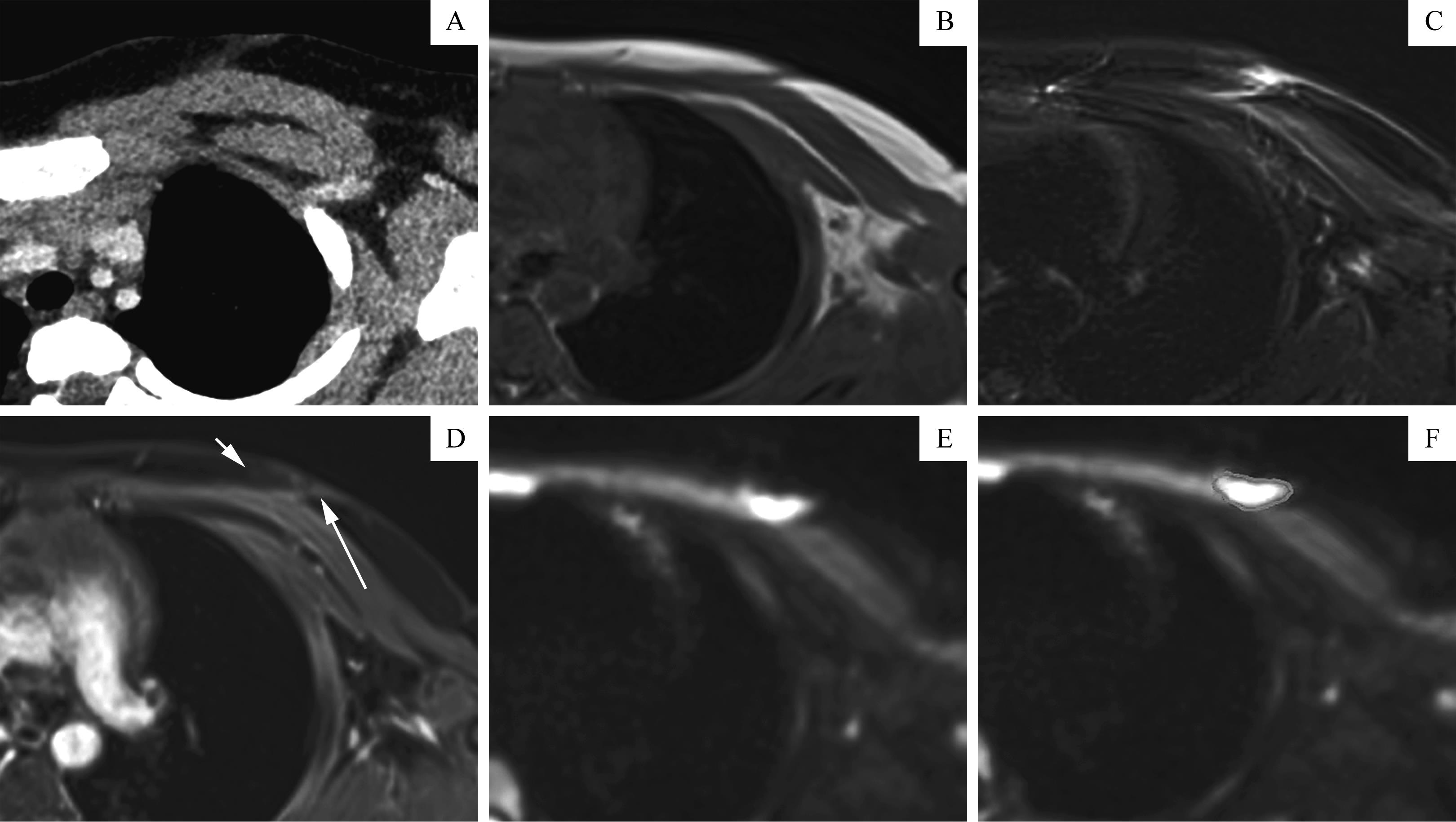
Fig 1 Example of a patient 's tumor images
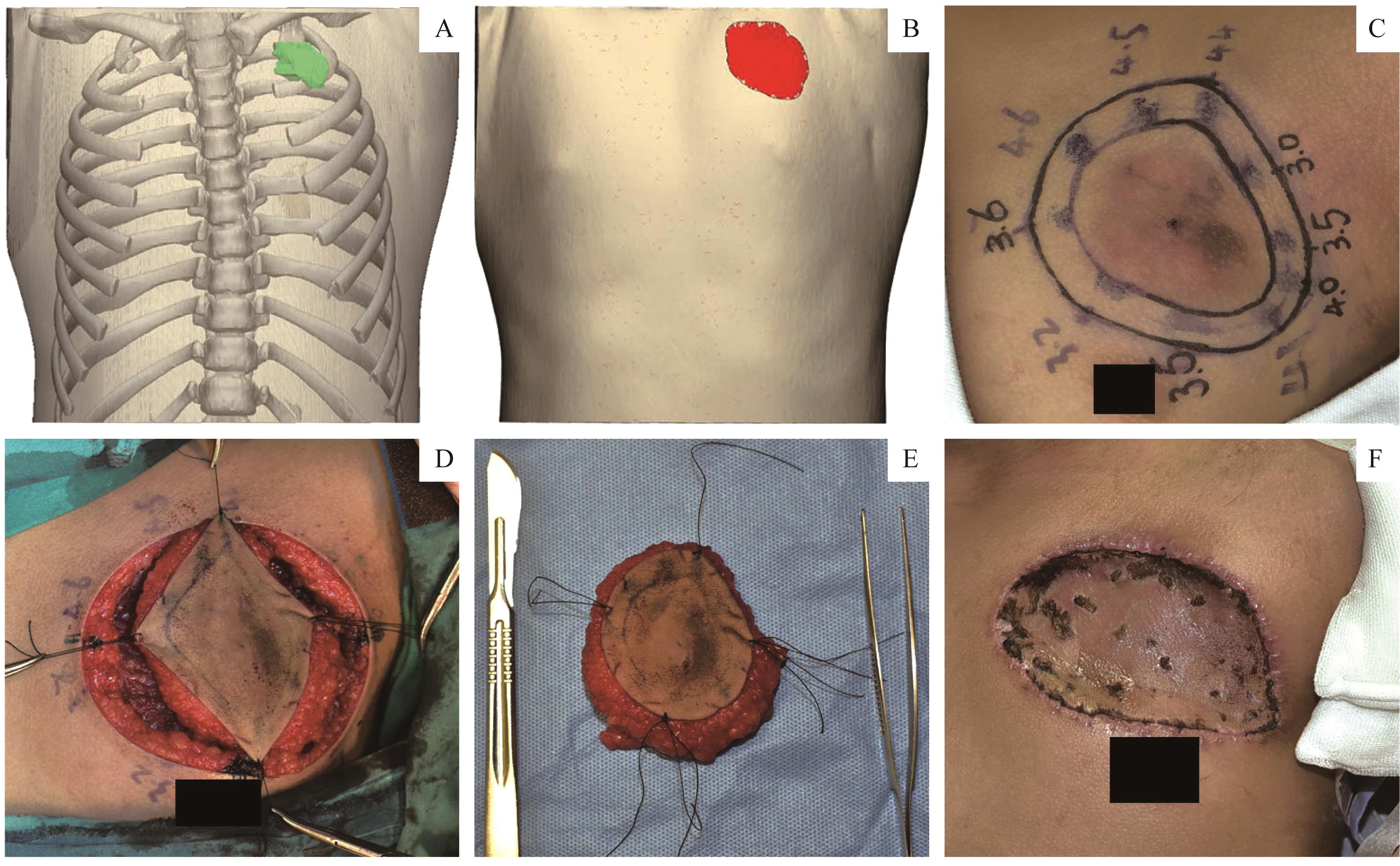
Fig 2 Example of image-assisted surgical procedure planning
| 1 | GLOSTER H M Jr. Dermatofibrosarcoma protuberans[J]. J Am Acad Dermatol, 1996, 35(3 Pt 1): 355-374. |
| 2 | MENDENHALL W M, ZLOTECKI R A, SCARBOROUGH M T. Dermatofibrosarcoma protuberans[J]. Cancer, 2004, 101(11): 2503-2508. |
| 3 | MUJTABA B, WANG F, TAHER A, et al. Dermatofibrosarcoma protuberans: pathological and imaging review[J]. Curr Probl Diagn Radiol, 2021, 50(2): 236-240. |
| 4 | CHANG C K, JACOBS I A, SALTI G I. Outcomes of surgery for dermatofibrosarcoma protuberans[J]. Eur J Surg Oncol, 2004, 30(3): 341-345. |
| 5 | THWAY K, NOUJAIM J, JONES R L, et al. Dermatofibrosarcoma protuberans: pathology, genetics, and potential therapeutic strategies[J]. Ann Diagn Pathol, 2016, 25: 64-71. |
| 6 | 刘珍如, 周园, 刘梦茜, 等. 隆突性皮肤纤维肉瘤的诊疗进展[J]. 中国美容医学, 2021, 30(3): 171-174. |
| LIU Z R, ZHOU Y, LIU M X, et al. Research progress in diagnosis and treatment of dermatofibrosarcoma protuberans[J]. Chin J Aesthet Med, 2021, 30(3): 171-174. | |
| 7 | SEDAGHAT S, SCHMITZ F, SEDAGHAT M, et al. Appearance of recurrent dermatofibrosarcoma protuberans in postoperative MRI follow-up[J]. J Plast Reconstr Aesthet Surg, 2020, 73(11): 1960-1965. |
| 8 | 王传彬, 韦超, 李乃玉, 等. 隆突性皮肤纤维肉瘤的CT和MRI表现[J]. 中国医学装备, 2021, 18(4): 58-61. |
| WANG C B, WEI C, LI N Y, et al. The manifestation of dermatofibrosarcoma protuberans on CT and MRI[J]. China Med Equip, 2021, 18(4): 58-61. | |
| 9 | 陈开良, 吴文婷. 隆突性皮肤纤维肉瘤的高频超声表现分析[J]. 肿瘤影像学, 2020, 29(6): 570-573. |
| CHEN K L, WU W T. Analysis of high-frequency ultrasound manifestations of dermatofibrosarcoma protuberan[J]. Oncoradiology, 2020, 29(6): 570-573. | |
| 10 | CHOONG P, LINDSAY D, KHOO M, et al. Dermatofibrosarcoma protuberans: the diagnosis of high-grade fibrosarcomatous transformation[J]. Skeletal Radiol, 2021, 50(4): 789-799. |
| 11 | HONG J H, JEE W H, JUNG C K, et al. Soft tissue sarcoma: adding diffusion-weighted imaging improves MR imaging evaluation of tumor margin infiltration[J]. Eur Radiol, 2019, 29(5): 2589-2597. |
| 12 | DOUIS H, DAVIES M A, SIAN P. The role of diffusion-weighted MRI (DWI) in the differentiation of benign from malignant skeletal lesions of the pelvis[J]. Eur J Radiol, 2016, 85(12): 2262-2268. |
| 13 | AMJAD G, ZEINALI ZADEH M, AZMOUDEH-ARDALAN F, et al. Evaluation of multimodal MR imaging for differentiating infiltrative versus reactive edema in brain gliomas[J]. Br J Neurosurg, 2020. DOI: 10.1080/02688697.2020.1849541. |
| 14 | KISHIMOTO A O, KATAOKA M, IIMA M, et al. The comparison of high-resolution diffusion weighted imaging (DWI) with high-resolution contrast-enhanced MRI in the evaluation of breast cancers[J]. Magn Reson Imaging, 2020, 71: 161-169. |
| 15 | 曲扬, 艾松涛, 杨飞, 等. CT和MRI图像配准融合联合3D打印技术在难治性骨盆肿瘤术前规划中的应用[J]. 上海交通大学学报(医学版), 2017, 37(9): 1239-1244, 1238. |
| QU Y, AI S T, YANG F, et al. Application of CT/MRI image registration and fusion combined with 3D printing technique in pre-surgical planning of refractory pelvic tumors[J]. J Shanghai Jiao Tong Univ (Med Sci), 2017, 37(9): 1239-1244, 1238. | |
| 16 | 潘文博, 蔡智慧, 石军荣, 等. 3D打印技术在卵巢癌手术中的应用价值[J]. 河北医药, 2022, 44(6): 938-942. |
| PAN W B, CAI Z H, SHI J R, et al. The application of 3D printing technique in ovarian cancer surgery[J]. Hebei Med J, 2022, 44(6): 938-942. | |
| 17 | 郑眉光, 刘正豪, 李文鹏, 等. 3D打印技术在复杂性颅底肿瘤手术中的应用[J]. 中华神经医学杂志, 2021, 20(9): 927-931. |
| ZHENG M G, LIU Z H, LI W P, et al. Three-dimensional printing technology in surgery of complex skull base tumors[J]. Chin J Neuromedicine, 2021, 20 (9): 927-931. | |
| 18 | ACOSTA A E, VÉLEZ C S. Dermatofibrosarcoma protuberans[J]. Curr Treat Options Oncol, 2017, 18(9): 56. |
| 19 | WONG E, AXIBAL E, BROWN M. Mohs micrographic surgery[J]. Facial Plast Surg Clin N Am, 2019, 27(1): 15-34. |
| 20 | 上海交通大学医学院附属第九人民医院皮肤梭形细胞肿瘤多学科精准诊疗团队. 隆突性皮肤纤维肉瘤多学科诊治实施规范: 上海交通大学医学院附属第九人民医院专家共识(2020年版)[J]. 上海交通大学学报(医学版), 2021, 41(12): 1669-1675. |
| Multi-Disciplinary Team for Cutaneous Spindle Cell Neoplasms, Shanghai Ninth People 's Hospital, Shanghai Jiao Tong University School of Medicine. Practice for multidisciplinary diagnosis and treatment of dermatofibrosarcoma protuberans:expert consensus of Shanghai Ninth People's Hospital, Shanghai Jiao Tong University School of Medicine (2020 edition)[J]. J Shanghai Jiao Tong Univ (Med Sci), 2021, 41(12): 1669-1675. | |
| 21 | HAO X P, BILLINGS S D, WU F B, et al. Dermatofibrosarcoma protuberans: update on the diagnosis and treatment[J]. J Clin Med, 2020, 9(6): 1752. |
| 22 | VELASCO ALBENDEA F J, DOÑA GIRÓN J, BLANCO VILLAR M L, et al. Perianal dermatofibrosarcoma protuberans: a case report, review and update[J]. Rev Esp Patol, 2019, 52(1): 62-68. |
| 23 | LIOU V, CHISHOLM S A, LOGUNOVA V, et al. Giant dermatofibrosarcoma protuberans with bilateral orbital involvement[J]. Ophthalmic Plast Reconstr Surg, 2019, 35(2): e36-e39. |
| 24 | LEE S J, MAHONEY M C, SHAUGHNESSY E. Dermatofibrosarcoma protuberans of the breast: imaging features and review of the literature[J]. AJR Am J Roentgenol, 2009, 193(1): W64-W69. |
| 25 | Expert Panel on Musculoskeletal Imaging. ACR appropriateness criteria ® soft-tissue masses[J]. J Am Coll Radiol, 2018, 15(5S): S189-S197. |
| 26 | HOURANI R, TASLAKIAN B, SHABB N S, et al. Fibroblastic and myofibroblastic tumors of the head and neck: comprehensive imaging-based review with pathologic correlation[J]. Eur J Radiol, 2015, 84(2): 250-260. |
| 27 | GARG M K, YADAV M K, GUPTA S, et al. Dermatofibrosarcoma protuberans with contiguous infiltration of the underlying bone[J]. Cancer Imaging, 2009, 9(1): 63-66. |
| 28 | 黄曦毅, 蔡金辉, 刘庆余, 等. 隆突性皮肤纤维肉瘤的CT和MRI影像学分析: 附10例报道[J]. 罕少疾病杂志, 2020, 27(4): 57-59, 70. |
| HUANG X Y, CAI J H, LIU Q Y,et al. The imaging findings of dermatofibrosarcoma protuberans on CT and MRI: 10 cases report[J]. J Rare Uncommon Dis, 2020, 27(4): 57-59, 70. | |
| 29 | YOON M A, CHEE C G, CHUNG H W, et al. Added value of diffusion-weighted imaging to conventional MRI for predicting fascial involvement of soft tissue sarcomas[J]. Eur Radiol, 2019, 29(4): 1863-1873. |
| [1] | CHEN Liqi, XUE Zhuowei, WU Qingkai. Review of MRI-based three-dimensional digital model reconstruction of female pelvic floor organs [J]. Journal of Shanghai Jiao Tong University (Medical Science), 2022, 42(3): 381-386. |
| [2] | Xuehong WANG, Xuzhuo CHEN, Yi MAO, Da SHEN, Shanyong ZHANG. Difference in recurrence rates after temporomandibular joint disc repositioning surgery with miniscrew anchor at different developmental stages in adolescents [J]. JOURNAL OF SHANGHAI JIAOTONG UNIVERSITY (MEDICAL SCIENCE), 2022, 42(2): 173-177. |
| [3] | Yihuan WANG, Ruokun LI, Huanhuan CHONG, Fuhua YAN. Research progress of Gd-EOB-DTPA-enhanced magnetic resonance imaging in the evaluation of biological behavior of hepatocellular carcinoma [J]. JOURNAL OF SHANGHAI JIAOTONG UNIVERSITY (MEDICAL SCIENCE), 2022, 42(1): 130-134. |
| [4] | Cui CHEN, Ye JIN, Lin WANG, Hongli LI, Caifeng WAN, Lixin JIANG. Comparative analysis of 30 cases of metaplastic carcinoma of the breast [J]. JOURNAL OF SHANGHAI JIAOTONG UNIVERSITY (MEDICAL SCIENCE), 2022, 42(1): 70-76. |
| [5] | . Practice for multidisciplinary diagnosis and treatment of dermatofibrosarcoma protuberans: expert consensus of Shanghai Ninth People's Hospital, Shanghai Jiao Tong University School of Medicine (2020 edition) [J]. JOURNAL OF SHANGHAI JIAOTONG UNIVERSITY (MEDICAL SCIENCE), 2021, 41(12): 1669-1675. |
| [6] | JI Ying-ying, XUE Bin, HUANG Yue, ZHANG Jian-wei. Efficacy and safety of oral midazolam in combination with intranasal dexmedetomidine for paediatric magnetic resonance imaging sedation [J]. JOURNAL OF SHANGHAI JIAOTONG UNIVERSITY (MEDICAL SCIENCE), 2020, 40(8): 1098-1102. |
| [7] | YANG Tao, CHEN Jun, FANG Yi-ru. Advances in magnetic resonance imaging study of bipolar Ⅰdisorder [J]. JOURNAL OF SHANGHAI JIAOTONG UNIVERSITY (MEDICAL SCIENCE), 2020, 40(12): 1660-1664. |
| [8] | YUE Xiu-hui, KONG Wei-dan, REN Ji-liang, YUAN Ying#, TAO Xiao-feng#. Value of 3.0-T MR diffusion-weighted imaging combined with dynamic contrast-enhanced imaging in differentiating benign and malignant thyroid nodules [J]. JOURNAL OF SHANGHAI JIAOTONG UNIVERSITY (MEDICAL SCIENCE), 2020, 40(10): 1393-1397. |
| [9] | LI Xiao-min1, QU Yang1, WU Wen2, ZHAO Liang3, ZHANG Shao-ting3, HAO Yong-qiang2, DAI Ke-rong2, AI Song-tao1. Preliminary application of MR imaging-pathology co-localization by 3D printing box in pelvic tumor assessment [J]. JOURNAL OF SHANGHAI JIAOTONG UNIVERSITY (MEDICAL SCIENCE), 2020, 40(10): 1408-1413. |
| [10] | WANG Tao, ZHANG Chen-cheng, LI Dian-you, SUN Bo-min, FU Meng. Imaging law of postoperative electrode locations in deep brain stimulation for Parkinsons disease [J]. , 2020, 40(1): 64-. |
| [11] | RUAN Jing-jing, LU Qing, TANG Hui, ZHU Zhen-ya, FAN Yu, ZHAO Xin-xin, NIU Xiao-yin. Value of multi-parameter magnetic resonance imaging of cartilage in evaluating efficacy of adipose-derived mesenchymal progenitor cells on knee osteoarthritis [J]. , 2019, 39(12): 1409-. |
| [12] | QU Yang, AI Song-tao, YANG Fei, ZHANG Heng-hui, WANG Yi-ping, TAO Xiao-feng, HAO Yong-qiang, DAI Ke-rong. Application of CT/MRI image registration and fusion combined with 3D printing technique in pre-surgical planning of refractory pelvic tumors#br# [J]. , 2017, 37(9): 1239-. |
| Viewed | ||||||
|
Full text |
|
|||||
|
Abstract |
|
|||||