人工耳蜗植入术后CT影像学评估的临床价值及进展
Clinical values and advances in computed tomography evaluation after cochlear implantation
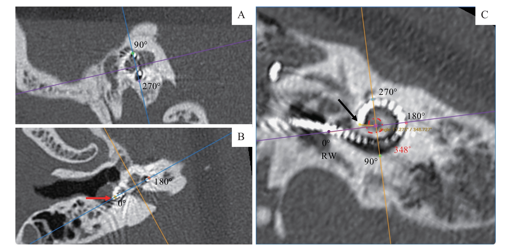
Note: A. Sagittal view; the blue line passing the cochlear turn at 90° and 270°. B. Axial view; the blue line passing the center of round window (0?) and cochlear turn at 180?; an extra-cochlear electrode (red arrow). C. Coronal view showing the full length of the electrode array; insertion depth angle of the electrode array was 348? with the center of the axes at the center of the cochlea modiolus. Black arrow—the most apical electrode; RW—round window.