川崎病合并冠状动脉病变患儿21例冠状动脉造影复查分析
Coronary angiography review in 21 children with Kawasaki disease complicated with coronary artery disease
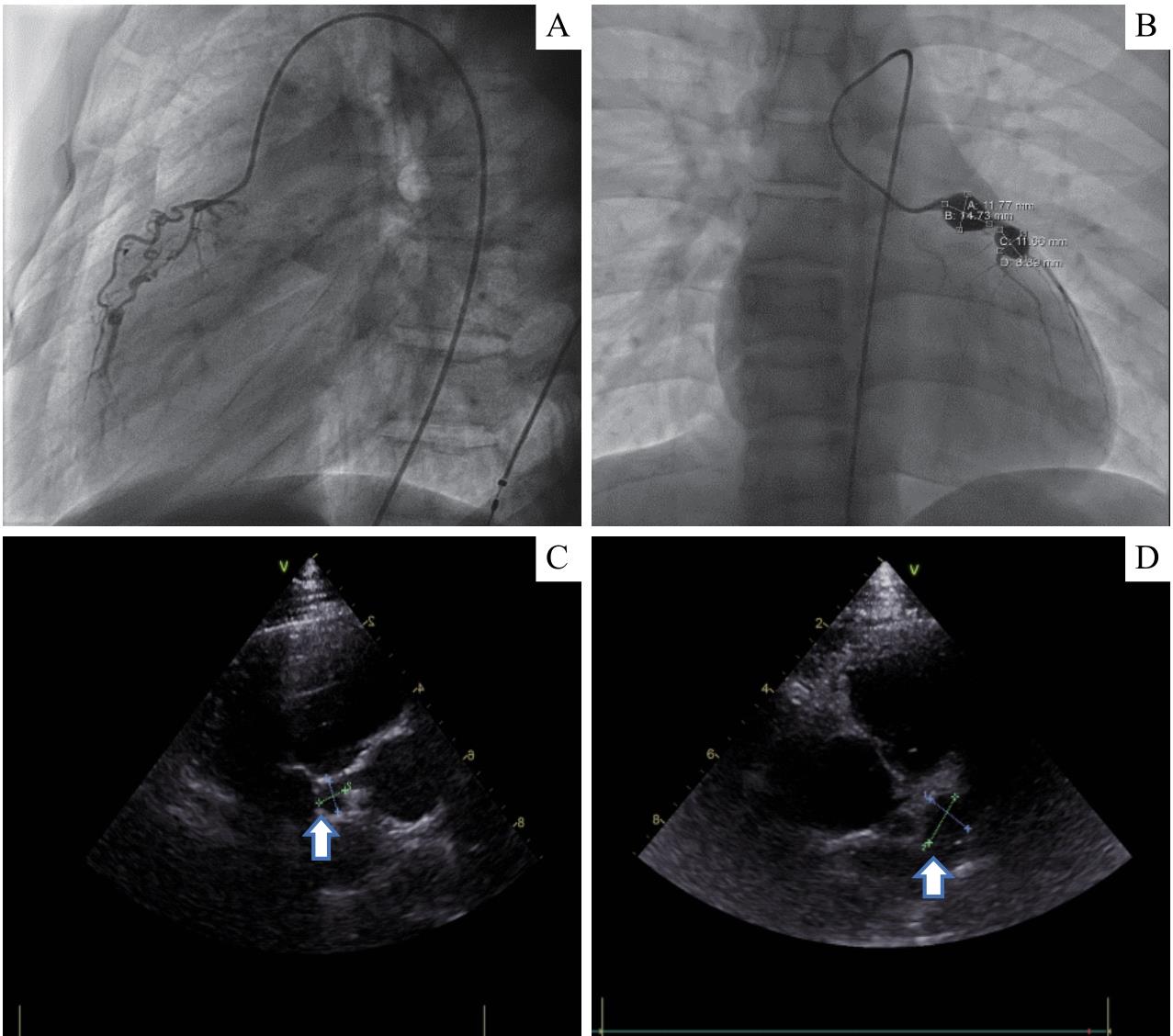
Note: A boy, aged 15 years and 8 months, had been found to have coronary artery aneurysm for 9 years. A. CAG showed stenosis of the proximal and middle segment of the RCA, close to atresia, and the formation of bridging collaterals. B. Two large coronary artery aneurysms with calcification were found by CAG in the proximal segment of the LAD, with the size of 14.73 mm×11.77 mm and 11.66 mm×8.89 mm, respectively. C/D. The corresponding echocardiography of this patient revealed 2 aneurysms in the LAD (arrows), while the RCA was not clearly visualized, and stenosis or atresia was not found.
