改良3D打印病理切片盒在骨肿瘤病理拼接中的应用初探
Preliminary application of improved 3D printed pathological section box to assisting stitching pathological images of bone tumor
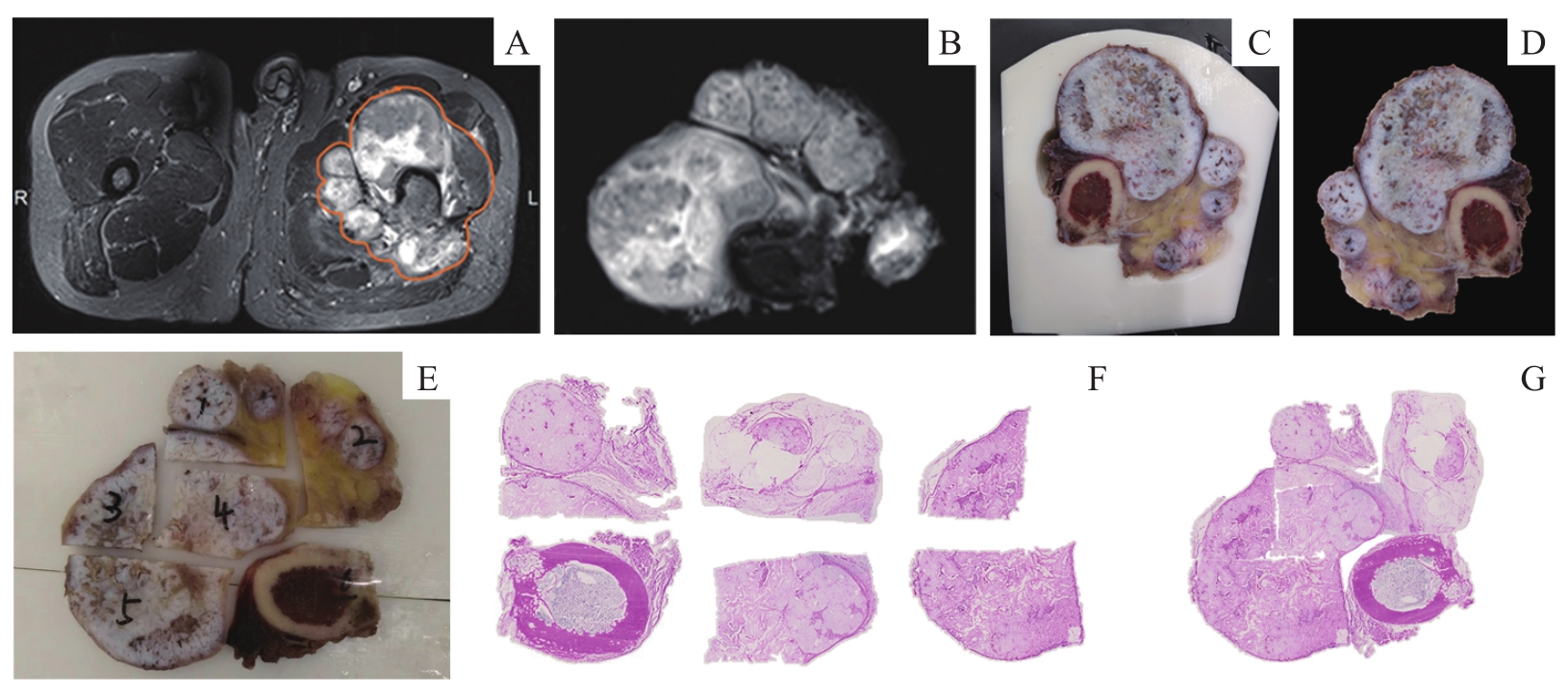
Note:A. Position outline of the bone tumor on preoperative MRI. B. MRI image of the postoperative specimen. C. The contour fitting of the specimen to the slice box. D. Gross view of the specimen in the corresponding section of Figure C. E. Grid-like segmentation of large slices at the same level. F. Scanned image of the segmented pathological fragments. G. AI-assisted stitching pathological images.