改良3D打印病理切片盒在骨肿瘤病理拼接中的应用初探
Preliminary application of improved 3D printed pathological section box to assisting stitching pathological images of bone tumor
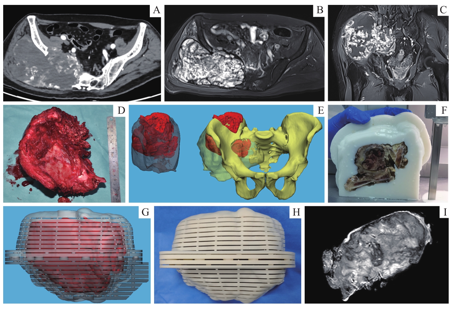
Note:A 62-year-old renal cell carcinoma bone metastasis patient, who underwent hemi-pelvic resection plus artificial hemi-pelvic replacement surgery. A. Preoperative CT scan of bone window image. B. Preoperative MR axial T2WI with fat suppression. C. Preoperative MR contrast-enhanced T1WI with fat suppression. D. Pathological specimen of bone tumor. E. Preoperative and postoperative 3D models of pelvic tumor. F. Section image. G. Virtual individualized 3D printed pathology section box image. H. Realistic individualized 3D printed pathology section box image. I. MR image of postoperative pelvic tumor specimen.