基于CRISPR/Cas9n技术建立携带mT-F2A-EGFP报告系统的小鼠胚胎干细胞系
Establishment of a mouse embryonic stem cell line carrying a reporter of mT-F2A-EGFP based on CRISPR/Cas9n technology
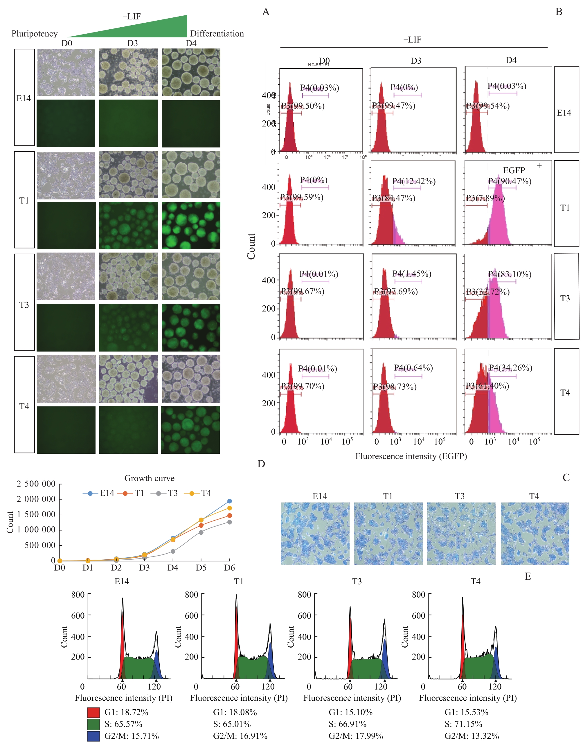
Note:A. Cell morphology and EGFP expression of E14, T1, T3 and T4 clones on Day 0 (D0), Day 3 (D3) and Day 4 (D4) of EB differentiation upon LIF removal (-LIF). All the microscopic pictures were taken under ×10 magnification. B. Flow cytometry analysis of E14, T1, T3 and T4 on D0, D3 and D4 of EB differentiation upon LIF removal (-LIF). Pink indicates EGFP+ cells, and red indicates EGFP- cells. C. AP staining of E14, T1, T3, and T4 cells. All the microscopic pictures were taken under ×10 magnification. D. The growth curve of E14, T1, T3 and T4. E. Cell cycle analysis of E14, T1, T3 and T4 cells.