宫颈机能不全孕妇早、中孕期盆底结构变化初探
Preliminary study of pelvic floor structural changes in early and middle pregnant women with cervical incompetence
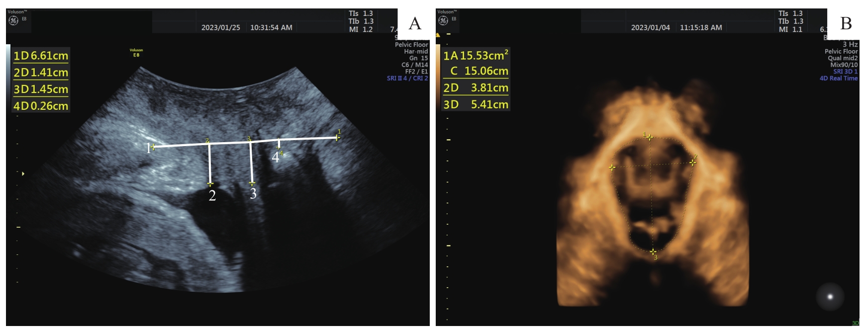
Note:A. Bladder neck position, lowest edge of cervix and rectal ampulla position observed by ultrasound at VM. 1—The reference line is a horizontal line placed at the inferoposterior margin of the symphysis pubis; 2—vertical distance of bladder neck; 3—vertical distance of cervix; 4—vertical distance of rectal ampulla position. B. HA measured by 4-dimensional ultrasound at VM.