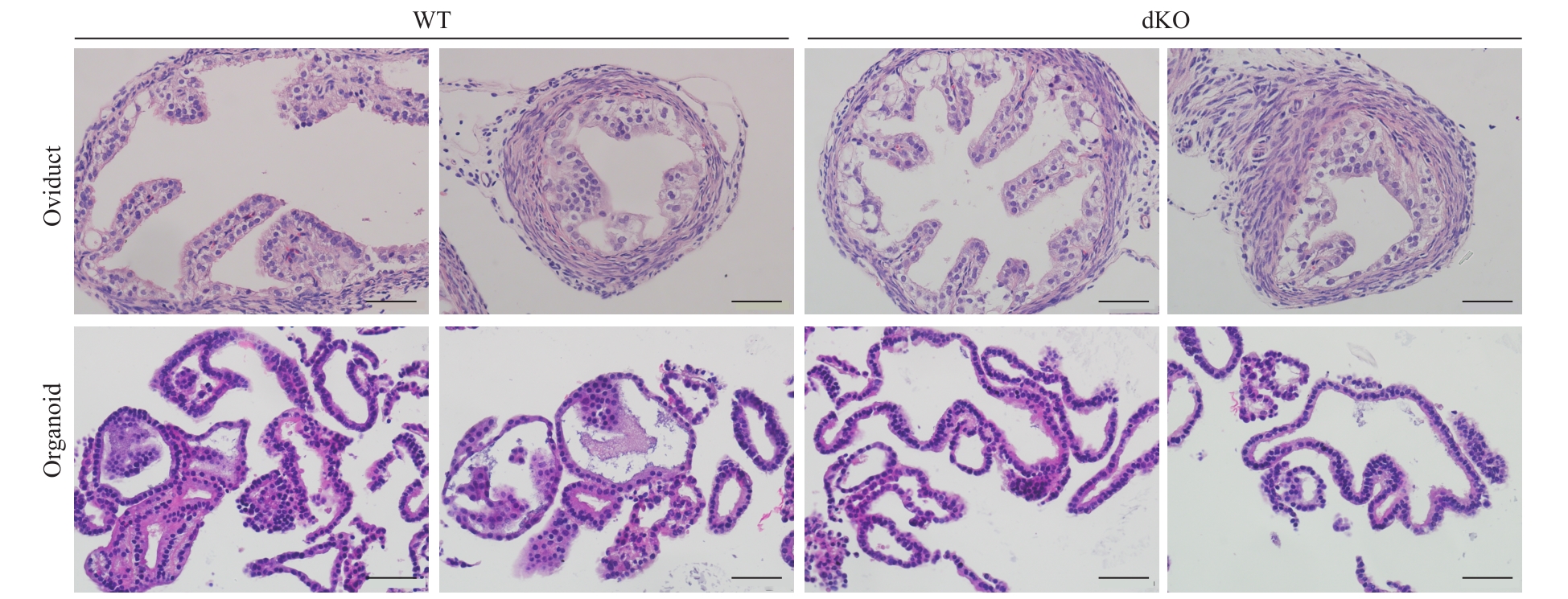
小鼠输卵管上皮类器官的构建及表型验证
Establishment and phenotype verification of mouse oviductal epithelial organoids
Note: Comparison of the histology structure of the oviducts and the oviductal epithelial organoids from WT mice and dKO mice after 2 months of culture (scale bar=50 μm).