小鼠输卵管上皮类器官的构建及表型验证
Establishment and phenotype verification of mouse oviductal epithelial organoids
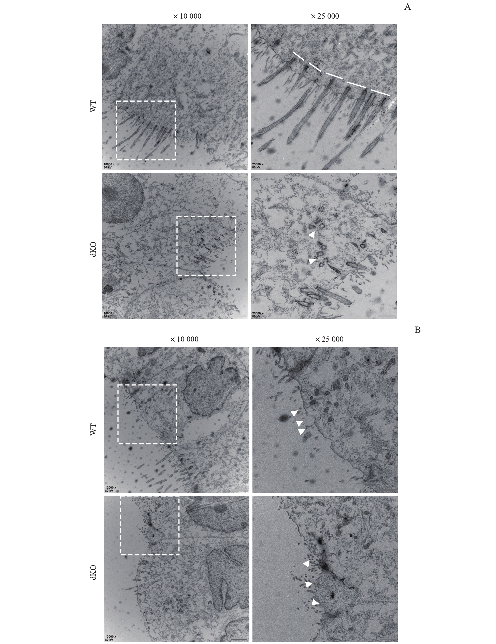
Note: A. Transmission electron microscopic images of the ciliated cells of oviductal epithelial organoids of WT mice and dKO mice after 2 months of culture (×10 000, scale bar=1 μm). The images on the right are the magnification ones in the dotted box on the left (×25 000, scale bar=500 nm). The white line showing the arrangement of the basal bodies, and the white arrows showing multiple disorganized centrioles. B. Transmission electron microscopic images of the secretory cells of oviductal epithelial organoids of WT mice and dKO mice after 2 months of culture (×10 000, scale bar=1 μm). The images on the right are the magnification ones in the dotted box on the left (×25 000, scale bar=500 nm). The white arrows showing the exocytosis granules by the secretory cells.
