小鼠输卵管上皮类器官的构建及表型验证
Establishment and phenotype verification of mouse oviductal epithelial organoids
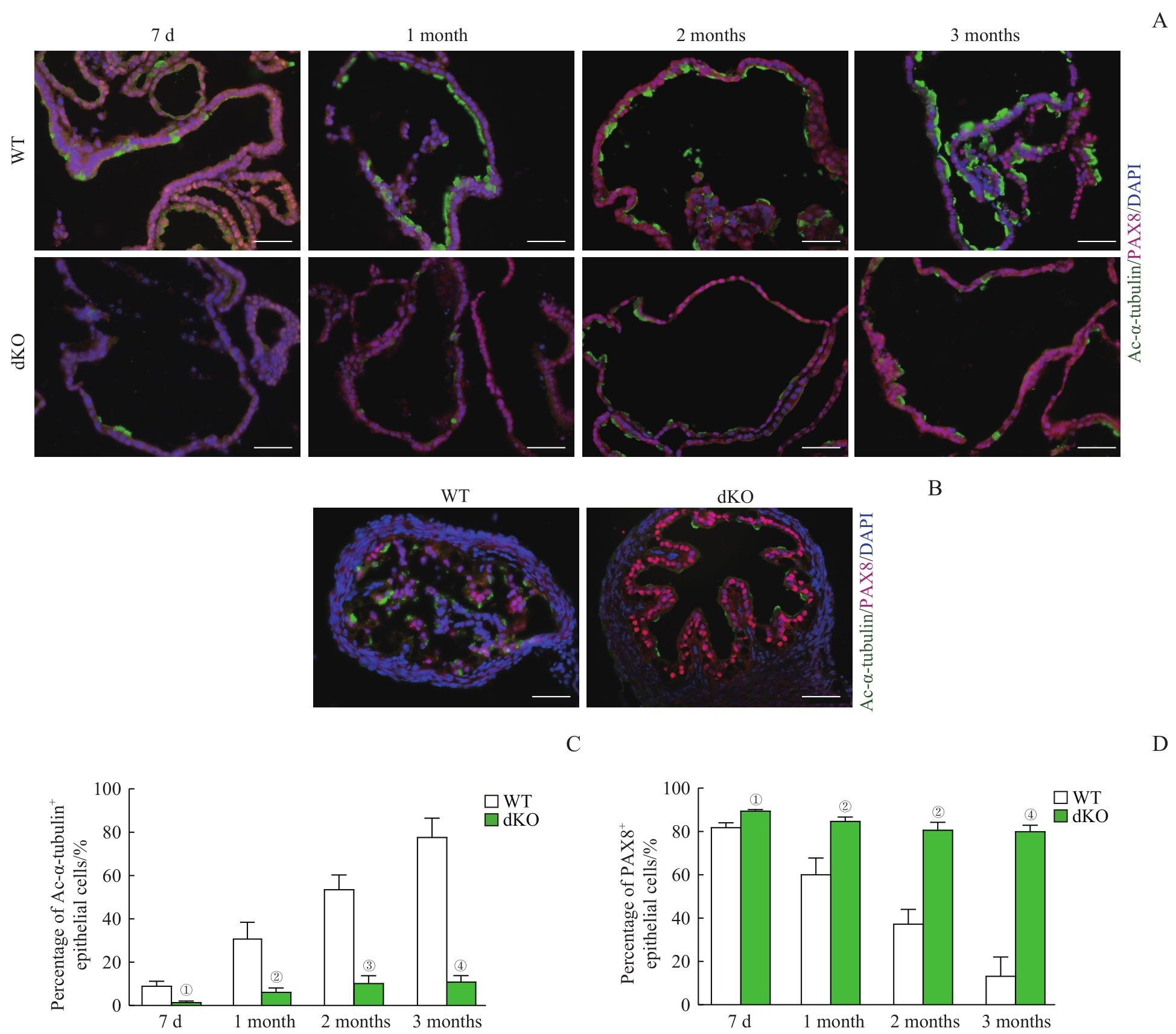
Note: A/B. Co-immunofluorescent staining of Ac-α-tubulin (showing ciliated cells) and PAX8 (showing secretory cells) in the organoid sections (A) and the oviducts (B) of WT mice and dKO mice (×400, scale bar=50 μm). C/D. Quantification of ciliated cells (C) and secretory cells (D) of the organoids from WT mice and dKO mice across different culture time. ①P=0.004, ②P=0.005, ③P=0.001, ④P=0.000, compared with the WT group.