18F-MD-PSMA PET/CT显像在中高危前列腺癌初始分期中的应用价值
Role of 18F-MD-PSMA PET/CT in initial stage of intermediate and high risk prostate cancer
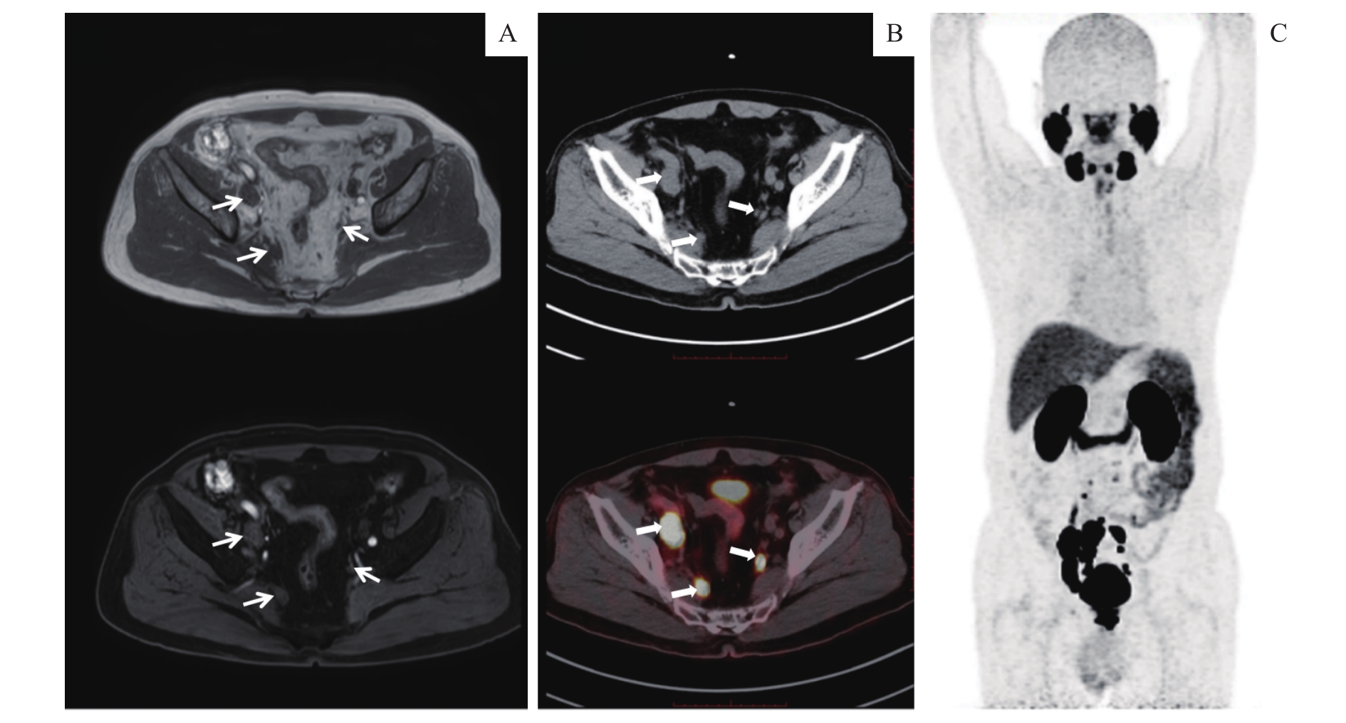
Note: A. Pelvic mp-MRI showed enlargement of the right iliac lymph nodes with abnormal signal which supported the diagnosis of metastasis lymph nodes and a small left iliac lymph node (white thin arrows). B. 18F-MD-PSMA PET/CT demonstrated abnormal 18F-MD-PSMA uptake not only at the right iliac lymph nodes, but also at the left iliac lymph node which was less than 1 cm in diameter (white thick arrows). C. Maximum intensity projection (MIP) image demonstrated more abnormal 18F-MD-PSMA uptake of metastatic lymph nodes in pelvic region.