18F-MD-PSMA PET/CT显像在中高危前列腺癌初始分期中的应用价值
Role of 18F-MD-PSMA PET/CT in initial stage of intermediate and high risk prostate cancer
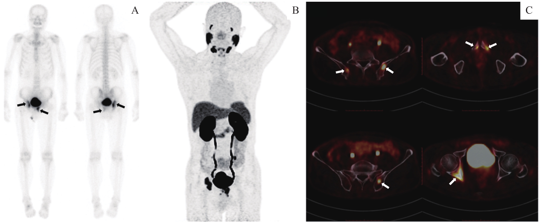
Note: A. BS image demonstrated oligometastasis status with 2 lesions in right hip joint and left pubic bone (black arrows). B. MIP image of 18F-MD-PSMA PET/CT demonstrated 6 lesions with high 18F-MD-PSMA uptake. C. Fusion images of PET and CT showed abnormal high uptake of 18F-MD-PSMA at bilateral ilium, right acetabulum and bilateral pubis (white arrows).