赖氨酸乙酰转移酶7的冷冻电镜全长结构分析
Structural analysis of full-length lysine acetyltransferase 7 by cryo-electron microscopy
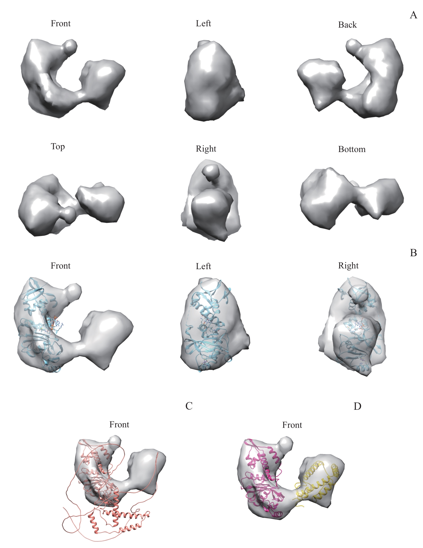
Note: A. Surface view of the electron micrograph density map of KAT7 shown in six orthogonal views. The three-dimensional model size of KAT7 is denoted on the left. B. The crystal structure of MYST motif (5GK9) was docked into corresponding mass of KAT7 shown in three orthogonal views. C. The AlphaFold prediction structure of KAT7 was docked into full-length KAT7 model. D. The N-terminal structural domain (amino acid residues 183?335, yellow) and MYST domain (amino acid residues 336?611, red) of the AlphaFold prediction model were docked to the full-length KAT7 model separately.