A-PRF促进兔膝关节骨软骨损伤愈合的观察
Observation on A-PRF promoting regeneration of osteochondral defects in rabbit knee joints
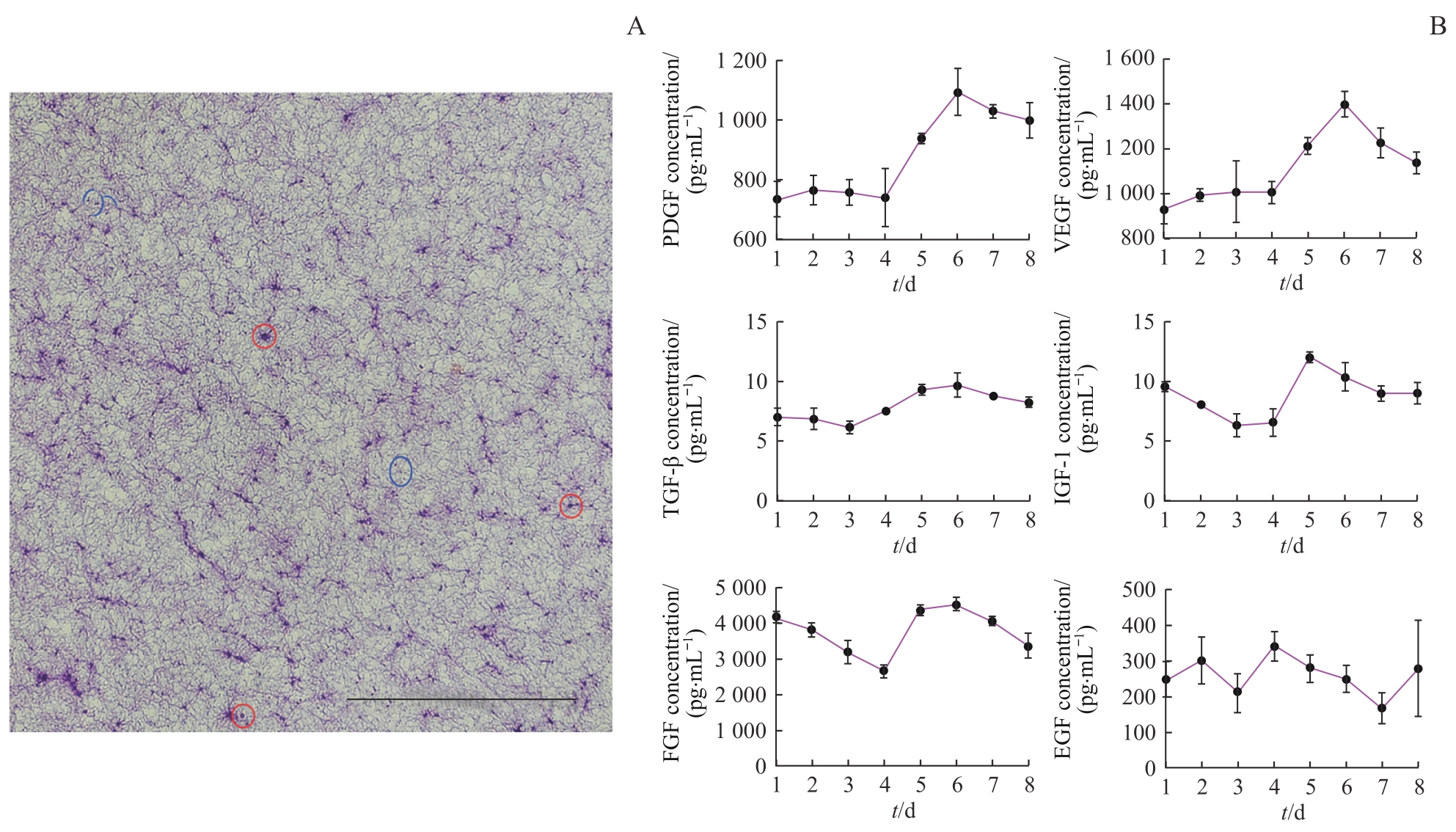
Note: A. H-E staining of A-PRF. Sparse fibrous reticular structures were shown, which trapped white blood cells (red circles) and platelets (blue circles). Scale bar=50 μm. B. Sustained release of growth factors of A-PRF in PBS.