牙周致病菌编码的Tannerpin-M对粒细胞释放的丝氨酸蛋白酶的抑制作用
Inhibition of Tannerpin-M encoded by periodontal pathogens on serine proteases released by granulocytes
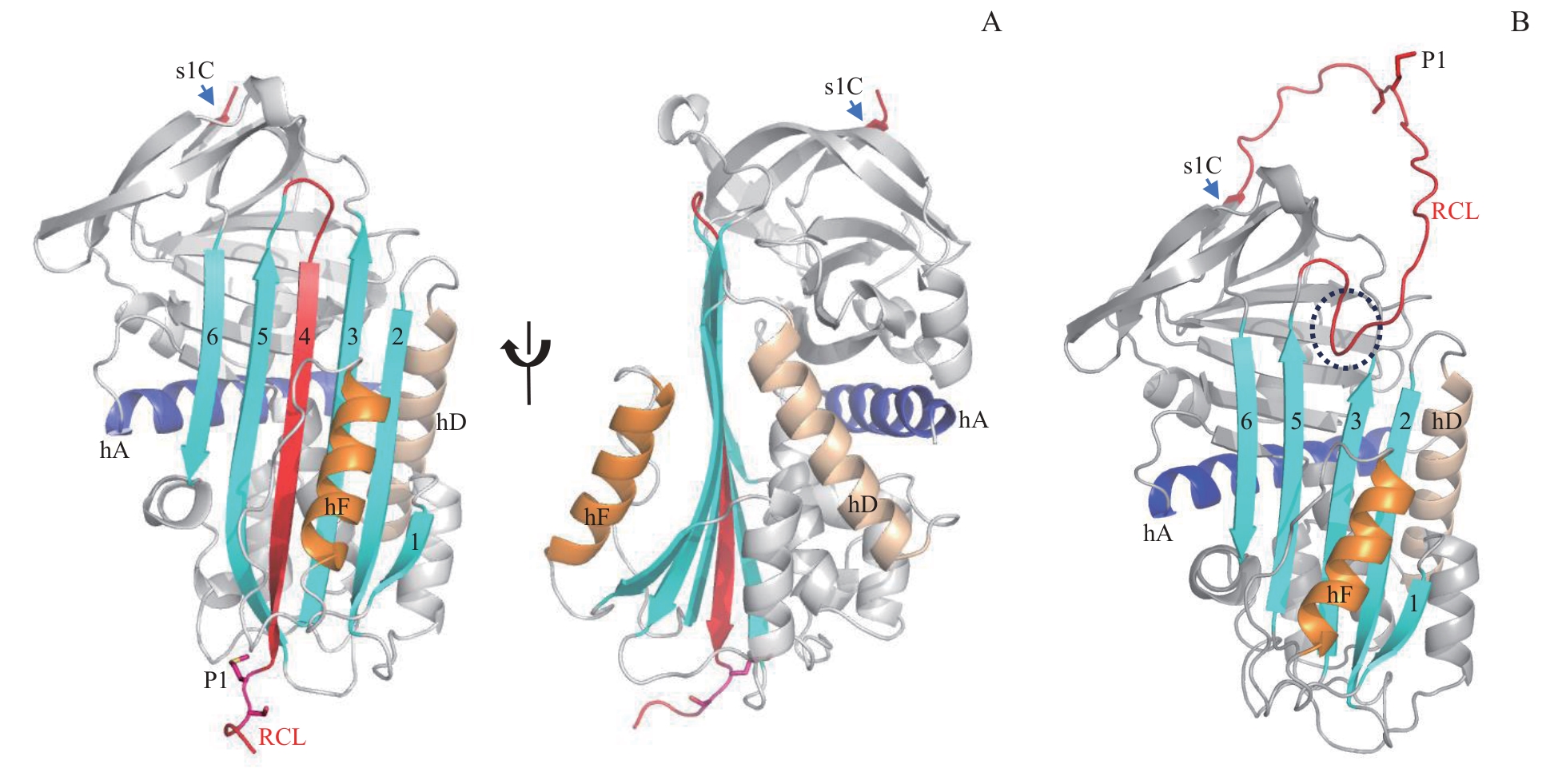
Note: A. Crystal structure of cleaved Tannerpin-M. B. Predicted structure of native Tannerpin-M. The central β-sheet A is colored in cyan with the strands traditionally numbered 1?6 from right to left. RCL is colored in red, helix A (hA) is colored in purple, helix F (hF) is colored in orange, and helix D (hD) is colored in gray. P1-P1' residue is shown in sticks.