先天缺牙相关EDAR基因突变报道及携带双突变位点的HED家系分析
Congenital tooth agenesis-related EDAR variants and pedigree analysis of HED patients with two variants
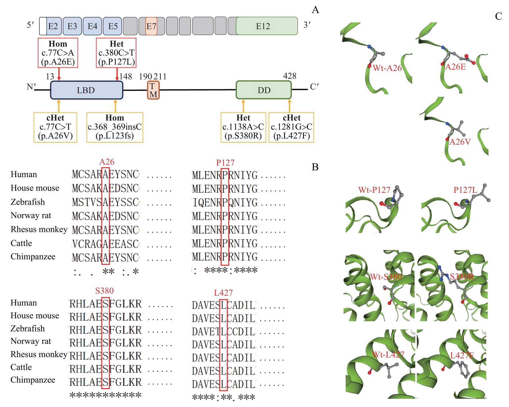
Note: A. Schematic diagram of wild-type EDAR protein and the localization of identified EDAR variants (red squares indicate variants found in NSTA patients, and yellow ones represent those in HED patients). B. Conservation analysis of affected amino acids among seven vertebrate species. C. Three-dimensional structure of wild-type EDAR and five missense variants (p.A26E, p.A26V, p.P127L, p.S380R, and p.L427F). E—exon; TM—transmembrane domain; Wt—wild-type.