结直肠癌中自然杀伤细胞表型及功能初探
Phenotype and function of NK cell in colorectal cancer
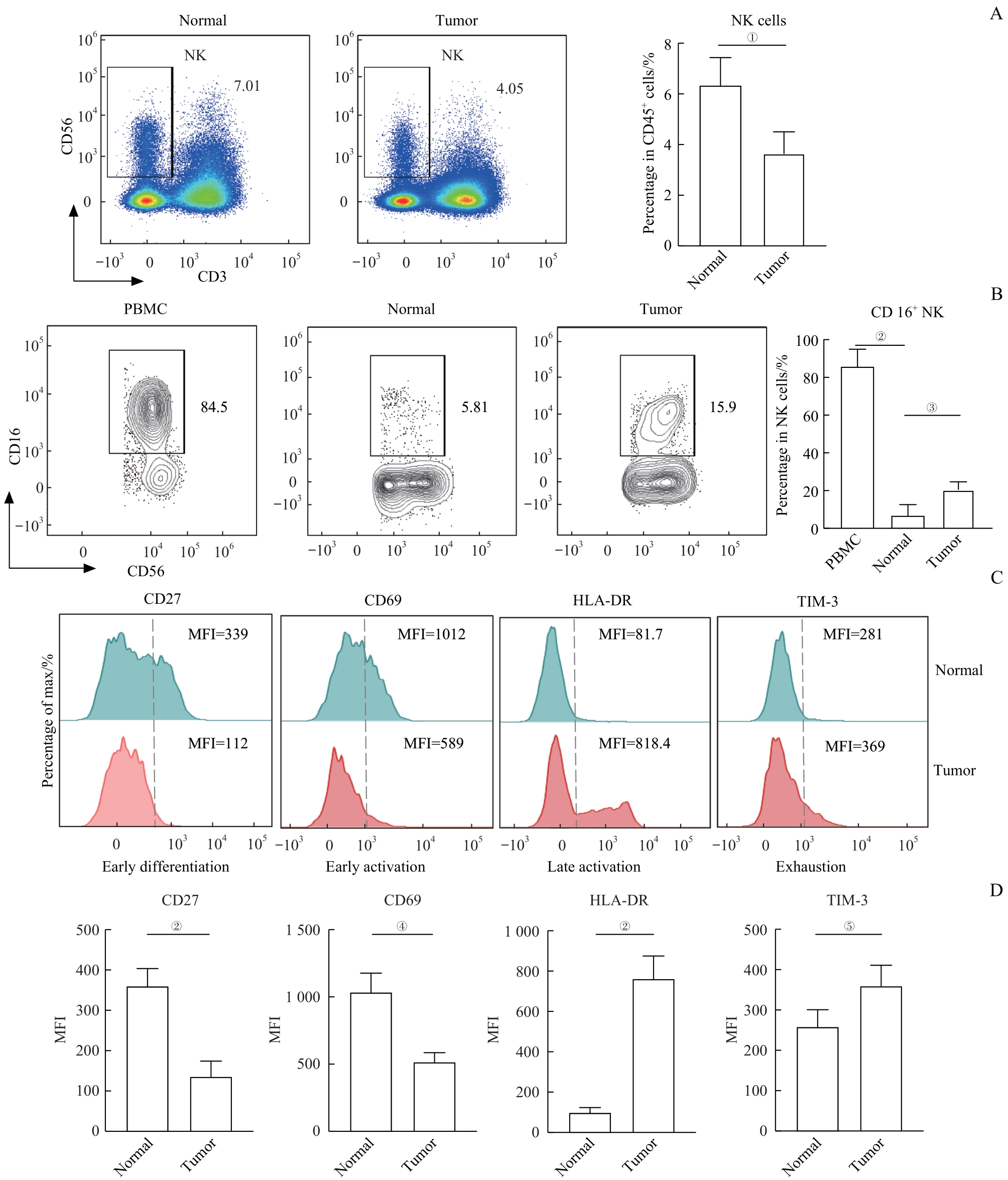
Note: A. Representative flow cytometry analysis and percentage of NK cells in normal and tumor tissues (n=25). B. Representative flow cytometry analysis and percentage of CD16+NK cells in PBMC, normal and tumor tissues (nPBMC=15, nnormal=25, and nTumor=25). C. Representative flow cytometry analysis of the expression of CD27, CD69, HLA-DR and TIM-3 on NK cells in normal and tumor tissues. D. The expression levels of CD27, CD69, HLA-DR and TIM-3 on NK cells in normal and tumor tissues (n=25). ①P=0.007, ②P=0.000, ③P=0.008, ④P=0.001, ⑤P=0.024. MFI—mean fluorenscence intensity.