原发性纵隔卵黄囊瘤临床和影像学分析
Clinical and imaging analyses of primary mediastinal yolk sac tumor
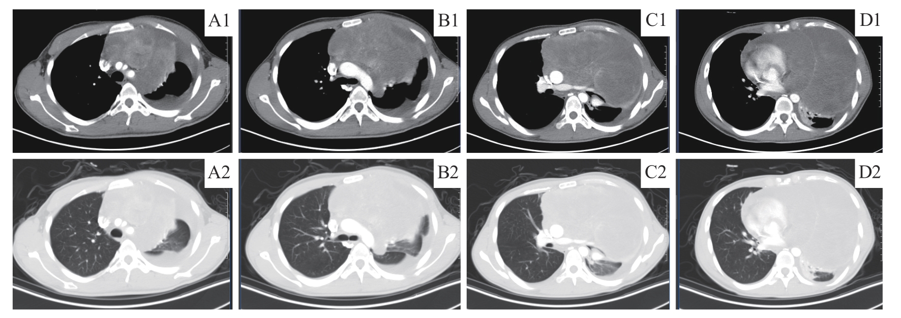
Note: A. Anterior mediastinal irregular mass with unevenly strengthened density encircled the brachiocephalic artery, left common carotid artery and left subclavian artery. B. The mass protruded into the left chest cavity and compressed the aortic arch. Pleural effusion was present. C. The mass caused compressive stenosis of main pulmonary artery, the left pulmonary artery and vein. D. Segmental atelectasis of the left lung and large pericardial effusion. A1?D1. Mediastinal window; A2?D2. Lung window.
