头颈部木村病的影像学特征研究
Study of imaging characteristics of Kimura disease in the head and neck
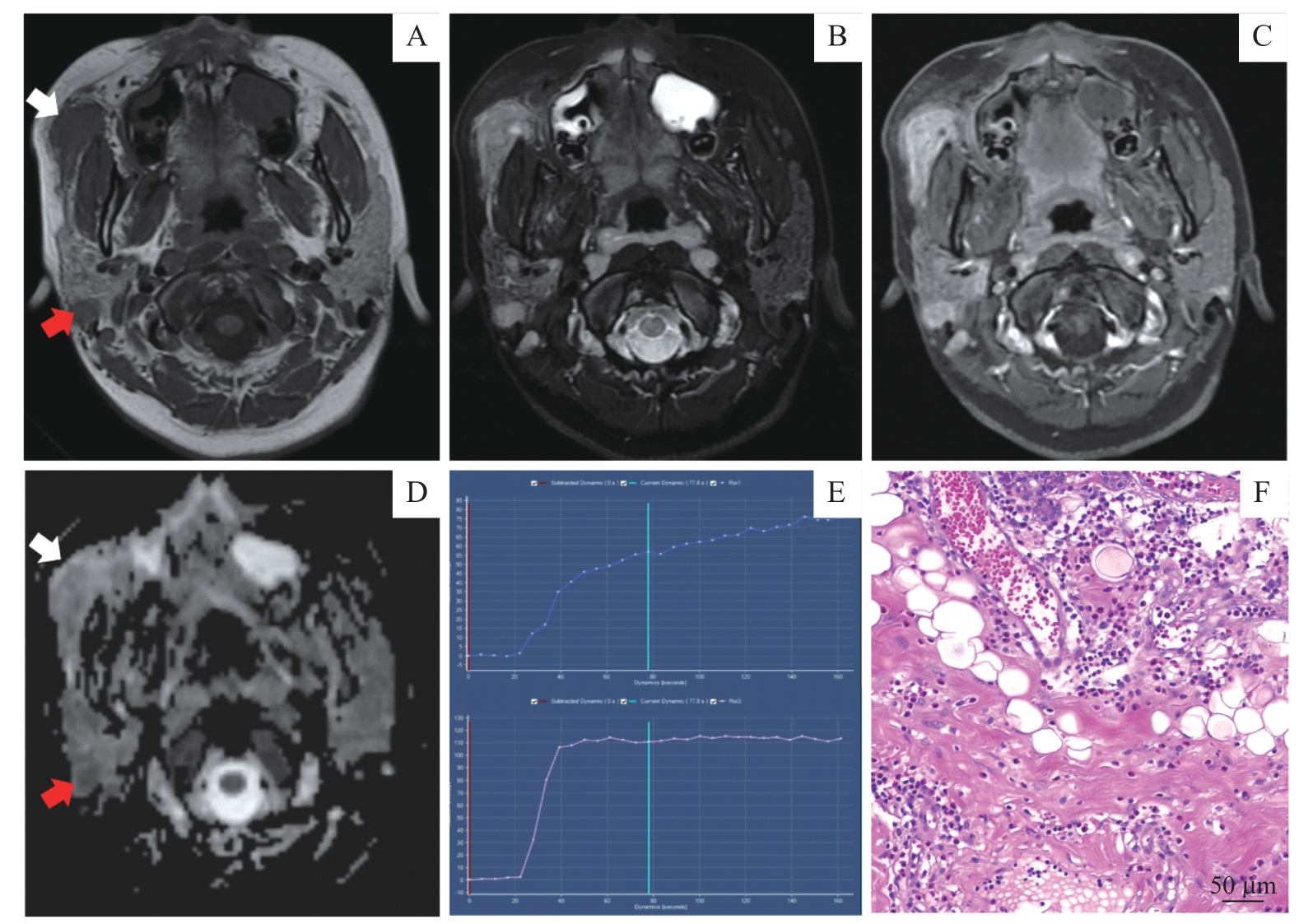
Note: A/B. Axial T1WI (A) and fat-suppressed T2WI (B) showing a well-defined nodular lesion (white arrow) and an enlarged lymph node (red arrow). C. Homogeneous hyperenhancement of the subcutaneous lesion on axial fat-suppressed enhanced T1WI. D. ADC map showing that the ADC value of the subcutaneous lesion (white arrow) was higher than that of the lymph node lesion (red arrow). E. TIC patterns of the subcutaneous lesion (top) and the lymph node lesion (bottom). F. The subcutaneous lesion showing hyperplastic follicles with diffused eosinophilic infiltration (hematoxylin-eosin staining, ×400).
