基于多尺度分辨率的耳蜗神经纤维三维成像
Three-dimensional imaging of cochlear nerve fibers based on multi-scale resolution
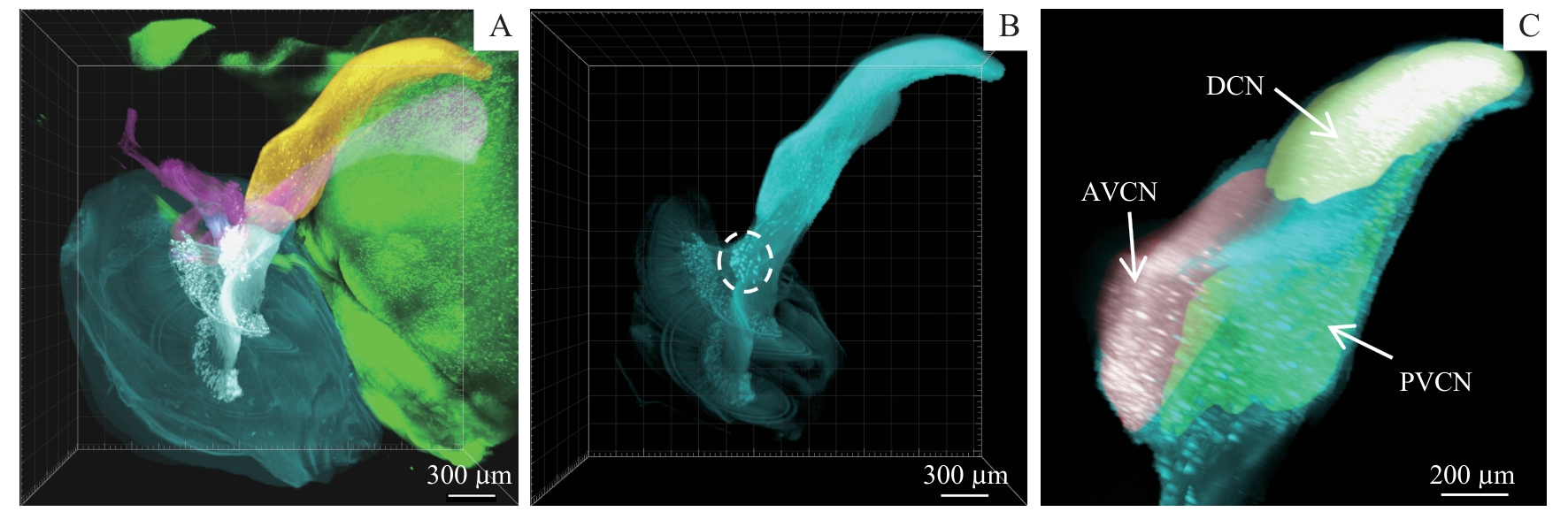
Note: A. 3D imaging of the cochlea and its surrounding tissues. The regions were segmented using different colours, including the cochlea (cyan), spiral ganglions and auditory nerves (off-white), cochlear nucleus (gold), vestibule and vestibular nerves (pink), and brainstem tissues (green). B. Cochlea-cochlear nucleus neural pathway. The circle indicates cochlear root neurons. C. Three subregions of the cochlear nucleus, including AVCN (red), PVCN (green), and DCN (gold).