基于多尺度分辨率的耳蜗神经纤维三维成像
Three-dimensional imaging of cochlear nerve fibers based on multi-scale resolution
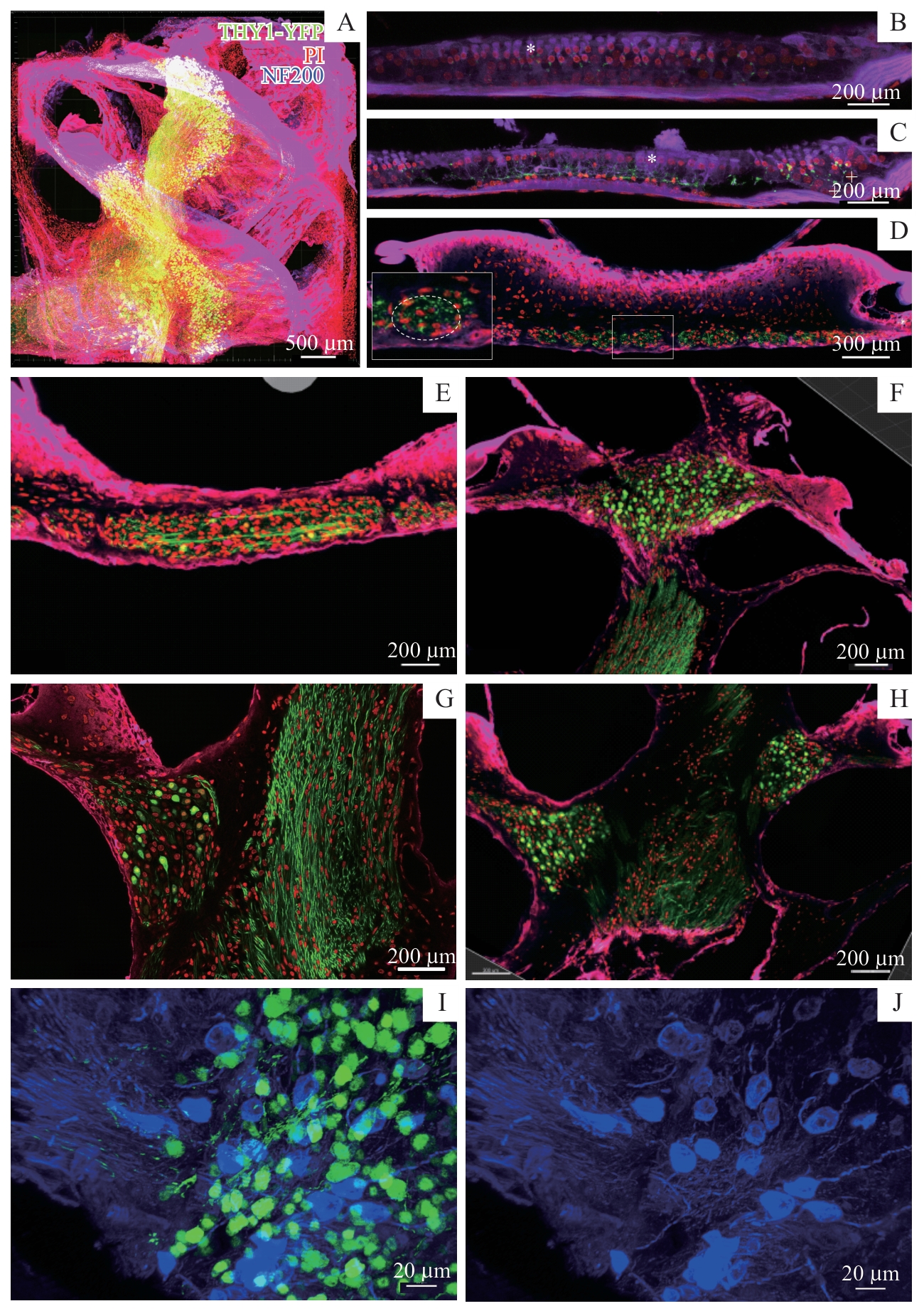
Note: A. 3D image of the cochlea. B. Distribution of outer hair cells (marked by *) in relation to THY1+ nerve fibers (green). C. Distribution of inner hair cells (marked by *) and supporting cells (marked by +) in relation to THY1+ nerve fibers (green). D. Position of nerve fiber bundles (green) in relation to the cochlear aperture. E‒H. Paths of THY1+ nerve fibers (green) in the cochlea. I/J. Distribution of NF200 expression (blue) in different parts of the cochlea. Red represents PI; green represents THY1-YFP;