基于多尺度分辨率的耳蜗神经纤维三维成像
Three-dimensional imaging of cochlear nerve fibers based on multi-scale resolution
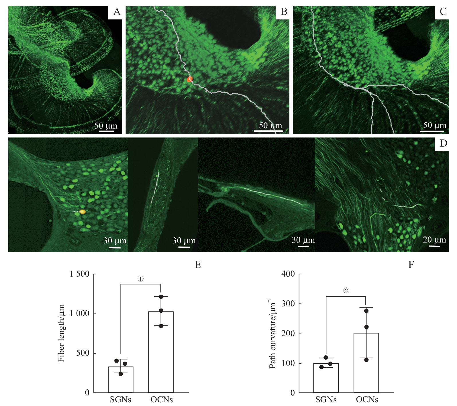
Note: A. 3D images of local spiral ganglia and fibers at a resolution of 0.24 μm×0.24 μm×1.50 μm. B/C. Two types of fiber pathways obtained by Imaris software tracing, an auditory nerve fiber (B) and MOC/LOC fibers (C). D. Local magnification of the 3D imaging of cochlear and labelling of fiber tracing at high resolution. E/F. Comparison of fiber length (E) and path curvature (F) between localized auditory nerve fibers and the MOC/LOC fibers within the cochlea. Neuronal designations were used to represent their corresponding fibers, with SGNs referring to auditory nerve fibers and OGNs (olivocochlear neurons) referring to MOC/LOC fibers. The orange/yellow dots represent the cell bodies of the tracked auditory nerves in B and D. ①P=0.002, ②P=0.113.