LINC01123通过结合ENO1促进胃癌的增殖和糖酵解
LINC01123 promotes proliferation and glycolysis of gastric cancer via binding to ENO1
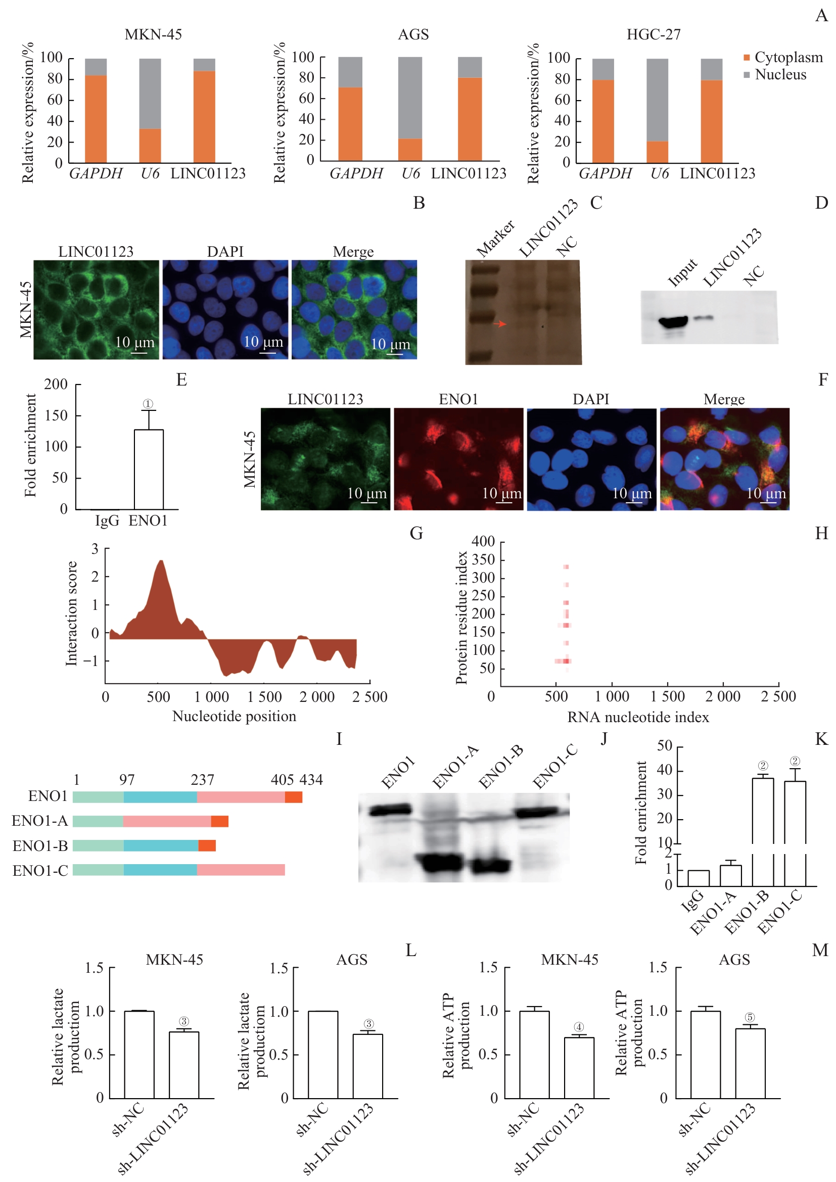
Note: A. Expression of LINC01123 in the cytoplasm and nucleus of gastric cancer cells. B. Fluorescence in situ hybridization (FISH) assay showing the localization of LINC01123 in MKN-45 cells. C. Silver-stained SDS-PAGE of proteins from RNA pull-down assay of MKN-45 cell extracts. The red arrow indicates the band used for mass spectrometry analysis. D. ENO1 pulled down by LINC01123 and negative control RNA was analyzed by Western blotting. Input represents the total protein used for RNA pull-down. E. PCR analysis of LINC01123 expression from RNA immunoprecipitation (RIP) assays. F. Co-staining of LINC01123 (green) and ENO1 (red) in MKN-45 cells by in FISH. G. Schematic map of the potential binding site of LINC01123 in ENO1 by catRAPID analysis. H. The interaction matrix between LINC01123 and ENO1 by catRAPID fragments analysis. I. Graphic display of ENO1 and its truncations. J. Flag-tagged ENO1 and its truncations were verified by Western blotting. K. Quantitative PCR of LINC01123 from purified RNAs of RIP experiments performed with 293T cell extracts transfected with Flag-tagged ENO1 or its truncations. L. Reduced lactate production in MKN-45 and AGS cells after LINC01123 knockdown. M. Reduced ATP production in MKN-45 and AGS cells after LINC01123 knockdown. ①P=0.002, ②P<0.001, compared with IgG; ③P<0.001, ④P=0.009, ⑤P=0.001, compared with sh-NC.