CD10+中性粒细胞通过SELPLG-SELL通路促进黏膜恶性黑色素瘤CD8+ T细胞耗竭
CD10+ neutrophils promote CD8+ T-cell depletion in mucosal malignant melanoma through the SELPG-SELL pathway
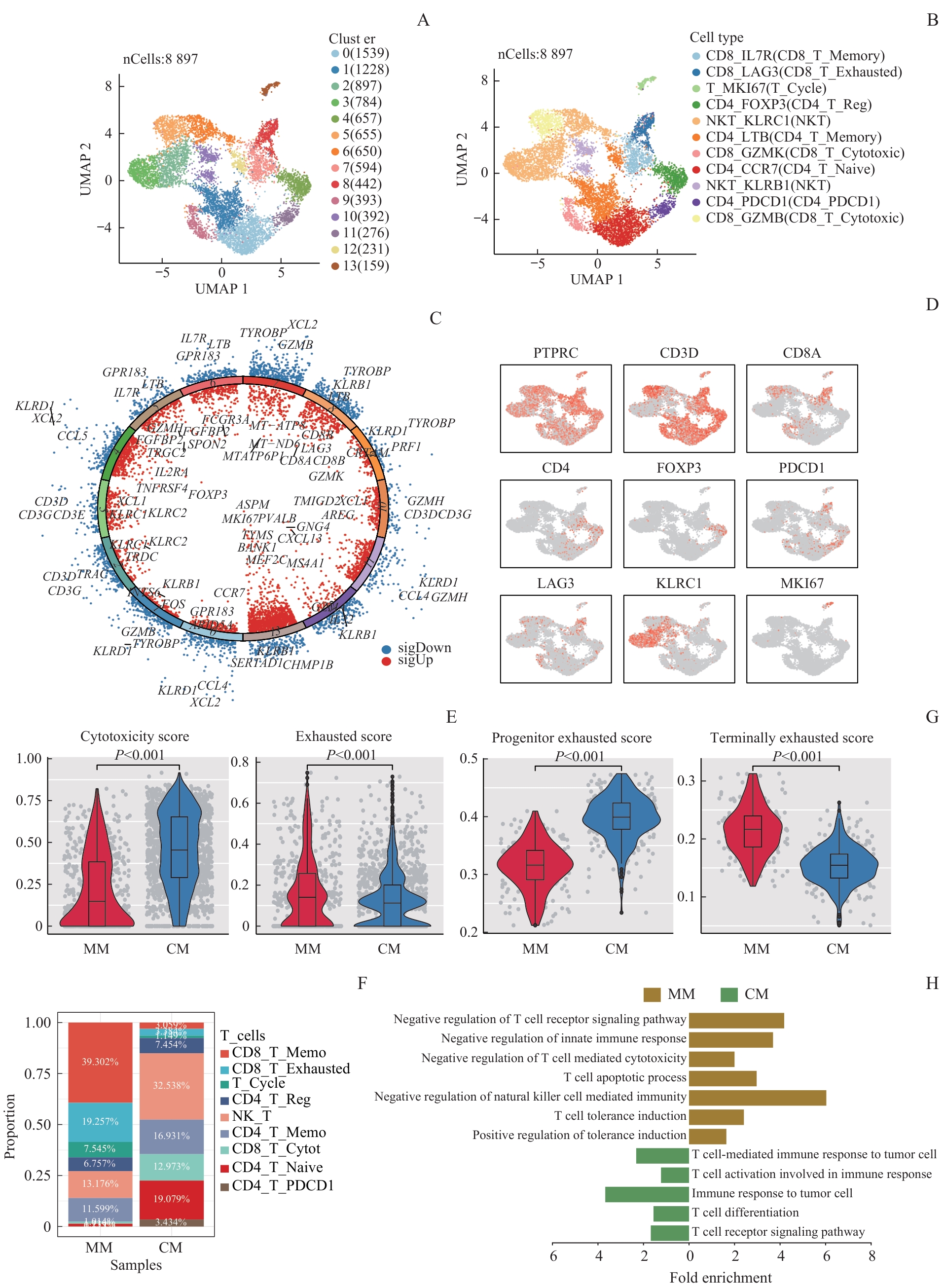
Note: A. UMAP map of T cell clustering in mucosal malignant melanoma and cutaneous malignant melanoma. B. UMAP map of T cell types in mucosal malignant melanoma and cutaneous malignant melanoma. C/D. volcano plot of the highly expressed genes of each T cell population (C) and marker expression maps of T cell types (D) in mucosal malignant melanoma and cutaneous malignant melanoma. E. Cytotoxicity and exhaustion gene set scores of CD8⁺ T cells in mucosal malignant melanoma and cutaneous malignant melanoma. F. Proportional distribution of different types of T cells. G. Progenitor exhaustion and terminal exhaustion gene set scores of exhausted T cells in mucosal malignant melanoma and cutaneous malignant melanoma. H. Differential gene enrichment analysis of exhausted T cells in mucosal malignant melanoma and cutaneous malignant melanoma.