2种参考体系下上颌前牙区牙槽骨改建特点的CBCT研究
A cone-beam computed tomographic study comparing characteristics of maxillary anterior regional alveolar bone remodeling under two reference systems
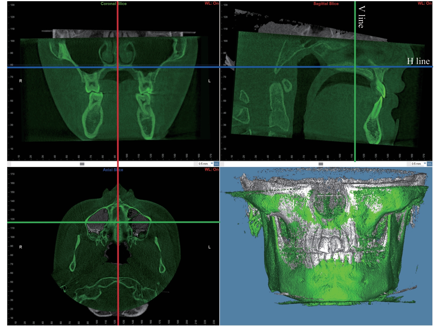
Note: White image refers to T0 (before treatment), while green image refers to T1 (after treatment). Orientation was performed based on T1 image. Upper left window shows the coronal view of the combined CBCT image of T0 and T1, while upper right window shows the sagittal view and lower left window shows the axial view. Lower right window shows the combined 3-dimensional reconstructed CBCT image. The red line represents the sagittal plane. The blue line (H line) represents the horizontal plane. The green line (V line) represents the coronal plane. Measurements were conducted on the sagittal slice (upper right window).