新一代基因编码5-羟色胺荧光探针优化及应用
Optimization of a genetically encoded fluorescent sensor for the detection of 5-HT
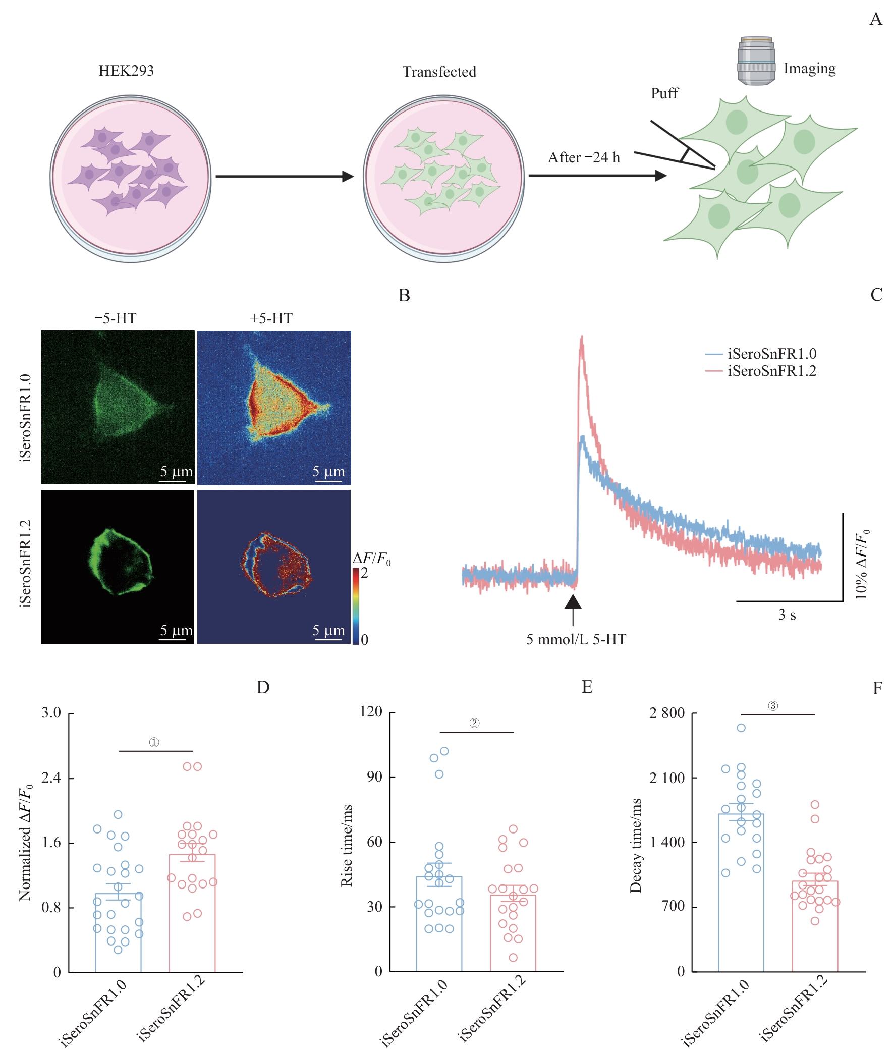
Note: A. Schematic drawing outlining the design of puff experiments in HEK293 cells. B. Representative fluorescence images of HEK293 cells expressing iSeroSnFR1.0 or iSeroSnFR1.2. Left: basal fluorescence; Right: heat maps showing fluorescence response to 5 mmol/L 5-HT perfusion. Scale bar: 5 μm. C. Normalized fluorescence response of HEK293 cells expressing iSeroSnFR1.0 or iSeroSnFR1.2 to a brief puff (10 ms) of 5 mmol/L 5-HT. D. Normalized values comparing fluorescence responses between iSeroSnFR1.0 and iSeroSnFR1.2. ①P=0.003 (n=20 cells from 3 independent experiments). E. Comparison of rise times of responses to the 5 mmol/L 5-HT puff between iSeroSnFR1.0 and iSeroSnFR1.2. ②P=0.461 (n=20 cells from 3 independent experiments). F. Comparison of decay times between iSeroSnFR1.0 and iSeroSnFR1.2. ③P<0.001 (n=22 cells from 3 independent experiments). Statistical analysis was performed using a Two-tailed unpaired Student′s t-test.