新一代基因编码5-羟色胺荧光探针优化及应用
Optimization of a genetically encoded fluorescent sensor for the detection of 5-HT
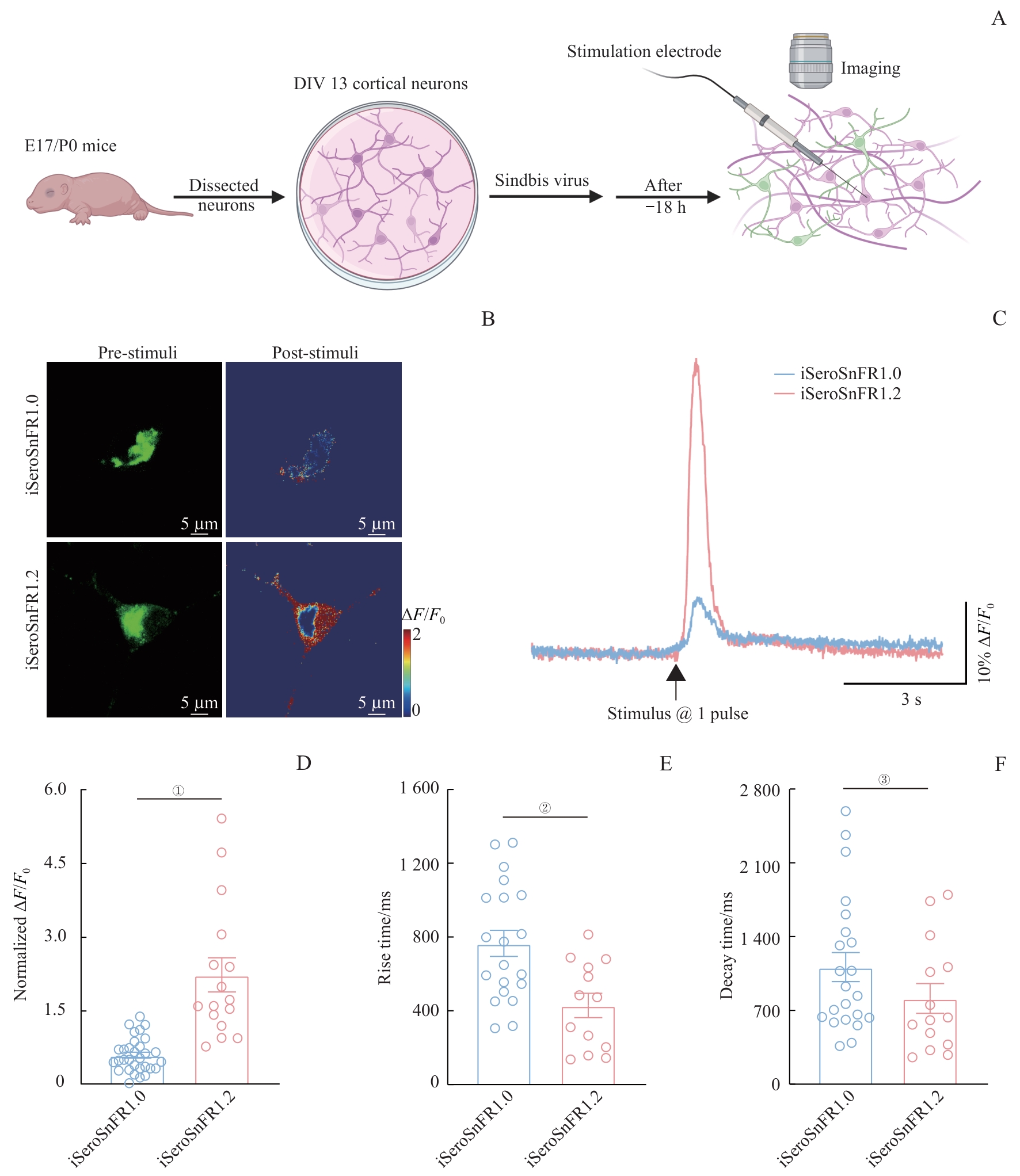
Note: A. Schematic illustration outlines the design of electrical stimulation experiments in mouse cortical neurons. B. Representative fluorescence imaging of cortical neurons expressing either iSeroSnFR1.0 or iSeroSnFR1.2. Images display GFP pseudocolor and corresponding heatmaps before (left) and after (right) a single 2-second electrical stimulus. Scale bar: 5 μm. The stimulus consisted of a single pulse. C/D. Simultaneous fluorescence responses (C) and normalized values (D) of iSeroSnFR1.0- and iSeroSnFR1.2-expressing mouse cortical neurons in response to electrical stimulation at a single pulse. ①P<0.001 (n=16 from 3 independent experiments). E. Comparison of rise times in responses to electrical stimulation between iSeroSnFR1.0 and iSeroSnFR1.2. ②P=0.003 (n=13 from 3 independent experiments). F. Comparison of decay times in response to electrical stimulation between iSeroSnFR1.0 and iSeroSnFR1.2. ③P=0.032 (n=14 from 3 independent experiments). Statistical analysis was performed using a two-tailed unpaired