颞下颌关节紊乱病患者关节盘与髁突距离的定量分析研究
Quantitative analysis of the distance between articular disc and condyle in patients with temporomandibular disorders
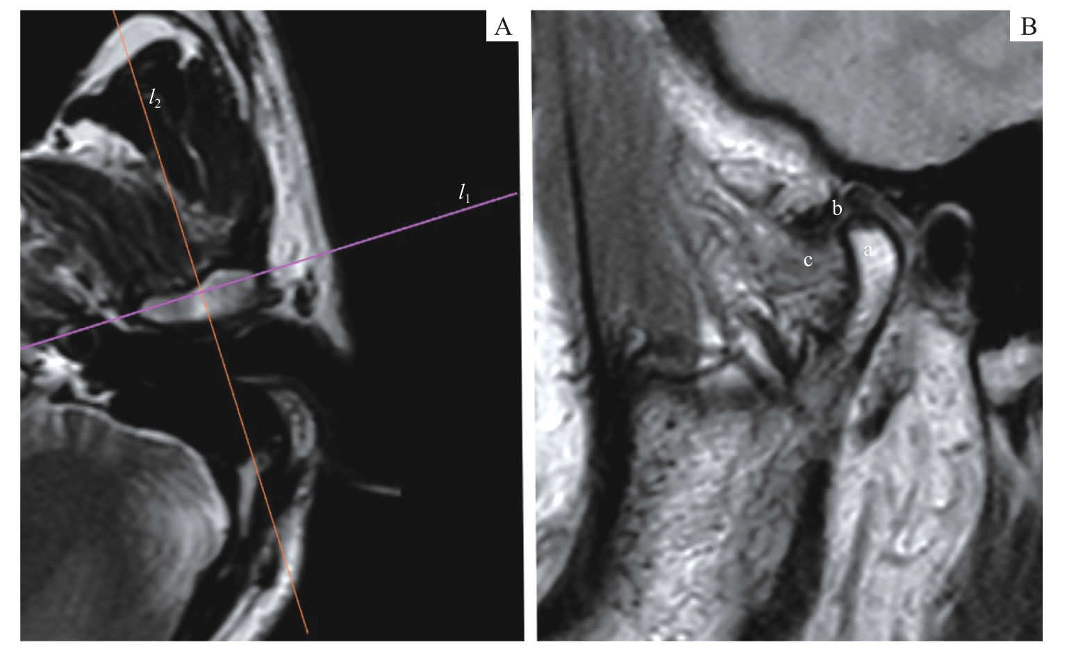
Note: PDWI images. A. Axial plane. l1—the transverse axis of the condyle (the line connecting the internal and the external poles of the condyle); l2—the positioning plane perpendicular to the condylar transverse axis. B. Oblique sagittal plane. a—condyle; b—articular disc; c—lateral pterygoid muscle.