颞下颌关节紊乱病患者关节盘与髁突距离的定量分析研究
Quantitative analysis of the distance between articular disc and condyle in patients with temporomandibular disorders
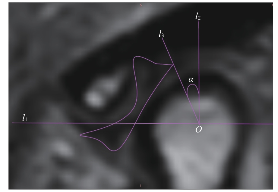
Note: An oblique sagittal PDWI image. l1—the line connecting the articular eminence and the vertex of the posterior tubercle; O—the midpoint of the intersection of line l1 with the condyle; l2—the perpendicular line to l1 through point O; l3—the line connecting the posterior edge of the articular disc with point O; α—the angle between l2 and l3, which is the angle of disc displacement.