基于磁敏感加权成像的终末期肾病患者脑影像特征与认知功能损伤的相关性
Correlation between brain imaging features and cognitive impairment in end-stage renal disease patients based on susceptibility-weighted imaging
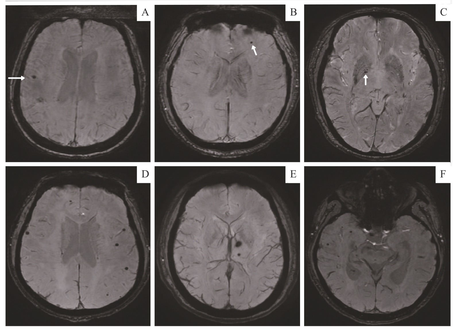
Note: A‒C. A 36-year-old female with ESRD who had undergone hemodialysis for 3 years had a few lesions in the right frontal lobe proximal cortex (A), the left frontal subcortex (B) and the right globus pallidus (C). The white arrows show the lesions in each brain region. This patient had no cognitive impairment, with a MoCA score of 28. D‒F. A 41-year-old male with ESRD who had undergone hemodialysis for 1.5 years showed multiple scattered CMBs in the frontal, temporal and occipital lobe (D and F), as well as in the left basal ganglia (E). This patient had cognitive impairment, with a MoCA score of 17.