| 1 |
PRINZ M, JUNG S, PRILLER J. Microglia biology: one century of evolving concepts[J]. Cell, 2019, 179(2): 292-311.
|
| 2 |
LI C Q, WANG Y, XING Y, et al. Regulation of microglia phagocytosis and potential involvement of exercise[J]. Front Cell Neurosci, 2022, 16: 953534.
|
| 3 |
HU S Y, LEE H, ZHAO H P, et al. Inflammation and severe cerebral venous thrombosis[J]. Front Neurol, 2022, 13: 873802.
|
| 4 |
YU F, WANG Y F, STETLER A R, et al. Phagocytic microglia and macrophages in brain injury and repair[J]. CNS Neurosci Ther, 2022, 28(9): 1279-1293.
|
| 5 |
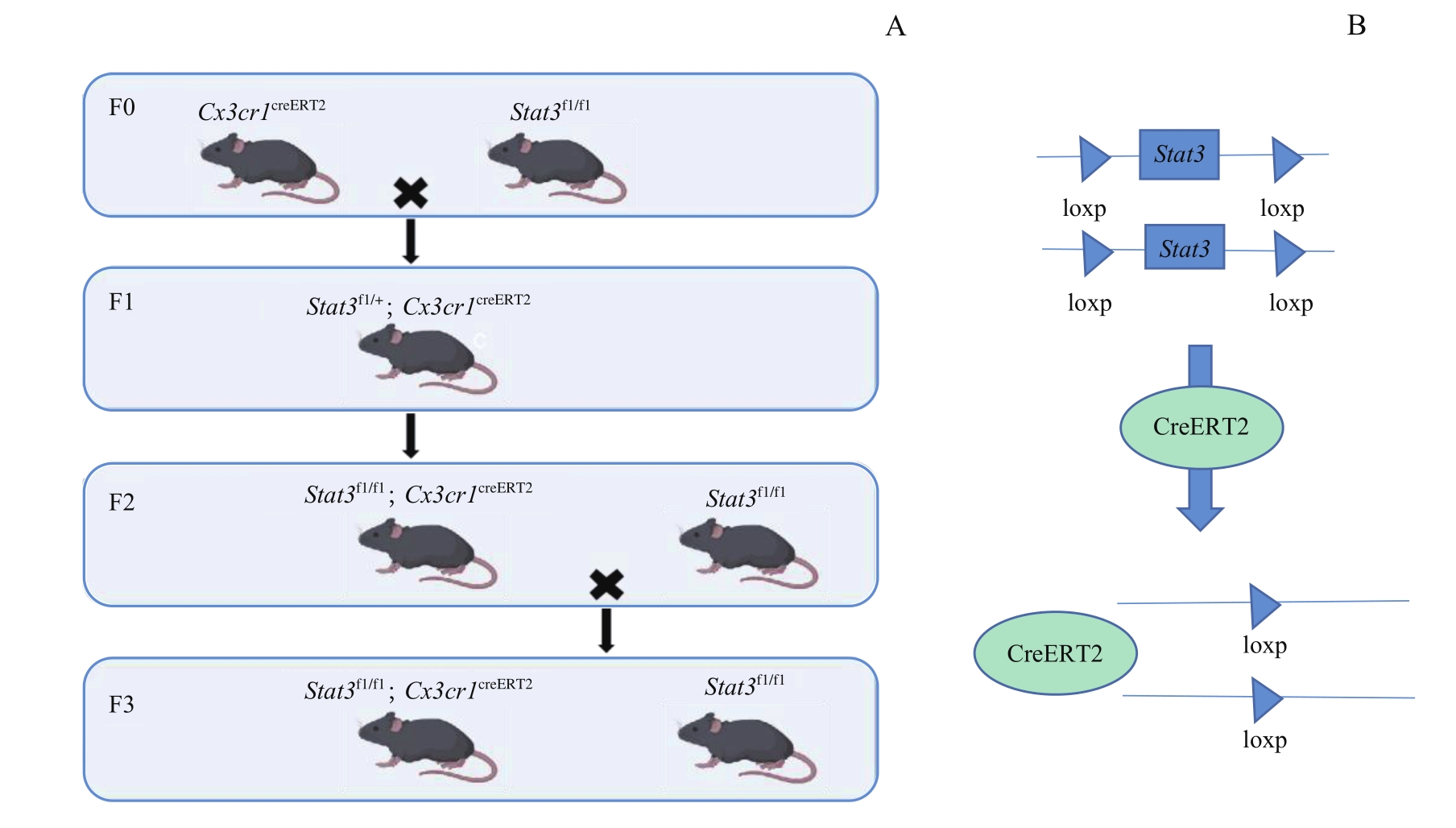
KIM H, KIM M, IM S K, et al. Mouse Cre-LoxP system: general principles to determine tissue-specific roles of target genes[J]. Lab Anim Res, 2018, 34(4): 147-159.
|
| 6 |
HU Y S, HAN X, LIU X H. STAT3: a potential drug target for tumor and inflammation[J]. Curr Top Med Chem, 2019, 19(15): 1305-1317.
|
| 7 |
TOŠIĆ I, FRANK D A. STAT3 as a mediator of oncogenic cellular metabolism: pathogenic and therapeutic implications[J]. Neoplasia, 2021, 23(12): 1167-1178.
|
| 8 |
ZHENG Z V, CHEN J F, LYU H, et al. Novel role of STAT3 in microglia-dependent neuroinflammation after experimental subarachnoid haemorrhage[J]. Stroke Vasc Neurol, 2022, 7(1): 62-70.
|
| 9 |
LUO L, AMBROZKIEWICZ M C, BENSELER F, et al. Optimizing nervous system-specific gene targeting with cre driver lines: prevalence of germline recombination and influencing factors[J]. Neuron, 2020, 106(1): 37-65.e5.
|
| 10 |
NAVABPOUR S, KWAPIS J L, JAROME T J. A neuroscientist's guide to transgenic mice and other genetic tools[J]. Neurosci Biobehav Rev, 2020, 108: 732-748.
|
| 11 |
SIMONETTI M, YILMAZER A, KRETSCHMER K. Genetic tools for analyzing Foxp3+ treg cells: fluorochrome-based transcriptional reporters and genetic fate-mapping[J]. Methods Mol Biol, 2023, 2559: 95-114.
|
| 12 |
ABUBAKAR M B, SANUSI K O, UGUSMAN A, et al. Alzheimer's disease: an update and insights into pathophysiology[J]. Front Aging Neurosci, 2022, 14: 742408.
|
| 13 |
SHEN Y N, ZHANG Y, DU J Y, et al. CXCR5 down-regulation alleviates cognitive dysfunction in a mouse model of sepsis-associated encephalopathy: potential role of microglial autophagy and the p38MAPK/NF-κB/STAT3 signaling pathway[J]. J Neuroinflammation, 2021, 18(1): 246.
|
| 14 |
REICHENBACH N, DELEKATE A, PLESCHER M, et al. Inhibition of Stat3-mediated astrogliosis ameliorates pathology in an Alzheimer's disease model[J]. EMBO Mol Med, 2019, 11(2): e9665.
|
| 15 |
MEHLA J, SINGH I, DIWAN D, et al. STAT3 inhibitor mitigates cerebral amyloid angiopathy and parenchymal amyloid plaques while improving cognitive functions and brain networks[J]. Acta Neuropathol Commun, 2021, 9(1): 193.
|
| 16 |
ZOU J, SHANG W L, YANG L, et al. Microglia activation in the mPFC mediates anxiety-like behaviors caused by Staphylococcus aureus strain USA300[J]. Brain Behav, 2022, 12(9): e2715.
|
| 17 |
HU Y, LI H X, ZHANG J, et al. Periodontitis induced by P. gingivalis-LPS is associated with neuroinflammation and learning and memory impairment in sprague-dawley rats[J]. Front Neurosci, 2020, 14: 658.
|
| 18 |
SHCHOLOK T, EFTEKHARPOUR E. Cre-recombinase systems for induction of neuron-specific knockout models: a guide for biomedical researchers[J]. Neural Regen Res, 2023, 18(2): 273-279.
|
| 19 |
JIA X N, GAO Z H, HU H L. Microglia in depression: current perspectives[J]. Sci China Life Sci, 2021, 64(6): 911-925.
|
 ), XIE Xinyi, ZHAO Xuri, XU Lina, HE Zhiyan, ZHOU Wei(