
Journal of Shanghai Jiao Tong University (Medical Science) ›› 2026, Vol. 46 ›› Issue (9): 1169-1178.doi: 10.3969/j.issn.1674-8115.2026.09.002
• Frontier review • Previous Articles
Yu Meixuan1, Sun Lin2,3, Fu Jingke2,3,#( ), Hao Yongqiang2,3,4,5,6,#(), Dai Kerong2,3,4,5,6
Received:2026-01-12
Accepted:2026-02-09
Online:2026-09-28
Published:2026-09-28
Contact:
Fu Jingke, Hao Yongqiang
E-mail:fujingke@sjtu.edu.cn;hao_yongqiang@hotmail.com
Supported by:CLC Number:
Yu Meixuan, Sun Lin, Fu Jingke, Hao Yongqiang, Dai Kerong. 3D printing biomimetic periosteum: from structural substitution to functional integration[J]. Journal of Shanghai Jiao Tong University (Medical Science), 2026,(9): 1169-1178.
Add to citation manager EndNote|Ris|BibTeX
URL: https://xuebao.shsmu.edu.cn/EN/10.3969/j.issn.1674-8115.2026.09.002
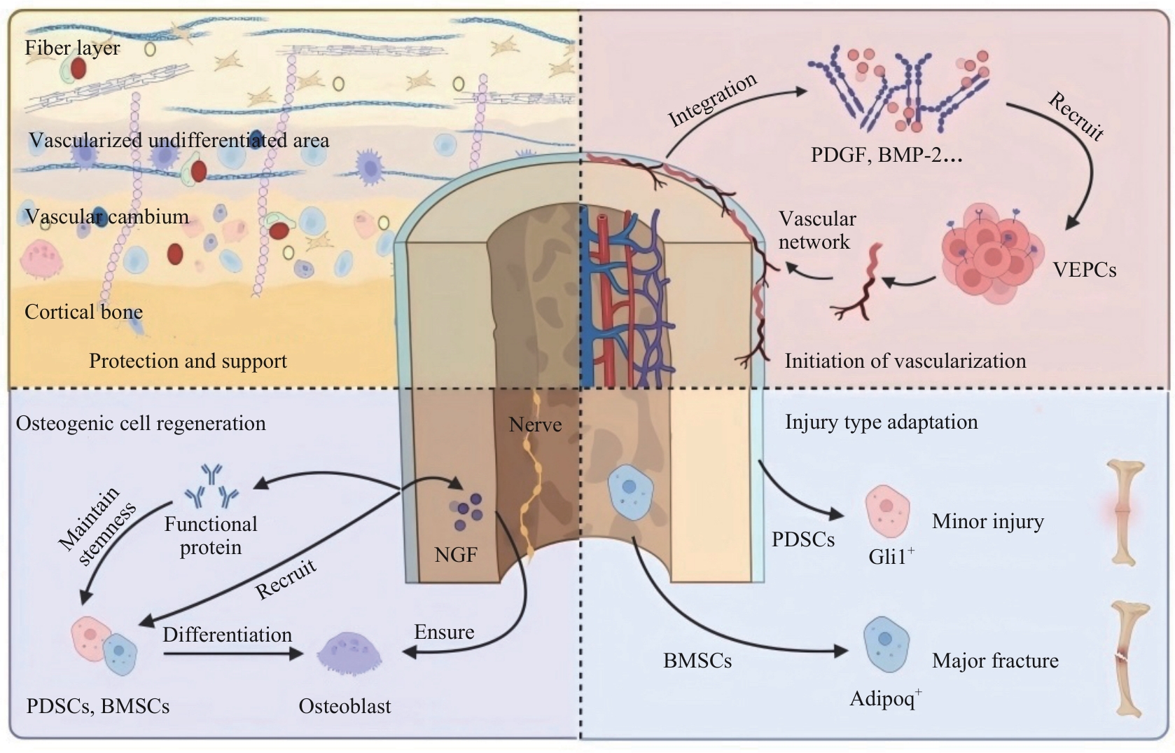
Fig 1 Clinical functional requirements of biomimetic periosteum
| Manufacturing | Model | Biomaterial in bioinks | OFC in bioinks | Main effect | Reference |
|---|---|---|---|---|---|
| EBP | Rat cranial defect | Fibrous layer: SFHGMA, SA Formation layer: SFP, SA, MCol | Mg2+ | Simulation of natural periosteal double-layer structure; promotion of cell proliferation and differentiation, angiogenesis, and PC12 cell growth | [ |
| EBP | Regeneration of critical-sized bone defects in long bones | GelMA | G-MBGN, VEGF-loaded liposomes | Simulation of periosteum, endosteum, and intermediate bone matrix; significant promotion of angiogenesis | [ |
| EBP | Critical-sized bone defect | Formation layer: GelMA, GelMA-H-nHA Fibrous layer: P(ACG-GelMA-L)-Mg2+ | Ca2+, Mg2+, nHA | Simulation of natural periosteal double-layer structure, with Mg²⁺-mediated immunomodulation and angiogenesis | [ |
| EBP | Bone defect | PLLA/HA, GelMA | rabBMSCs, rabPDSCs | Excellent cell encapsulation and bioactivity; co-culture of BMSCs and PDSCs | [ |
| EBP | Bone defect | GelMA, SilMA, GelDA | GO nanosheets, BMSCs | Excellent thermosensitivity and printability; high cell viability and adhesion | [ |
| MEW | Critical-sized segmental rat femoral defect | MEW core: PCL FDM Shell: PCL Coating: nnHA | BMP2 | High rigidity, structural integrity, sustained BMP2 release, and significant angiogenesis enhancement | [ |
| MEW | Large bone defect | PCL membrane, PCL sheet | rhBMP-2, PLGA-PEG-COOH microparticles | Controlled rhBMP-2 release and significant enhancement of in vivo bone regeneration | [ |
| SLA | Bone defect | Fibrous layer: PEGDA, TCP Formation layer: GelMA, PEGDA | Mo, Chitosan | Fibrous layer resistance to muscle traction and soft tissue invasion; promotion of osteogenic differentiation by the formation layer | [ |
Tab 1 Representative construction strategies for 3D-printed biomimetic periosteum
| Manufacturing | Model | Biomaterial in bioinks | OFC in bioinks | Main effect | Reference |
|---|---|---|---|---|---|
| EBP | Rat cranial defect | Fibrous layer: SFHGMA, SA Formation layer: SFP, SA, MCol | Mg2+ | Simulation of natural periosteal double-layer structure; promotion of cell proliferation and differentiation, angiogenesis, and PC12 cell growth | [ |
| EBP | Regeneration of critical-sized bone defects in long bones | GelMA | G-MBGN, VEGF-loaded liposomes | Simulation of periosteum, endosteum, and intermediate bone matrix; significant promotion of angiogenesis | [ |
| EBP | Critical-sized bone defect | Formation layer: GelMA, GelMA-H-nHA Fibrous layer: P(ACG-GelMA-L)-Mg2+ | Ca2+, Mg2+, nHA | Simulation of natural periosteal double-layer structure, with Mg²⁺-mediated immunomodulation and angiogenesis | [ |
| EBP | Bone defect | PLLA/HA, GelMA | rabBMSCs, rabPDSCs | Excellent cell encapsulation and bioactivity; co-culture of BMSCs and PDSCs | [ |
| EBP | Bone defect | GelMA, SilMA, GelDA | GO nanosheets, BMSCs | Excellent thermosensitivity and printability; high cell viability and adhesion | [ |
| MEW | Critical-sized segmental rat femoral defect | MEW core: PCL FDM Shell: PCL Coating: nnHA | BMP2 | High rigidity, structural integrity, sustained BMP2 release, and significant angiogenesis enhancement | [ |
| MEW | Large bone defect | PCL membrane, PCL sheet | rhBMP-2, PLGA-PEG-COOH microparticles | Controlled rhBMP-2 release and significant enhancement of in vivo bone regeneration | [ |
| SLA | Bone defect | Fibrous layer: PEGDA, TCP Formation layer: GelMA, PEGDA | Mo, Chitosan | Fibrous layer resistance to muscle traction and soft tissue invasion; promotion of osteogenic differentiation by the formation layer | [ |
| Multi-technology integration | Biomaterial in ink | Bioactive substance in ink | Highlight | Reference |
|---|---|---|---|---|
| Electrospinning+3D printing | Electrospun collagen-dense scaffold, IMCS | BMSCs | Upper-layer blockade of fibrous tissue invasion; lower-layer promotion of BMSC osteogenic differentiation | [ |
| IMT+3D printing | mPCL-TCP | ABG, rhBMP-2, Cerament G®, Cerament V® | Excellent clinical adaptability, superior surgical manoeuvrability, and enhanced angiogenesis and osteogenesis | [ |
| IMT+3D printing | PLA scaffold, dECM | Bone marrow cells, BMP-2 | High-dose, sustained release of BMP-2; reduced fibrosis and macrophage infiltration; optimized BMP-2 loading capacity | [ |
Tab 2 Multi-technology integration for biomimetic periosteum construction
| Multi-technology integration | Biomaterial in ink | Bioactive substance in ink | Highlight | Reference |
|---|---|---|---|---|
| Electrospinning+3D printing | Electrospun collagen-dense scaffold, IMCS | BMSCs | Upper-layer blockade of fibrous tissue invasion; lower-layer promotion of BMSC osteogenic differentiation | [ |
| IMT+3D printing | mPCL-TCP | ABG, rhBMP-2, Cerament G®, Cerament V® | Excellent clinical adaptability, superior surgical manoeuvrability, and enhanced angiogenesis and osteogenesis | [ |
| IMT+3D printing | PLA scaffold, dECM | Bone marrow cells, BMP-2 | High-dose, sustained release of BMP-2; reduced fibrosis and macrophage infiltration; optimized BMP-2 loading capacity | [ |
| [1] | De Pace R, Molinari S, Mazzoni E, et al. Bone regeneration: a review of current treatment strategies[J]. J Clin Med, 2025, 14(6): 1838. |
| [2] | 中华医学会骨科学分会创伤骨科学组, 中华医学会骨科学分会外固定与肢体重建学组, 北京医学会显微外科分会. 中国自体骨移植临床实践指南(2024 版)[J]. 中华医学杂志, 2024, 104(35): 3307-3322. |
| The Trauma Orthopedic Branch of Chinese Orthopedic Association, the External Fixation and Limb Reconstruction Branch of Chinese Orthopedic Association, the Microsurgery Branch of Beijing Orthopedic Association. Clinical practice guideline for autologous bone grafting in China (2024 edition)[J]. National Medical Journal of China, 2024, 104(35): 3307-3322. | |
| [3] | Li C, Fennessy P. The periosteum: a simple tissue with many faces, with special reference to the antler-lineage periostea[J]. Biol Direct, 2021, 16(1): 17. |
| [4] | Yang G, Liu H, Cui Y, et al. Bioinspired membrane provides periosteum-mimetic microenvironment for accelerating vascularized bone regeneration[J]. Biomaterials, 2021, 268: 120561. |
| [5] | Zhang Z, Shen J, Xu R, et al. Dual-functional biomimetic periosteum incorporating engineered small extracellular vesicles for treating critical bone defect with soft tissue fenestration via TGF-β1/SMAD pathway[J]. Mater Today Bio, 2025, 32: 101892. |
| [6] | Duda G N, Geissler S, Checa S, et al. The decisive early phase of bone regeneration[J]. Nat Rev Rheumatol, 2023, 19(2): 78-95. |
| [7] | March A, Wu H, Choe R, et al. Optimizing tissue-engineered periosteum biochemical cues to hasten bone allograft healing[J]. J Biomed Mater Res A, 2025, 113(3): e37890. |
| [8] | March A, Castillo Aguirre S H, Eliseev R, et al. Long-term tissue engineered periosteum-mediated allograft healing is hindered due to persistent fibrosis and limited allograft remodeling[J]. Bone Rep, 2025, 26: 101865. |
| [9] | Ling F, Bai J, Xie J, et al. Biomimetic periosteum combining BMP-2-loaded M2 macrophage-derived exosomes for enhanced bone defect repair[J]. Front Bioeng Biotechnol, 2025, 13: 1639394. |
| [10] | Zhan Z, Li R, Wu Y, et al. Biomimetic periosteum-bone scaffolds with codelivery of BMP-2 and PDGF-BB for skull repair[J]. Bone, 2025, 190: 117315. |
| [11] | Durand M, Mathieu L, Venant J, et al. Engineering the bone reconstruction surgery: the case of the Masquelet-induced membrane technique[J]. Eur J Trauma Emerg Surg, 2025, 51(1): 138. |
| [12] | Tripathi S, Mandal S S, Bauri S, et al. 3D bioprinting and its innovative approach for biomedical applications[J]. MedComm, 2023, 4(1): e194. |
| [13] | Zhao X, Li N, Zhang Z, et al. Beyond hype: unveiling the real challenges in clinical translation of 3D printed bone scaffolds and the fresh prospects of bioprinted organoids[J]. J Nanobiotechnol, 2024, 22(1): 500. |
| [14] | Li Z, Wang Q, Liu G. A review of 3D printed bone implants[J]. Micromachines, 2022, 13(4): 528. |
| [15] | Wang J, Chen G, Chen Z M, et al. Current strategies in biomaterial-based periosteum scaffolds to promote bone regeneration: a review[J]. J Biomater Appl, 2023, 37(7): 1259-1270. |
| [16] | Zhang W, Wang N, Yang M, et al. Periosteum and development of the tissue-engineered periosteum for guided bone regeneration[J]. J Orthop Translat, 2022, 33: 41-54. |
| [17] | McLoughlin S T, Wilcox P, Caccamese J F, et al. Osteoblast-mesenchymal stem cell coculture drives in vitro osteogenesis in 3D bioprinted periosteum[J]. Tissue Eng Part A, 2025, 31(21/22): 1239-1252. |
| [18] | Yang Y, Rao J, Liu H, et al. Biomimicking design of artificial periosteum for promoting bone healing[J]. J Orthop Translat, 2022, 36: 18-32. |
| [19] | Zhang X, Deng C, Qi S. Periosteum containing implicit stem cells: a progressive source of inspiration for bone tissue regeneration[J]. Int J Mol Sci, 2024, 25(4): 2162. |
| [20] | Cao R, Chen B, Li Q, et al. Potential of periosteal cells in bone and cartilage regeneration: a systematic review[J]. Front Bioeng Biotechnol, 2023, 11: 1292483. |
| [21] | Li X, Yang S, Li S, et al. Reverse biogradient biomimetic periosteum with osteogenic and angiogenic characteristics for bone regeneration[J]. Mater Today Bio, 2025, 33: 101967. |
| [22] | Hua X, Hou M, Deng L, et al. Irisin-loaded electrospun core-shell nanofibers as calvarial periosteum accelerate vascularized bone regeneration by activating the mitochondrial SIRT3 pathway[J]. Regen Biomater, 2024, 11: rbad096. |
| [23] | Zhou Z, Liu Y, Li W, et al. A self-adaptive biomimetic periosteum employing nitric oxide release for augmenting angiogenesis in bone defect regeneration[J]. Adv Healthc Mater, 2024, 13(3): e2302153. |
| [24] | Su Y, Zeng L, Deng R, et al. Endogenous electric field-coupled PD@BP biomimetic periosteum promotes bone regeneration through sensory nerve via Fanconi anemia signaling pathway[J]. Adv Healthc Mater, 2023, 12(12): e2203027. |
| [25] | Ono N. The mechanism of bone repair: stem cells in the periosteum dedicated to bridging a large gap[J]. Cell Rep Med, 2022, 3(11): 100807. |
| [26] | Yin B, Shen F, Ma Q, et al. Identification of Postn + periosteal progenitor cells with bone regenerative potential[J]. JCI Insight, 2024, 9(19): e182524. |
| [27] | Alshebly Y S, Nafea M, Mohamed Ali M S, et al. Review on recent advances in 4D printing of shape memory polymers[J]. Eur Polym J, 2021, 159: 110708. |
| [28] | He C, He J, Wu C, et al. 3D printing for tissue/organ regeneration in China[J]. Bio-Des Manuf, 2025, 8(2): 169-242. |
| [29] | Feng H, Lian X, Lv S, et al. Bioinspired bilayer 3D printing periosteum scaffold with hierarchical structure based on silk fibroin and sodium alginate for bone regeneration[J]. Int J Biol Macromol, 2025, 310(Pt 2): 143175. |
| [30] | Sun Y, Gao Z, Zhang X, et al. 3D-printed, bi-layer, biomimetic artificial periosteum for boosting bone regeneration[J]. Bio-Des Manuf, 2022, 5(3): 540-555. |
| [31] | Zhao D, Wang Y, Yu Z, et al. Co-culture bioprinting of tissue-engineered bone-periosteum biphasic complex for repairing critical-sized skull defects in rabbits[J]. Int J Bioprint, 2023, 9(3): 698. |
| [32] | Eichholz K F, Pitacco P, Burdis R, et al. Integrating melt electrowriting and fused deposition modeling to fabricate hybrid scaffolds supportive of accelerated bone regeneration[J]. Adv Healthc Mater, 2024, 13(3): e2302057. |
| [33] | Zhou X, Zou B, Chen Q, et al. Construction of bilayer biomimetic periosteum based on SLA-3D printing for bone regeneration[J]. Colloids Surf B Biointerfaces, 2025, 246: 114368. |
| [34] | Beheshtizadeh N, Farzin A, Rezvantalab S, et al. 3D printing of complicated GelMA-coated alginate/tri-calcium silicate scaffold for accelerated bone regeneration[J]. Int J Biol Macromol, 2023, 229: 636-653. |
| [35] | Romero-Torrecilla J A, Echanove-González de Anleo M, Martínez-Ohárriz C, et al. 3D-printed polycaprolactone scaffolds functionalized with poly(lactic-co-glycolic) acid microparticles enhance bone regeneration through tunable drug release[J]. Acta Biomater, 2025, 198: 219-233. |
| [36] | Xu H, Li X, Wang W, et al. Strontium-doped marine collagen membranes promote osteogenesis by inducing M2 macrophage polarization[J]. Tissue Eng Regen Med, 2025, 22(6): 847-864. |
| [37] | Zhao D W, Liu C, Zuo K Q, et al. Strontium-zinc phosphate chemical conversion coating improves the osseointegration of titanium implants by regulating macrophage polarization[J]. Chem Eng J, 2021, 408: 127362. |
| [38] | Qi H, Zhang B, Lian F. 3D-printed bioceramic scaffolds for bone defect repair: bone aging and immune regulation[J]. Front Bioeng Biotechnol, 2025, 13: 1557203. |
| [39] | Wang L, Mao J, Cai F, et al. A structured scaffold featuring biomimetic heterogeneous architecture for the regeneration of critical-size bone defects[J]. Front Bioeng Biotechnol, 2022, 10: 927050. |
| [40] | Sun X, Yang J, Ma J, et al. Three-dimensional bioprinted BMSCs-laden highly adhesive artificial periosteum containing gelatin-dopamine and graphene oxide nanosheets promoting bone defect repair[J]. Biofabrication, 2023, 15(2): 025010. |
| [41] | Khan A R, Grewal N S, Jun Z, et al. Raising the bar: progress in 3D-printed hybrid bone scaffolds for clinical applications: a review[J]. Cell Transplant, 2024, 33: 9636897241273562. |
| [42] | Gharibshahian M, Salehi M, Beheshtizadeh N, et al. Recent advances on 3D-printed PCL-based composite scaffolds for bone tissue engineering[J]. Front Bioeng Biotechnol, 2023, 11: 1168504. |
| [43] | Sun F, Sun X, Wang H, et al. Application of 3D-printed, PLGA-based scaffolds in bone tissue engineering[J]. Int J Mol Sci, 2022, 23(10): 5831. |
| [44] | Leu Alexa R, Cucuruz A, Ghitulicǎ C D, et al. 3D printable composite biomaterials based on GelMA and hydroxyapatite powders doped with cerium ions for bone tissue regeneration[J]. Int J Mol Sci, 2022, 23(3): 1841. |
| [45] | Geevarghese R, Żur-Pińska J, Parisi D, et al. A comprehensive protocol for hydrogel-based bioink design: balancing printability, stability, and biocompatibility[J]. J Mater Chem B, 2025, 13(42): 13750-13768. |
| [46] | Ghafouri Azar M, Wiesnerova L, Dvorakova J, et al. Optimizing PCL/PLGA scaffold biocompatibility using gelatin from bovine, porcine, and fish origin[J]. Gels, 2023, 9(11): 900. |
| [47] | Zhao X, Zhuang Y, Cao Y, et al. Electrospun biomimetic periosteum capable of controlled release of multiple agents for programmed promoting bone regeneration[J]. Adv Healthc Mater, 2024, 13(12): e2303134. |
| [48] | Xu Z, Wu L, Tang Y, et al. Spatiotemporal regulation of the bone immune microenvironment via dam-like biphasic bionic periosteum for bone regeneration[J]. Adv Healthc Mater, 2023, 12(1): e2201661. |
| [49] | Li Z, Li S, Gao C, et al. Continuous manufacturing of bioinspired bone-periosteum integrated scaffold to promote bone regeneration[J]. Adv Funct Mater, 2024, 34(41): 2403235. |
| [50] | Laubach M, Suresh S, Herath B, et al. Clinical translation of a patient-specific scaffold-guided bone regeneration concept in four cases with large long bone defects[J]. J Orthop Translat, 2022, 34: 73-84. |
| [51] | Jiang C, Miao T, Xing X, et al. Masquelet inspired in vivo engineered extracellular matrix as functional periosteum for bone defect repair and reconstruction[J]. Adv Healthc Mater, 2025, 14(7): e2404975. |
| [52] | Zhang X, Yang Y, Yang Z, et al. Four-dimensional printing and shape memory materials in bone tissue engineering[J]. Int J Mol Sci, 2023, 24(1): 814. |
| [53] | Liu C, Lou Y, Sun Z, et al. 4D printing of personalized-tunable biomimetic periosteum with anisotropic microstructure for accelerated vascularization and bone healing[J]. Adv Healthc Mater, 2023, 12(22): e2202868. |
| [54] | McLoughlin S T, McKenna A R, Fisher J P. 4D bioprinting via molecular network contraction for membranous tissue fabrication[J]. Adv Healthc Mater, 2023, 12(27): e2300642. |
| [55] | Mohammadnabi S, Moslemy N, Taghvaei H, et al. Role of artificial intelligence in data-centric additive manufacturing processes for biomedical applications[J]. J Mech Behav Biomed Mater, 2025, 166: 106949. |
| [56] | Lin C H, Wang H L, Yu L W, et al. Deep learning for the identification of ridge deficiency around dental implants[J]. Clin Implant Dent Relat Res, 2024, 26(2): 376-384. |
| [57] | Yuan X, Zhu W, Yang Z, et al. Recent advances in 3D printing of smart scaffolds for bone tissue engineering and regeneration[J]. Adv Mater, 2024, 36(34): e2403641. |
| [58] | 邱健, 李昂, 欧阳龙. 三维生物打印甲基丙烯酰化明胶-透明质酸水凝胶应用于组织工程的初步研究[J]. 中华整形外科杂志, 2025, 41(8): 847-854. |
| Qiu J, Li A, Ouyang L. Preliminary study on three-dimensional bioprinted gelatin methacryloyl-hyaluronic acid hydrogel for tissue engineering[J]. Chinese Journal of Plastic Surgery, 2025, 41(8): 847-854. |
| [1] | QU Yang1, 2, AI Song-tao2, WU Wen1, WANG Lei1, LI Xiao-min2, JIANG Wen-bo3, HAO Yong-qiang1, DAI Ke-rong1. Preliminary application of 3D printed customized guide in bone tumor biopsy [J]. , 2018, 38(9): 1053-. |
| Viewed | ||||||
|
Full text |
|
|||||
|
Abstract |
|
|||||
