Journal of Shanghai Jiao Tong University (Medical Science) ›› 2025, Vol. 45 ›› Issue (5): 585-596.doi: 10.3969/j.issn.1674-8115.2025.05.007
• Clinical research • Previous Articles Next Articles
ZHANG Zhengjia, LI Xiaomin, ZHOU Xin, MA Hairong, AI Songtao( )
)
Received:2024-12-30
Accepted:2025-02-26
Online:2025-05-28
Published:2025-05-21
Contact:
AI Songtao
E-mail:ai.songtao@qq.com
Supported by:CLC Number:
ZHANG Zhengjia, LI Xiaomin, ZHOU Xin, MA Hairong, AI Songtao. Preliminary study on the value of high-order functional magnetic resonance imaging in the evaluation of bone and soft tissue tumors[J]. Journal of Shanghai Jiao Tong University (Medical Science), 2025, 45(5): 585-596.
Add to citation manager EndNote|Ris|BibTeX
URL: https://xuebao.shsmu.edu.cn/EN/10.3969/j.issn.1674-8115.2025.05.007
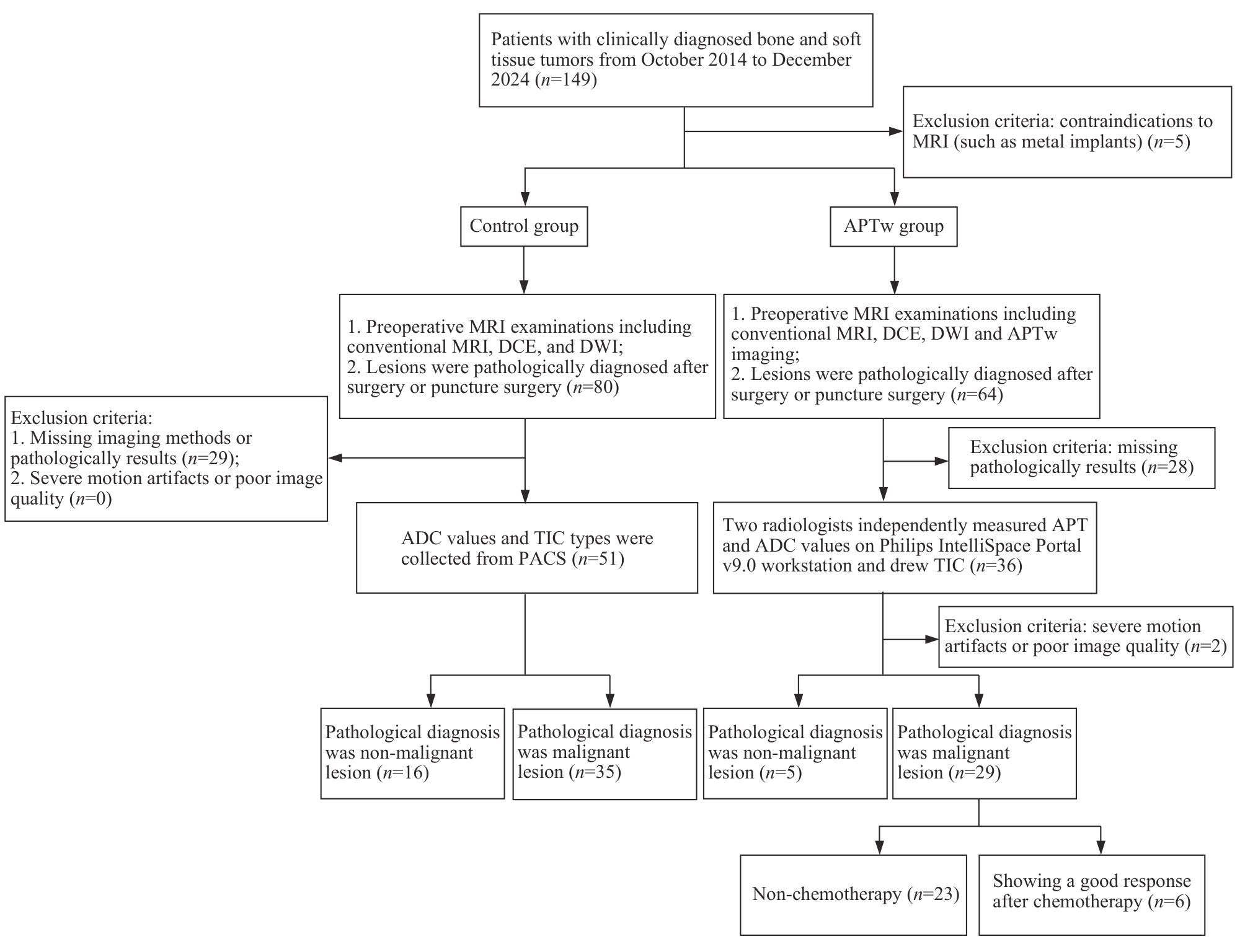
Fig 1 Flowchart summarizing participant selection
| Parameter | T1WI | T2WI | T2-SPAIR | DWI | DCE | APTw |
|---|---|---|---|---|---|---|
| Sequence | TSE | TSE | TSE | EPI | TSE | TSE |
| Plane | Axial | Axial | Coronal | Axial | Axial | Axial |
| Fat saturation | NO | NO | SPAIR | SPAIR | NO | SPIR |
| Time to repeat/ms | 672 | 3 000 | 3 200 | 3 048 | 638 | 7 300 |
| Time to echo/ms | 13 | 75 | 60 | 56 | 15 | 8.3 |
| Thickness/mm | 4 | 4 | 4 | 5 | 4 | 6 |
| FOV/mm | 250×400 | 300×379 | 280×350 | 300×450 | 300×379 | 230×180 |
| Matrix | 312×418 | 352×375 | 352×368 | 152×197 | 352×402 | 128×100 |
| Slice gap/mm | 0.4 | 0.4 | 0.4 | 0.5 | 0.4 | 0 |
| NSA | 1 | 1 | 1 | 1 | 1 | 1 |
| b value/(s·mm -2) | ‒ | ‒ | ‒ | 0, 1 000 | ‒ | ‒ |
| Scan time | 2 min 5 s | 2 min 30 s | 2 min 14 s | 1 min 41 s | 2 min 32 s | 3 min 37 s |
Tab 1 MRI scanning parameters
| Parameter | T1WI | T2WI | T2-SPAIR | DWI | DCE | APTw |
|---|---|---|---|---|---|---|
| Sequence | TSE | TSE | TSE | EPI | TSE | TSE |
| Plane | Axial | Axial | Coronal | Axial | Axial | Axial |
| Fat saturation | NO | NO | SPAIR | SPAIR | NO | SPIR |
| Time to repeat/ms | 672 | 3 000 | 3 200 | 3 048 | 638 | 7 300 |
| Time to echo/ms | 13 | 75 | 60 | 56 | 15 | 8.3 |
| Thickness/mm | 4 | 4 | 4 | 5 | 4 | 6 |
| FOV/mm | 250×400 | 300×379 | 280×350 | 300×450 | 300×379 | 230×180 |
| Matrix | 312×418 | 352×375 | 352×368 | 152×197 | 352×402 | 128×100 |
| Slice gap/mm | 0.4 | 0.4 | 0.4 | 0.5 | 0.4 | 0 |
| NSA | 1 | 1 | 1 | 1 | 1 | 1 |
| b value/(s·mm -2) | ‒ | ‒ | ‒ | 0, 1 000 | ‒ | ‒ |
| Scan time | 2 min 5 s | 2 min 30 s | 2 min 14 s | 1 min 41 s | 2 min 32 s | 3 min 37 s |
| Variable | Control group ( n=51) | APTw group ( n=34) | P value | ||||
|---|---|---|---|---|---|---|---|
| Non-malignant lesion ( n=16) | Malignant lesion ( n=35) | P value | Non-malignant lesion ( n=5) | Malignant lesion ( n=29) | P value | ||
| Gender/ n(%) | 0.068 | 0.618 | 0.119 | ||||
| Male | 5 (31.3) | 22 (62.9) | 3 (60.0) | 21 (72.4) | |||
| Female | 11 (68.8) | 13 (37.1) | 2 (40.0) | 8 (27.6) | |||
| Age/year | 39.00±12.87 | 47.40±17.57 | 0.111 | 41.80±12.46 | 40.24±20.10 | 0.925 | 0.317 |
| Maximum diameter/cm | 4.80 (5.28) | 7.69±3.13 | 0.082 | 4.24±0.83 | 8.14±4.33 | 0.038 | 0.986 |
| Margin/ n(%) | 0.001 | 0.146 | 0.509 | ||||
| Clear | 14 (87.5) | 13 (37.1) | 4 (80.0) | 11 (37.9) | |||
| Unclear | 2 (12.5) | 22 (62.9) | 1 (20.0) | 18 (62.1) | |||
| Signal/ n(%) | 0.187 | 0.570 | 0.624 | ||||
| Homogeneous | 7 (43.8) | 8 (22.9) | 2 (40.0) | 6 (20.7) | |||
| Heterogeneous | 9 (56.3) | 27 (77.1) | 3 (60.0) | 23 (79.3) | |||
| Shape/ n(%) | 0.157 | 0.205 | 0.595 | ||||
| Regular | 6 (37.5) | 6 (17.1) | 2 (40.0) | 4 (13.8) | |||
| Irregular | 10 (62.5) | 29 (82.9) | 3 (60.0) | 25 (86.2) | |||
| Peritumoral edema/ n(%) | 0.115 | 0.591 | 0.632 | ||||
| No | 8 (50.0) | 9 (25.7) | 2 (40.0) | 7 (24.1) | |||
| Yes | 8 (50.0) | 26 (74.3) | 3 (60.0) | 22 (75.9) | |||
| Cortical bone destruction/ n(%) | 1.000 | 1.000 | 0.257 | ||||
| No | 1 (6.3) | 2 (5.7) | 1 (20.0) | 4 (13.8) | |||
| Yes | 15 (93.8) | 33 (94.3) | 4 (80.0) | 25 (86.2) | |||
Tab 2 Patients′ clinical and conventional imaging characteristics
| Variable | Control group ( n=51) | APTw group ( n=34) | P value | ||||
|---|---|---|---|---|---|---|---|
| Non-malignant lesion ( n=16) | Malignant lesion ( n=35) | P value | Non-malignant lesion ( n=5) | Malignant lesion ( n=29) | P value | ||
| Gender/ n(%) | 0.068 | 0.618 | 0.119 | ||||
| Male | 5 (31.3) | 22 (62.9) | 3 (60.0) | 21 (72.4) | |||
| Female | 11 (68.8) | 13 (37.1) | 2 (40.0) | 8 (27.6) | |||
| Age/year | 39.00±12.87 | 47.40±17.57 | 0.111 | 41.80±12.46 | 40.24±20.10 | 0.925 | 0.317 |
| Maximum diameter/cm | 4.80 (5.28) | 7.69±3.13 | 0.082 | 4.24±0.83 | 8.14±4.33 | 0.038 | 0.986 |
| Margin/ n(%) | 0.001 | 0.146 | 0.509 | ||||
| Clear | 14 (87.5) | 13 (37.1) | 4 (80.0) | 11 (37.9) | |||
| Unclear | 2 (12.5) | 22 (62.9) | 1 (20.0) | 18 (62.1) | |||
| Signal/ n(%) | 0.187 | 0.570 | 0.624 | ||||
| Homogeneous | 7 (43.8) | 8 (22.9) | 2 (40.0) | 6 (20.7) | |||
| Heterogeneous | 9 (56.3) | 27 (77.1) | 3 (60.0) | 23 (79.3) | |||
| Shape/ n(%) | 0.157 | 0.205 | 0.595 | ||||
| Regular | 6 (37.5) | 6 (17.1) | 2 (40.0) | 4 (13.8) | |||
| Irregular | 10 (62.5) | 29 (82.9) | 3 (60.0) | 25 (86.2) | |||
| Peritumoral edema/ n(%) | 0.115 | 0.591 | 0.632 | ||||
| No | 8 (50.0) | 9 (25.7) | 2 (40.0) | 7 (24.1) | |||
| Yes | 8 (50.0) | 26 (74.3) | 3 (60.0) | 22 (75.9) | |||
| Cortical bone destruction/ n(%) | 1.000 | 1.000 | 0.257 | ||||
| No | 1 (6.3) | 2 (5.7) | 1 (20.0) | 4 (13.8) | |||
| Yes | 15 (93.8) | 33 (94.3) | 4 (80.0) | 25 (86.2) | |||
| Variable | Radiologist 1 | Radiologist 2 | ICC/ κ | 95% CI |
|---|---|---|---|---|
| APT | 2.50% (1.23%) | 2.63% (1.09%) | 0.977 | 0.954‒0.988 |
| ADC/(10 -3·mm 2·s -1) | 1.01 (0.67) | 1.08 (0.69) | 0.964 | 0.929‒0.982 |
| TIC (Ⅰ/Ⅱ/Ⅲ)/ n(%) | 17 (50.0)/14 (41.2)/3 (8.8) | 16 (47.1)/15 (44.1)/3 (8.8) | 0.847 | 0.678‒1.000 |
Tab 3 ICCs between the two radiologists in the APTw group
| Variable | Radiologist 1 | Radiologist 2 | ICC/ κ | 95% CI |
|---|---|---|---|---|
| APT | 2.50% (1.23%) | 2.63% (1.09%) | 0.977 | 0.954‒0.988 |
| ADC/(10 -3·mm 2·s -1) | 1.01 (0.67) | 1.08 (0.69) | 0.964 | 0.929‒0.982 |
| TIC (Ⅰ/Ⅱ/Ⅲ)/ n(%) | 17 (50.0)/14 (41.2)/3 (8.8) | 16 (47.1)/15 (44.1)/3 (8.8) | 0.847 | 0.678‒1.000 |
| Variable | Non-malignant lesions group ( n=5) | Malignant lesions group ( n=29) | U/ χ2 | P value |
|---|---|---|---|---|
| APT | 4.52%±0.76% | 2.47% (0.56%) | 5.00 | <0.001 |
| ADC/(10 -3·mm 2·s -1) | 1.37±0.52 | 1.03 (0.66) | 44.00 | 0.527 |
| TIC(Ⅰ/Ⅱ/Ⅲ)/ n(%) | 3 (60.0)/1 (20.1)/1 (20.0) | 14 (48.3)/13 (44.8)/2 (6.9) | 1.59 | 0.453 |
Tab 4 APT and ADC values, and TIC classifications between non-malignant and malignant lesions in the APTw group
| Variable | Non-malignant lesions group ( n=5) | Malignant lesions group ( n=29) | U/ χ2 | P value |
|---|---|---|---|---|
| APT | 4.52%±0.76% | 2.47% (0.56%) | 5.00 | <0.001 |
| ADC/(10 -3·mm 2·s -1) | 1.37±0.52 | 1.03 (0.66) | 44.00 | 0.527 |
| TIC(Ⅰ/Ⅱ/Ⅲ)/ n(%) | 3 (60.0)/1 (20.1)/1 (20.0) | 14 (48.3)/13 (44.8)/2 (6.9) | 1.59 | 0.453 |
| Variable | Non-chemotherapy group ( n=23) | Chemotherapy group ( n=6) | U/ χ2 | P value |
|---|---|---|---|---|
| APT | 2.43%±0.23% | 3.67%±0.24% | 138.00 | <0.001 |
| ADC/(10 -3·mm 2·s -1) | 1.00 (0.66) | 1.31±0.65 | 75.00 | 0.643 |
| TIC(Ⅰ/Ⅱ/Ⅲ)/ n(%) | 13 (56.5)/9 (39.1)/1 (4.3) | 1 (16.7)/4 (66.7)/1 (16.7) | 3.42 | 0.181 |
Tab 5 APT and ADC values, and TIC classifications in APTw group patients with malignant lesions, comparing those who received chemotherapy with those who did not
| Variable | Non-chemotherapy group ( n=23) | Chemotherapy group ( n=6) | U/ χ2 | P value |
|---|---|---|---|---|
| APT | 2.43%±0.23% | 3.67%±0.24% | 138.00 | <0.001 |
| ADC/(10 -3·mm 2·s -1) | 1.00 (0.66) | 1.31±0.65 | 75.00 | 0.643 |
| TIC(Ⅰ/Ⅱ/Ⅲ)/ n(%) | 13 (56.5)/9 (39.1)/1 (4.3) | 1 (16.7)/4 (66.7)/1 (16.7) | 3.42 | 0.181 |
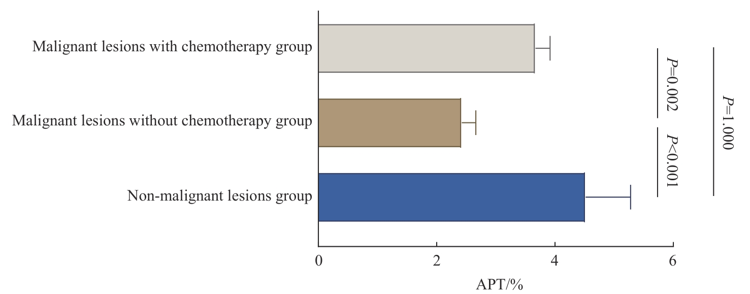
Fig 2 Multiple comparisons of APT values in the APTw group
| Variable | Non-malignant lesions group ( n=16) | Malignant lesions group ( n=35) | U/ χ2 | P value |
|---|---|---|---|---|
| ADC/(10 -3·mm 2·s -1) | 1.33±0.55 | 1.00 (0.89) | 260.50 | 0.690 |
| TIC(Ⅰ/Ⅱ/Ⅲ)/ n(%) | 10 (62.5)/4 (25.0)/2 (12.5) | 17 (48.6)/13 (37.1)/5 (14.3) | 0.91 | 0.633 |
Tab 6 ADC values and TIC classification between non-malignant and malignant lesions in the control group
| Variable | Non-malignant lesions group ( n=16) | Malignant lesions group ( n=35) | U/ χ2 | P value |
|---|---|---|---|---|
| ADC/(10 -3·mm 2·s -1) | 1.33±0.55 | 1.00 (0.89) | 260.50 | 0.690 |
| TIC(Ⅰ/Ⅱ/Ⅲ)/ n(%) | 10 (62.5)/4 (25.0)/2 (12.5) | 17 (48.6)/13 (37.1)/5 (14.3) | 0.91 | 0.633 |
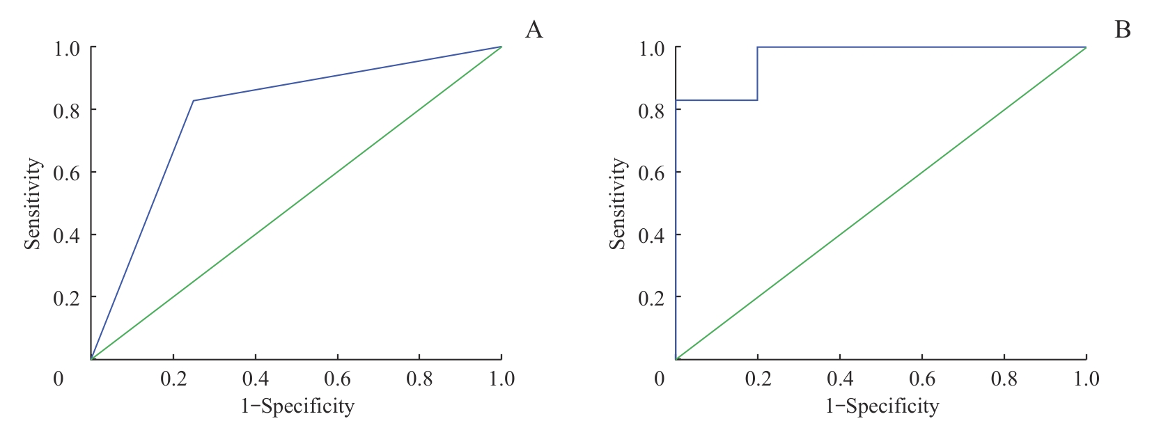
Fig 3 ROC curves showing the diagnostic efficacy of different models for distinguishing malignant from non-malignant bone and soft tissue lesions
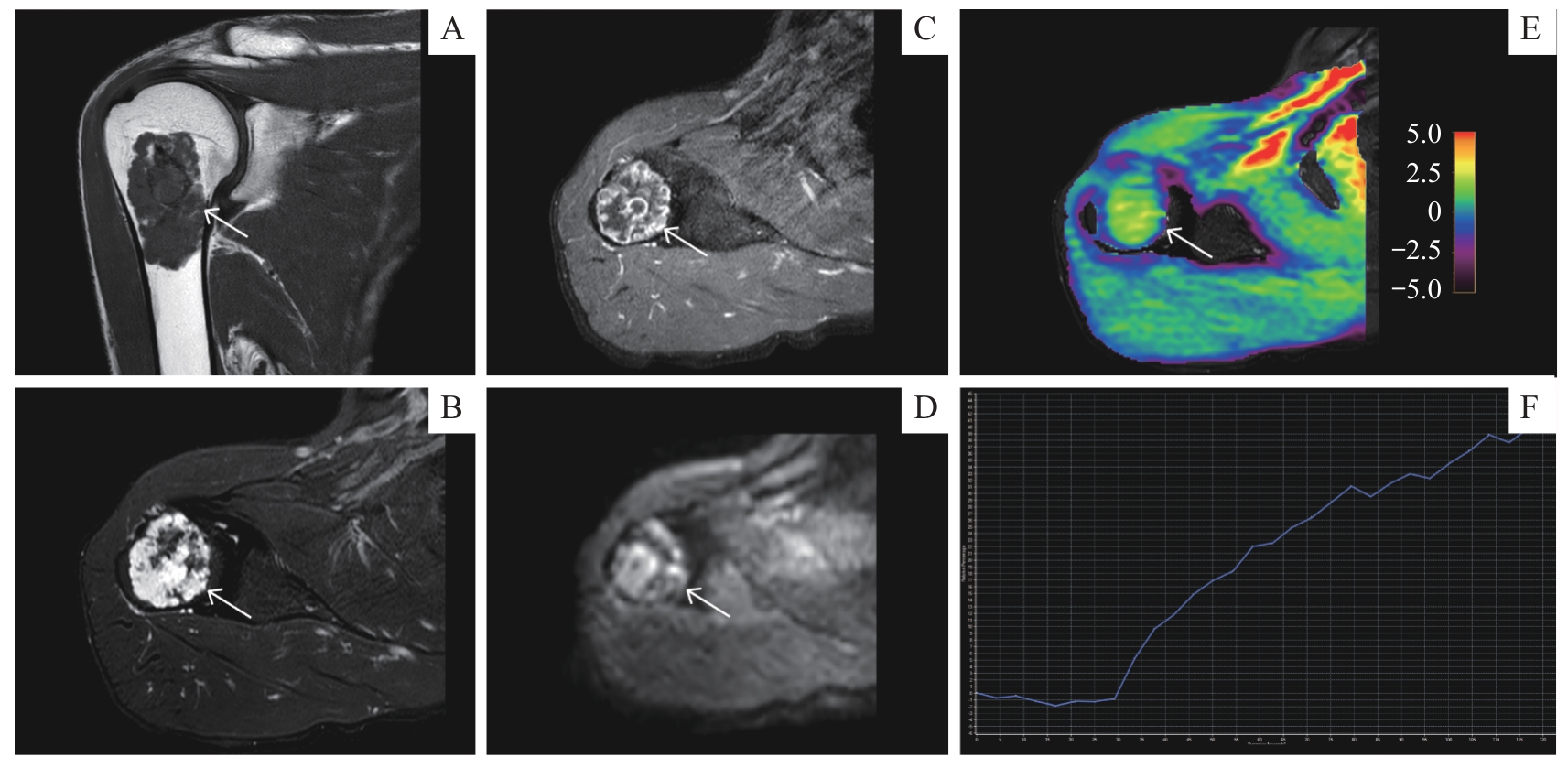
Fig 4 Application of APTw imaging to malignant bone tumor
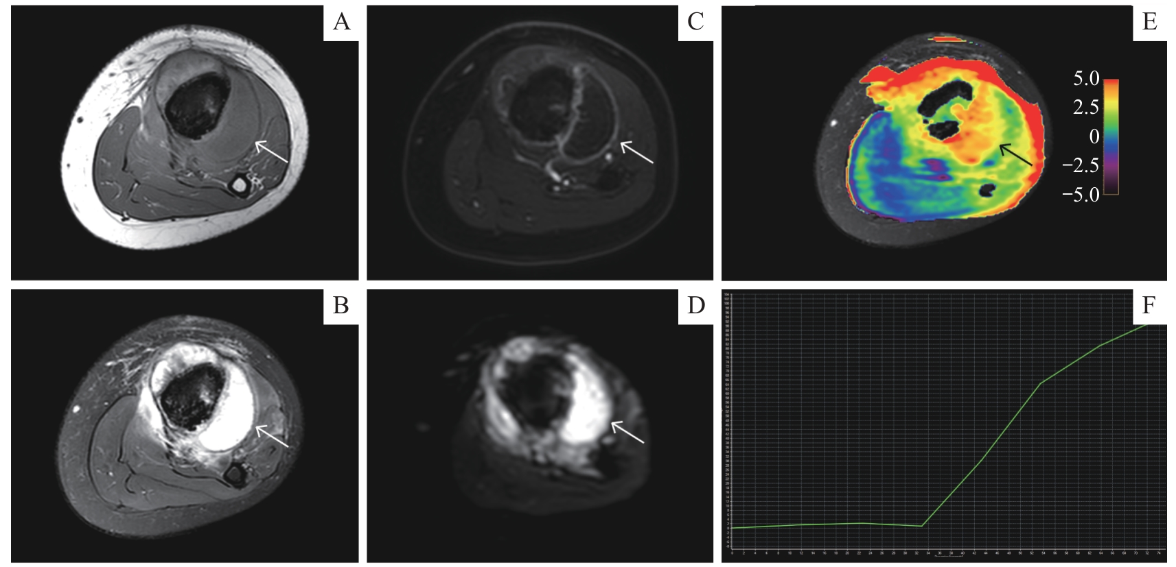
Fig 5 Application of APTw imaging in neoadjuvant chemotherapy for malignant bone tumor
| 1 | CHOI J H, RO J Y. The 2020 WHO classification of tumors of bone: an updated review[J]. Adv Anat Pathol, 2021, 28(3): 119-138. |
| 2 | CHOI J H, RO J Y. The 2020 WHO classification of tumors of soft tissue: selected changes and new entities[J]. Adv Anat Pathol, 2021, 28(1): 44-58. |
| 3 | WANG Y A, YAN Q J, FAN C M, et al. Overview and countermeasures of cancer burden in China[J]. Sci China Life Sci, 2023, 66(11): 2515-2526. |
| 4 | COLE S, MATTHEW GIANFERANTE D, ZHU B, et al. Osteosarcoma: a surveillance, epidemiology, and end results program-based analysis from 1975 to 2017[J]. Cancer, 2022, 128(11): 2107-2118. |
| 5 | JAFARI F, JAVDANSIRAT S, SANAIE S, et al. Osteosarcoma: a comprehensive review of management and treatment strategies[J]. Ann Diagn Pathol, 2020, 49: 151654. |
| 6 | KUNISADA T, NAKATA E, FUJIWARA T, et al. Soft-tissue sarcoma in adolescents and young adults[J]. Int J Clin Oncol, 2023, 28(1): 1-11. |
| 7 | CHOI J H, RO J Y. The recent advances in molecular diagnosis of soft tissue tumors[J]. Int J Mol Sci, 2023, 24(6): 5934. |
| 8 | TANAKA K, OZAKI T. Adjuvant and neoadjuvant chemotherapy for soft tissue sarcomas: JCOG Bone and Soft Tissue Tumor Study Group[J]. Jpn J Clin Oncol, 2021, 51(2): 180-184. |
| 9 | YUAN W Z, YU Q Y, WANG Z, et al. Efficacy of diffusion-weighted imaging in neoadjuvant chemotherapy for osteosarcoma: a systematic review and meta-analysis[J]. Acad Radiol, 2022, 29(2): 326-334. |
| 10 | HABRE C, DABADIE A, LOUNDOU A D, et al. Diffusion-weighted imaging in differentiating mid-course responders to chemotherapy for long-bone osteosarcoma compared to the histologic response: an update[J]. Pediatr Radiol, 2021, 51(9): 1714-1723. |
| 11 | GUEDES A, NAKAGAWA S A. Biopsy of bone tumors: a literature review[J]. Rev Assoc Med Bras (1992), 2024, 70(suppl 1): e2024S131. |
| 12 | WHELAN J S, DAVIS L E. Osteosarcoma, chondrosarcoma, and chordoma[J]. J Clin Oncol, 2018, 36(2): 188-193. |
| 13 | LI J L, XU Y, XIANG Y S, et al. The value of amide proton transfer MRI in the diagnosis of malignant and benign urinary bladder lesions: comparison with diffusion-weighted imaging[J]. J Magn Reson Imaging, 2024, 60(3): 1124-1133. |
| 14 | SETIAWATI R, NOVARIYANTO B, RAHARDJO P, et al. Characteristic of apparent diffusion coefficient and time intensity curve analysis of dynamic contrast enhanced MRI in osteosarcoma histopathologic subtypes[J]. Int J Med Sci, 2023, 20(2): 163-171. |
| 15 | COSTA F M, CANELLA C, GASPARETTO E. Advanced magnetic resonance imaging techniques in the evaluation of musculoskeletal tumors[J]. Radiol Clin North Am, 2011, 49(6): 1325-1358, Ⅶ-Ⅷ. |
| 16 | KHALIFA F, SOLIMAN A, EL-BAZ A, et al. Models and methods for analyzing DCE-MRI: a review[J]. Med Phys, 2014, 41(12): 124301. |
| 17 | 赵傲, 武新英, 付彤, 等. DWI、DCE联合APT成像在鉴别乳腺良恶性病变中的价值研究[J]. 中国医疗设备, 2024, 39(6): 124-130, 155. |
| ZHAO A, WU X Y, FU T, et al. Value of DWI, DCE and APT imaging in identifying benign and malignant breast lesions[J]. China Medical Devices, 2024, 39(6): 124-130, 155. | |
| 18 | KUANG F, YAN Z P, LI H L, et al. Diagnostic accuracy of diffusion-weighted MRI for differentiation of cervical cancer and benign cervical lesions at 3.0T: comparison with routine MRI and dynamic contrast-enhanced MRI[J]. J Magn Reson Imaging, 2015, 42(4): 1094-1099. |
| 19 | ZHOU J Y, HEO H Y, KNUTSSON L, et al. APT-weighted MRI: techniques, current neuro applications, and challenging issues[J]. J Magn Reson Imaging, 2019, 50(2): 347-364. |
| 20 | WU M H, JIANG T L, GUO M, et al. Amide proton transfer-weighted imaging and derived radiomics in the classification of adult-type diffuse gliomas[J]. Eur Radiol, 2024, 34(5): 2986-2996. |
| 21 | LI Y, LIN C Y, QI Y F, et al. Non-invasive differentiation of endometrial adenocarcinoma from benign lesions in the uterus by utilization of amide proton transfer-weighted MRI[J]. Mol Imaging Biol, 2021, 23(3): 446-455. |
| 22 | LIU Z, WEN J, WANG M, et al. Breast amide proton transfer imaging at 3 T: diagnostic performance and association with pathologic characteristics[J]. J Magn Reson Imaging, 2023, 57(3): 824-833. |
| 23 | DENG H Z, ZHANG H W, HUANG B, et al. Advances in diffuse glioma assessment: preoperative and postoperative applications of chemical exchange saturation transfer[J]. Front Neurosci, 2024, 18: 1424316. |
| 24 | QAMAR S, KING A D, AI Q H, et al. Pre-treatment amide proton transfer imaging predicts treatment outcome in nasopharyngeal carcinoma[J]. Eur Radiol, 2020, 30(11): 6339-6347. |
| 25 | ZHANG N, SONG Q W, LIANG H B, et al. Early prediction of pathological response to neoadjuvant chemotherapy of breast tumors: a comparative study using amide proton transfer-weighted, diffusion weighted and dynamic contrast enhanced MRI[J]. Front Med (Lausanne), 2024, 11: 1295478. |
| 26 | 史天亮, 杨崇双, 刘元早, 等. APT成像在宫颈癌同步根治性放化疗疗效评估中的价值[J]. 磁共振成像, 2024, 15(1): 132-136. |
| SHI T L, YANG C S, LIU Y Z, et al. The value of APT imaging in evaluating the therapeutic efficacy of concurrent chemoradiotherapy for cervical cancer[J]. Chinese Journal of Magnetic Resonance Imaging, 2024, 15(1): 132-136. | |
| 27 | 刘斯润, 蔡香然, 邱麟. 新版(2020)WHO骨肿瘤分类解读[J]. 磁共振成像, 2020, 11(12): 1086-1091. |
| LIU S R, CAI X R, QIU L. Interpretation of the new WHO classification of bone tumors (2020) [J]. Chinese Journal of Magnetic Resonance Imaging, 2020, 11(12): 1086-1091. | |
| 28 | 方三高, 魏建国, 陈真伟. WHO(2020)软组织肿瘤分类[J]. 临床与实验病理学杂志, 2020, 36(9): 1132-1134. |
| FANG S G, WEI J G, CHEN Z W. WHO classification of soft tissue tumors (2020) [J]. Chinese Journal of Clinical and Experimental Pathology, 2020, 36(9): 1132-1134. | |
| 29 | YABUUCHI H, FUKUYA T, TAJIMA T, et al. Salivary gland tumors: diagnostic value of gadolinium-enhanced dynamic MR imaging with histopathologic correlation[J]. Radiology, 2003, 226(2): 345-354. |
| 30 | CHOI Y J, LEE I S, SONG Y S, et al. Diagnostic performance of diffusion-weighted (DWI) and dynamic contrast-enhanced (DCE) MRI for the differentiation of benign from malignant soft-tissue tumors[J]. J Magn Reson Imaging, 2019, 50(3): 798-809. |
| 31 | HAYAKAWA K, MATSUMOTO S, AE K, et al. Definitive surgery of primary lesion should be prioritized over preoperative chemotherapy to treat high-grade osteosarcoma in patients aged 41‒65 years[J]. J Orthop Traumatol, 2020, 21(1): 13. |
| 32 | CAO Z Y, ZHANG Y L, XU Q, et al. The role of chemotherapy in the survival benefits of patients aged older than 40 years with osteosarcoma[J]. Technol Cancer Res Treat, 2021, 20: 15330338211066195. |
| 33 | GUO Z X, QIN X Y, MU R H, et al. Amide proton transfer could provide more accurate lesion characterization in the transition zone of the prostate[J]. J Magn Reson Imaging, 2022, 56(5): 1311-1319. |
| 34 | GAO Y T, ZHOU H, LIU G G, et al. Tumor microenvironment: lactic acid promotes tumor development[J]. J Immunol Res, 2022, 2022: 3119375. |
| 35 | TOGAO O, YOSHIURA T, KEUPP J, et al. Amide proton transfer imaging of adult diffuse gliomas: correlation with histopathological grades[J]. Neuro Oncol, 2014, 16(3): 441-448. |
| 36 | MENG N, WANG X J, SUN J, et al. Comparative study of amide proton transfer-weighted imaging and intravoxel incoherent motion imaging in breast cancer diagnosis and evaluation[J]. J Magn Reson Imaging, 2020, 52(4): 1175-1186. |
| 37 | KOIKE H, MORIKAWA M, ISHIMARU H, et al. Amide proton transfer-chemical exchange saturation transfer imaging of intracranial brain tumors and tumor-like lesions: our experience and a review[J]. Diagnostics (Basel), 2023, 13(5): 914. |
| 38 | YANG L, WANG L, TAN Y C, et al. Amide Proton Transfer-weighted MRI combined with serum prostate-specific antigen levels for differentiating malignant prostate lesions from benign prostate lesions: a retrospective cohort study[J]. Cancer Imaging, 2023, 23(1): 3. |
| 39 | JIA G, ABAZA R, WILLIAMS J D, et al. Amide proton transfer MR imaging of prostate cancer: a preliminary study[J]. J Magn Reson Imaging, 2011, 33(3): 647-654. |
| 40 | ZHANG H W, LIU X L, ZHANG H B, et al. Differentiation of meningiomas and gliomas by amide proton transfer imaging: a preliminary study of brain tumour infiltration[J]. Front Oncol, 2022, 12: 886968. |
| 41 | 李莹, 程敬亮, 任翠萍, 等. 3D氨基质子转移加权成像联合弥散加权成像鉴别良、恶性骨与软组织肿瘤[J]. 中国医学影像技术, 2024, 40(10): 1572-1576. |
| LI Y,CHENG J L, REN C P, et al. 3D amide proton transfer weighted imaging combined with diffusion weighted imaging for differentiating benign and malignant bone and soft tissue tumors[J]. Chinese Journal of Medical Imaging Technology, 2024, 40(10): 1572-1576. | |
| 42 | LI Y, LIN L J, ZHANG Y, et al. Preliminary exploration of amide proton transfer weighted imaging in differentiation between benign and malignant bone tumors[J]. Front Oncol, 2024, 14: 1402628. |
| 43 | 刘记存, 崔建岭, 李石玲, 等. 表观扩散系数值鉴别良恶性骨肿瘤及肿瘤样病变的价值[J]. 中华放射学杂志, 2009, 43(6): 567-570. |
| LIU J C, CUI J L, LI S L, et al. The role of apparent diffusion coefficient in the differentiation between benign and malignant bone tumors[J]. Chinese Journal of Radiology, 2009, 43(6): 567-570. | |
| 44 | HAYASHIDA Y, HIRAI T, YAKUSHIJI T, et al. Evaluation of diffusion-weighted imaging for the differential diagnosis of poorly contrast-enhanced and T2-prolonged bone masses: initial experience[J]. J Magn Reson Imaging, 2006, 23(3): 377-382. |
| 45 | WANG T T, WU X R, CUI Y F, et al. Role of apparent diffusion coefficients with diffusion-weighted magnetic resonance imaging in differentiating between benign and malignant bone tumors[J]. World J Surg Oncol, 2014, 12: 365. |
| 46 | 王绍武, 孙美玉, 张丽娜, 等. 骨肿瘤的MR灌注和扩散加权成像研究[J]. 国际医学放射学杂志, 2008, 31(1): 6-10. |
| WANG S W, SUN M Y, ZHANG L N, et al. MR perfusion and diffusion imaging in diagnosing benign and malignant bone tumors[J]. International Journal of Medical Radiology, 2008, 31(1): 6-10. | |
| 47 | HISATOMI M, ASAUMI J I, YANAGI Y, et al. Diagnostic value of dynamic contrast-enhanced MRI in the salivary gland tumors[J]. Oral Oncol, 2007, 43(9): 940-947. |
| 48 | ZHOU J Y, ZAISS M, KNUTSSON L, et al. Review and consensus recommendations on clinical APT-weighted imaging approaches at 3T: application to brain tumors[J]. Magn Reson Med, 2022, 88(2): 546-574. |
| 49 | GUO H, LIU J, HU J J, et al. Diagnostic performance of gliomas grading and IDH status decoding A comparison between 3D amide proton transfer APT and four diffusion-weighted MRI models[J]. J Magn Reson Imaging, 2022, 56(6): 1834-1844. |
| [1] | JIANG Yi, HUANG Chenhao, LI Zhiliang, WU Junwei, ZHAO Ren, ZHANG Tao. Effect of preoperative chemotherapy combined with immunotherapy in a colorectal cancer patient with KRAS mutation [J]. Journal of Shanghai Jiao Tong University (Medical Science), 2025, 45(9): 1256-1260. |
| [2] | WU Qizhen, LIU Qiming, CHAI Yezi, TAO Zhengyu, WANG Yinan, GUO Xinning, JIANG Meng, PU Jun. Evaluation of machine learning prediction of altered inflammatory metabolic state after neoadjuvant therapy for breast cancer [J]. Journal of Shanghai Jiao Tong University (Medical Science), 2024, 44(9): 1169-1181. |
| [3] | XING Zhengwen, WU Ying, WANG Xueli, WANG Qingyu, WANG Wenting, LI Zhi, ZHANG Bin, JIN Jing. Clinicopathological characteristics of CIC-rearranged sarcoma in children [J]. Journal of Shanghai Jiao Tong University (Medical Science), 2022, 42(8): 1151-1157. |
| [4] | HE Fang-zhou, ZHANG Wei-bin, SHEN Yu-hui. A report on 8 cases of total elbow arthroplasty with customized constrained elbow prosthesis [J]. , 2016, 36(8): 1205-. |
| Viewed | ||||||
|
Full text |
|
|||||
|
Abstract |
|
|||||