
Journal of Shanghai Jiao Tong University (Medical Science) ›› 2026, Vol. 46 ›› Issue (10): 1339-1354.doi: 10.3969/j.issn.1674-8115.2026.10.004
• Basic research • Previous Articles
Deng Rui1,2, Fang Houshun1,2, Zhou Jiquan1,2, Chen Yao1,2( ), Li Hui1,2()
Received:2026-04-15
Accepted:2026-06-20
Online:2026-10-15
Published:2026-10-15
Contact:
Chen Yao, Li Hui
E-mail:chenyao01@scmc.com.cn;lihui@scmc.com.cn
Supported by:CLC Number:
Deng Rui, Fang Houshun, Zhou Jiquan, Chen Yao, Li Hui. USP37-mediated deubiquitination maintains c-MYC protein homeostasis and contributes to hepatoblastoma progression[J]. Journal of Shanghai Jiao Tong University (Medical Science), 2026,(10): 1339-1354.
Add to citation manager EndNote|Ris|BibTeX
URL: https://xuebao.shsmu.edu.cn/EN/10.3969/j.issn.1674-8115.2026.10.004
| shRNA name | shRNA sequence (5'→3') | |
|---|---|---|
| USP37 shRNA-1 | Forward | CCGGTCCGGGTAGAGGATCGATTAACTCGAGTTAATCGATCCTCTACCCGGATTTTTG |
| Reverse | AATTCAAAAATCCGGGTAGAGGATCGATTAACTCGAGTTAATCGATCCTCTACCCGGA | |
| USP37 shRNA-2 | Forward | CCGGCCGGATTTGCAGAAGATGATACTCGAGTATCATCTTCTGCAAATCCGGTTTTTG |
| Reverse | AATTCAAAAACCGGATTTGCAGAAGATGATACTCGAGTATCATCTTCTGCAAATCCGG |
Tab 1 shRNA sequences
| shRNA name | shRNA sequence (5'→3') | |
|---|---|---|
| USP37 shRNA-1 | Forward | CCGGTCCGGGTAGAGGATCGATTAACTCGAGTTAATCGATCCTCTACCCGGATTTTTG |
| Reverse | AATTCAAAAATCCGGGTAGAGGATCGATTAACTCGAGTTAATCGATCCTCTACCCGGA | |
| USP37 shRNA-2 | Forward | CCGGCCGGATTTGCAGAAGATGATACTCGAGTATCATCTTCTGCAAATCCGGTTTTTG |
| Reverse | AATTCAAAAACCGGATTTGCAGAAGATGATACTCGAGTATCATCTTCTGCAAATCCGG |
| Primer | Application | Forward | Reverse |
|---|---|---|---|
| USP37-Flag | Plasmid construction | CTAGAGGATCCCCGGGTACCGGTCGCCACCATGTCTCCTCTGAAGATACATGGT | CCGTCATGGTCTTTGTAGTCGCTAGCCAAGGCCTGACGGGTAGTCTT |
| USP37-HA | Plasmid construction | CTAGAGGATCCCCGGGTACCGGTCGCCACCATGTCTCCTCTGAAGATACATGGT | TCTGGGACATCATAAGGGTAGCTAGCCAAGGCCTGACGGGTAGTCTT |
| USP37-shRNA-resistant | Plasmid construction | CCCTGGCCGTGGCTCAATCAAGACTGTAGCAGGAAGTGGAATAGCTCGGA | GATTGAGCCACGGCCAGGGTTACCAAGAACTTTTCGAAATGG |
| USP37 | qPCR | GGAACACTCTTCTGGTGGCA | GTGAGAGGAAAGGGGCACTC |
| c-MYC | qPCR | GTGGTCTTCCCCTACCCTCT | TGTTCCTCCTCAGAGTCGCT |
| β-actin | qPCR | AACTGGGACGACATGGAGAAAA | GGATAGCACAGCCTGGATAGCA |
Tab 2 Primer sequences and applications (5′→3′)
| Primer | Application | Forward | Reverse |
|---|---|---|---|
| USP37-Flag | Plasmid construction | CTAGAGGATCCCCGGGTACCGGTCGCCACCATGTCTCCTCTGAAGATACATGGT | CCGTCATGGTCTTTGTAGTCGCTAGCCAAGGCCTGACGGGTAGTCTT |
| USP37-HA | Plasmid construction | CTAGAGGATCCCCGGGTACCGGTCGCCACCATGTCTCCTCTGAAGATACATGGT | TCTGGGACATCATAAGGGTAGCTAGCCAAGGCCTGACGGGTAGTCTT |
| USP37-shRNA-resistant | Plasmid construction | CCCTGGCCGTGGCTCAATCAAGACTGTAGCAGGAAGTGGAATAGCTCGGA | GATTGAGCCACGGCCAGGGTTACCAAGAACTTTTCGAAATGG |
| USP37 | qPCR | GGAACACTCTTCTGGTGGCA | GTGAGAGGAAAGGGGCACTC |
| c-MYC | qPCR | GTGGTCTTCCCCTACCCTCT | TGTTCCTCCTCAGAGTCGCT |
| β-actin | qPCR | AACTGGGACGACATGGAGAAAA | GGATAGCACAGCCTGGATAGCA |
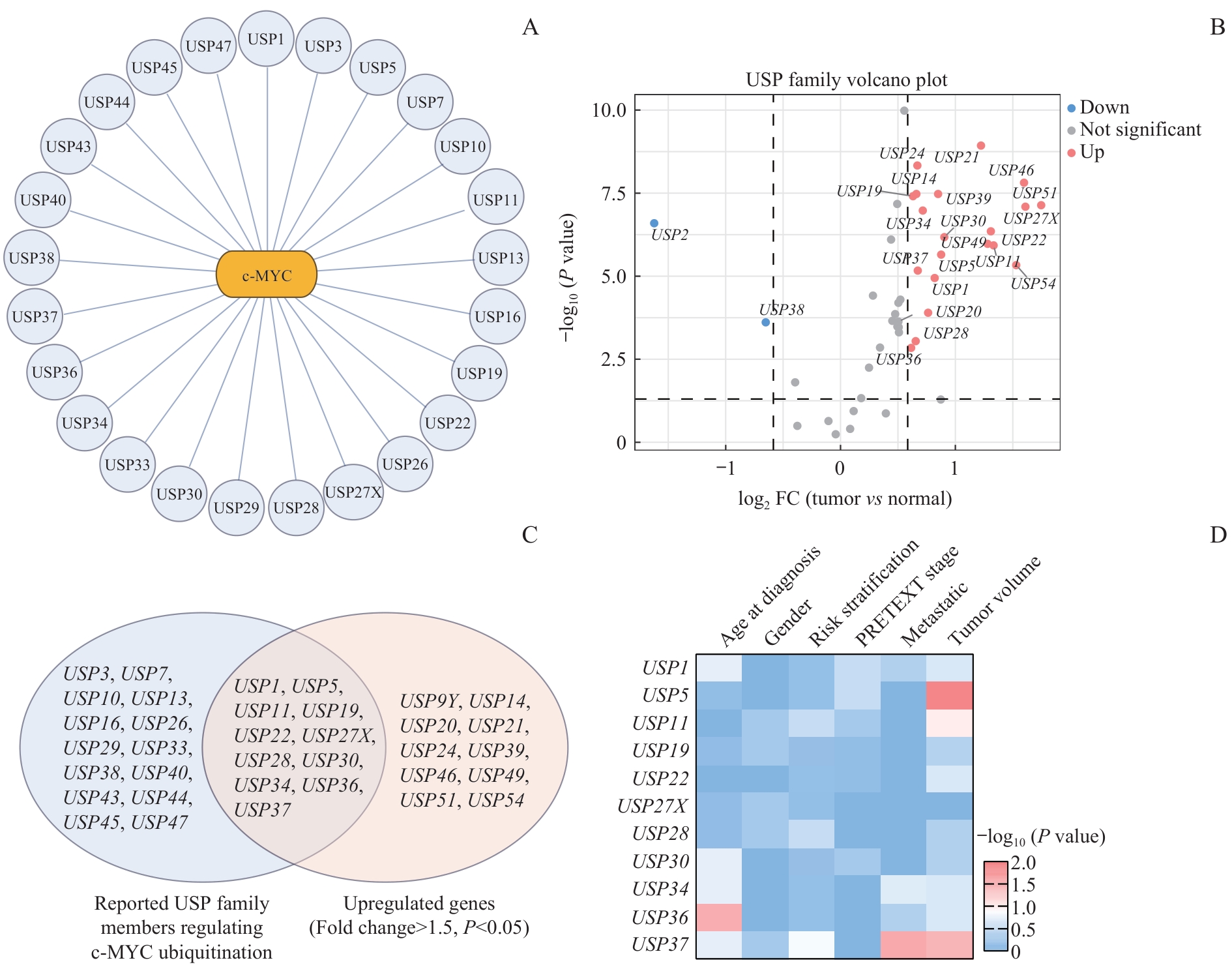
Fig 1 Systematic screening of c-MYC-associated USP family members in HB
| USP | Cell model | Reference |
|---|---|---|
| USP1 | Bladder cancer cell lines (T24, UM-UC-3) | [ |
| USP3 | Clear cell renal cell carcinoma cell lines (786-O, Caki-2) | [ |
| USP5 | Hepatocellular carcinoma cell lines (Huh-7, HepG2) | [ |
| USP7 | Pancreatic ductal adenocarcinoma cell lines (PANC1, CFPAC-1, AsPC1) | [ |
| USP10 | Osteosarcoma cell lines (143B, HOS) | [ |
| USP11 | Prostate cancer cell line (22Rv1) | [ |
| USP13 | Glioblastoma stem cells | [ |
| USP16 | Prostate cancer cell lines (PC3, DU145) | [ |
| USP19 | Hepatocellular carcinoma cell lines (Hep3b, Huh-7) | [ |
| USP22 | Breast cancer cell lines (MCF7, T47D) | [ |
| USP26 | Gastric cancer cell lines (SNU-216, HGC-27) | [ |
| USP27X | Breast cancer cell lines (MCF-7, MDA-MB-231); mouse embryonic fibroblasts | [ |
| USP28 | Osteosarcoma cell line (U2OS), cervical cancer cell line (HeLa); mouse intestinal model, MEFs, colorectal cancer model | [ |
| USP29 | Neuroblastoma cell line (SK-N-BE2), burkitt lymphoma cell line (Ramos), non-small cell lung cancer cell line (A549) | [ |
| USP30 | Oral squamous cell carcinoma cell lines (SCC4, HSC4) | [ |
| USP33 | Prostate cancer cell lines (22Rv1, DU145, PC3); ovarian cancer cell lines (SKOV3, ES2) | [ |
| USP34 | Hepatocellular carcinoma cell lines (Huh-7, HepG2, HCCLM3, MHCC97H) | [ |
| USP36 | Cervical cancer cell line (HeLa), non-small cell lung cancer cell line (H1299), breast cancer cell line (MDA-MB-231), non-tumorigenic breast epithelial cell line (MCF10A) | [ |
| USP37 | Non-small cell lung cancer cell lines (H1299) | [ |
| USP38 | Non-small cell lung cancer cell lines (A549), cervical cancer cell lines (HeLa) | [ |
| USP40 | Acute myeloid leukemia cell lines (HL-60, Kasumi-1) | [ |
| USP43 | Bladder cancer cell lines (T24, 5637, UM-UC-3) | [ |
| USP44 | Meningioma cell lines (IOMM-Lee, CH157) | [ |
| USP45 | Cervical cancer cell lines (HeLa, SiHa) | [ |
| USP47 | Non-small cell lung cancer cell lines (A549, Calu-1, H358) | [ |
Tab 3 USP family members involved in the regulation of c-MYC ubiquitination and stability
| USP | Cell model | Reference |
|---|---|---|
| USP1 | Bladder cancer cell lines (T24, UM-UC-3) | [ |
| USP3 | Clear cell renal cell carcinoma cell lines (786-O, Caki-2) | [ |
| USP5 | Hepatocellular carcinoma cell lines (Huh-7, HepG2) | [ |
| USP7 | Pancreatic ductal adenocarcinoma cell lines (PANC1, CFPAC-1, AsPC1) | [ |
| USP10 | Osteosarcoma cell lines (143B, HOS) | [ |
| USP11 | Prostate cancer cell line (22Rv1) | [ |
| USP13 | Glioblastoma stem cells | [ |
| USP16 | Prostate cancer cell lines (PC3, DU145) | [ |
| USP19 | Hepatocellular carcinoma cell lines (Hep3b, Huh-7) | [ |
| USP22 | Breast cancer cell lines (MCF7, T47D) | [ |
| USP26 | Gastric cancer cell lines (SNU-216, HGC-27) | [ |
| USP27X | Breast cancer cell lines (MCF-7, MDA-MB-231); mouse embryonic fibroblasts | [ |
| USP28 | Osteosarcoma cell line (U2OS), cervical cancer cell line (HeLa); mouse intestinal model, MEFs, colorectal cancer model | [ |
| USP29 | Neuroblastoma cell line (SK-N-BE2), burkitt lymphoma cell line (Ramos), non-small cell lung cancer cell line (A549) | [ |
| USP30 | Oral squamous cell carcinoma cell lines (SCC4, HSC4) | [ |
| USP33 | Prostate cancer cell lines (22Rv1, DU145, PC3); ovarian cancer cell lines (SKOV3, ES2) | [ |
| USP34 | Hepatocellular carcinoma cell lines (Huh-7, HepG2, HCCLM3, MHCC97H) | [ |
| USP36 | Cervical cancer cell line (HeLa), non-small cell lung cancer cell line (H1299), breast cancer cell line (MDA-MB-231), non-tumorigenic breast epithelial cell line (MCF10A) | [ |
| USP37 | Non-small cell lung cancer cell lines (H1299) | [ |
| USP38 | Non-small cell lung cancer cell lines (A549), cervical cancer cell lines (HeLa) | [ |
| USP40 | Acute myeloid leukemia cell lines (HL-60, Kasumi-1) | [ |
| USP43 | Bladder cancer cell lines (T24, 5637, UM-UC-3) | [ |
| USP44 | Meningioma cell lines (IOMM-Lee, CH157) | [ |
| USP45 | Cervical cancer cell lines (HeLa, SiHa) | [ |
| USP47 | Non-small cell lung cancer cell lines (A549, Calu-1, H358) | [ |
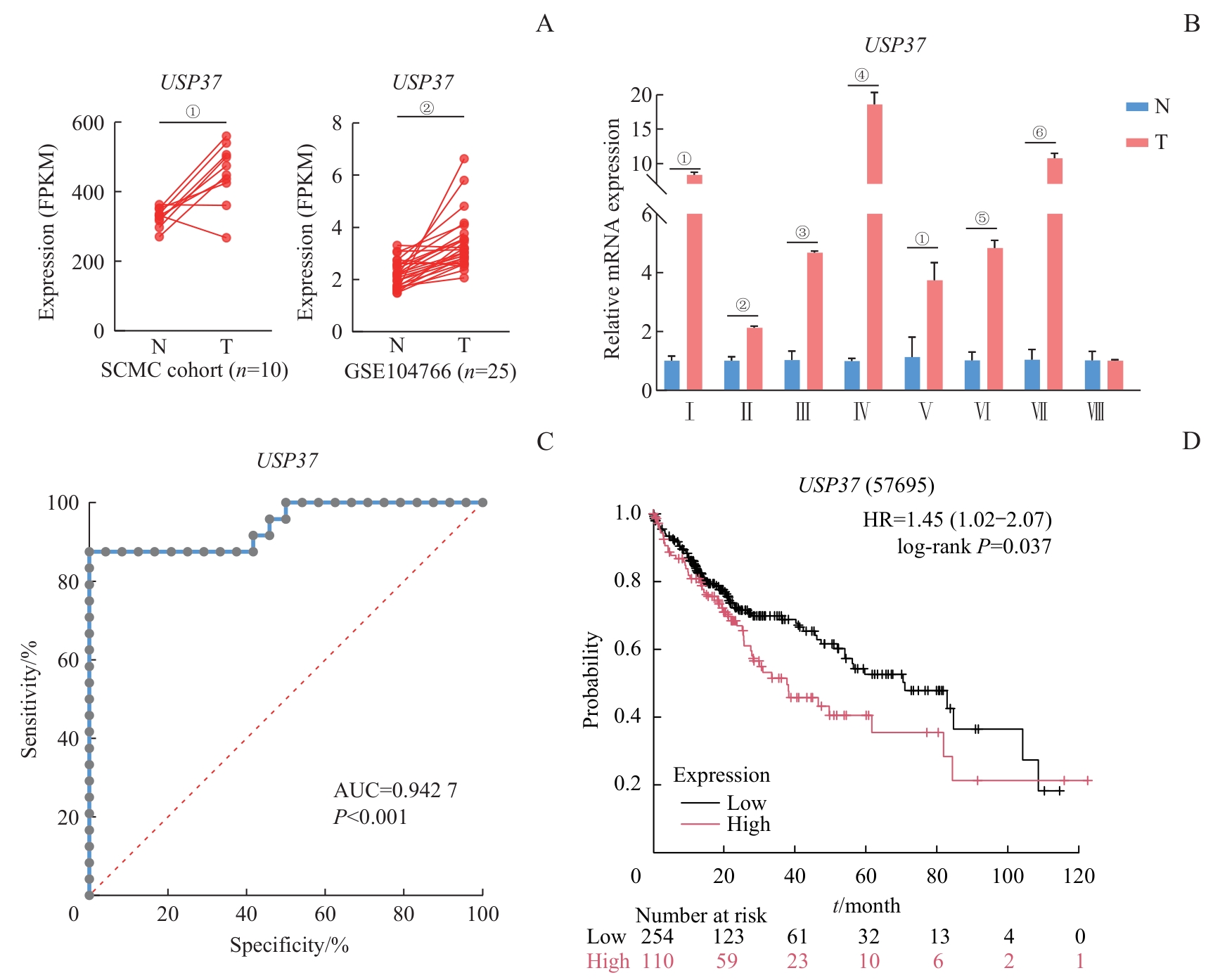
Fig 2 Expression analysis and prognostic evaluation of USP37 in HB
| Characteristic | USP37 Expression | P value | |
|---|---|---|---|
| Low (n=17) | High (n=16) | ||
| Age at diagnosis | 0.176 | ||
| ≤24 months | 11 (33.33) | 6 (18.18) | |
| >24 months | 6 (18.18) | 10 (30.30) | |
| Gender | 0.514 | ||
| Male | 9 (27.27) | 11 (33.33) | |
| Female | 8 (24.24) | 5 (15.15) | |
| Risk stratification | 0.142 | ||
| Low/intermediate-risk | 14 (42.42) | 9 (27.27) | |
| High-risk | 3 (9.09) | 7 (21.21) | |
| PRETEXT stage | 1.000 | ||
| Ⅰ/Ⅱ | 7 (21.21) | 7 (21.21) | |
| Ⅲ/Ⅳ | 10 (30.30) | 9 (27.27) | |
| Metastatic | 0.028 | ||
| Non-metastatic | 16 (48.48) | 8 (24.24) | |
| Metastatic | 1 (3.03) | 6 (18.18) | |
| NA | 0 | 2 (6.06) | |
| Tumor volume | 0.031 | ||
| ≤500 cm3 | 11 (33.33) | 3 (9.09) | |
| >500 cm3 | 2 (6.06) | 7 (21.21) | |
| NA | 4 (12.12) | 6 (18.18) | |
Tab 4 Association between USP37 expression and clinical features in HB [n(%)]
| Characteristic | USP37 Expression | P value | |
|---|---|---|---|
| Low (n=17) | High (n=16) | ||
| Age at diagnosis | 0.176 | ||
| ≤24 months | 11 (33.33) | 6 (18.18) | |
| >24 months | 6 (18.18) | 10 (30.30) | |
| Gender | 0.514 | ||
| Male | 9 (27.27) | 11 (33.33) | |
| Female | 8 (24.24) | 5 (15.15) | |
| Risk stratification | 0.142 | ||
| Low/intermediate-risk | 14 (42.42) | 9 (27.27) | |
| High-risk | 3 (9.09) | 7 (21.21) | |
| PRETEXT stage | 1.000 | ||
| Ⅰ/Ⅱ | 7 (21.21) | 7 (21.21) | |
| Ⅲ/Ⅳ | 10 (30.30) | 9 (27.27) | |
| Metastatic | 0.028 | ||
| Non-metastatic | 16 (48.48) | 8 (24.24) | |
| Metastatic | 1 (3.03) | 6 (18.18) | |
| NA | 0 | 2 (6.06) | |
| Tumor volume | 0.031 | ||
| ≤500 cm3 | 11 (33.33) | 3 (9.09) | |
| >500 cm3 | 2 (6.06) | 7 (21.21) | |
| NA | 4 (12.12) | 6 (18.18) | |
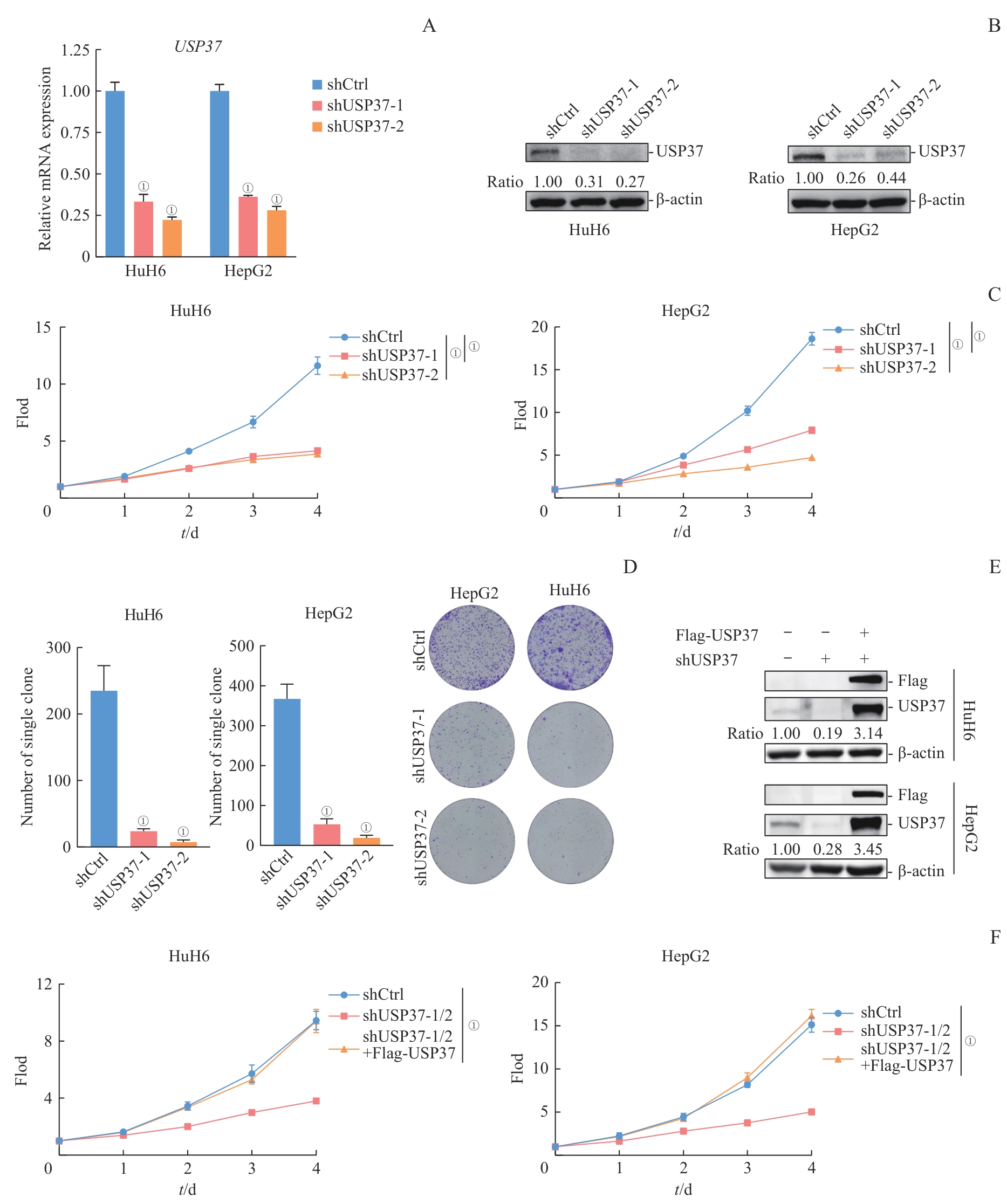
Fig 3 Effects of USP37 knockdown on HB cell proliferation
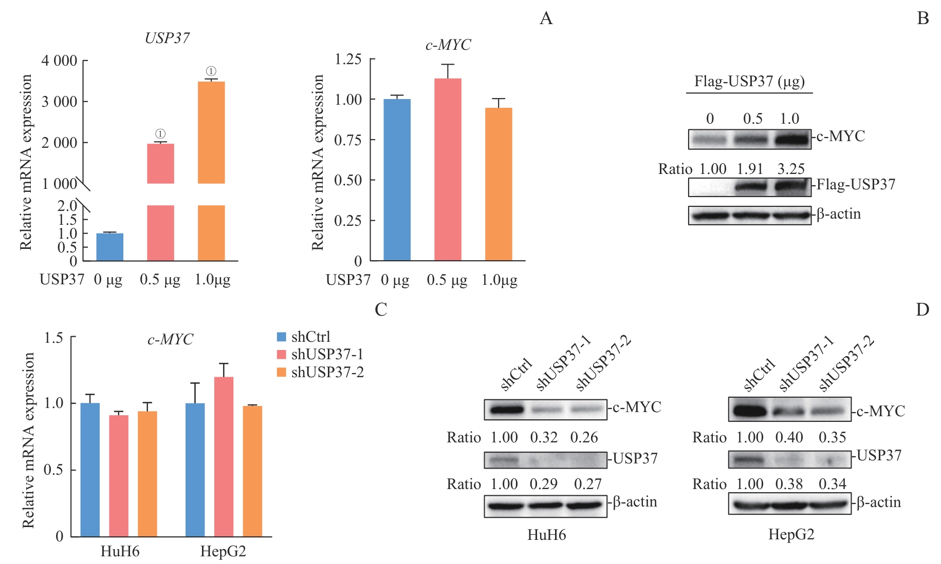
Fig 4 Effects of USP37 expression modulation on c-MYC mRNA and protein levels
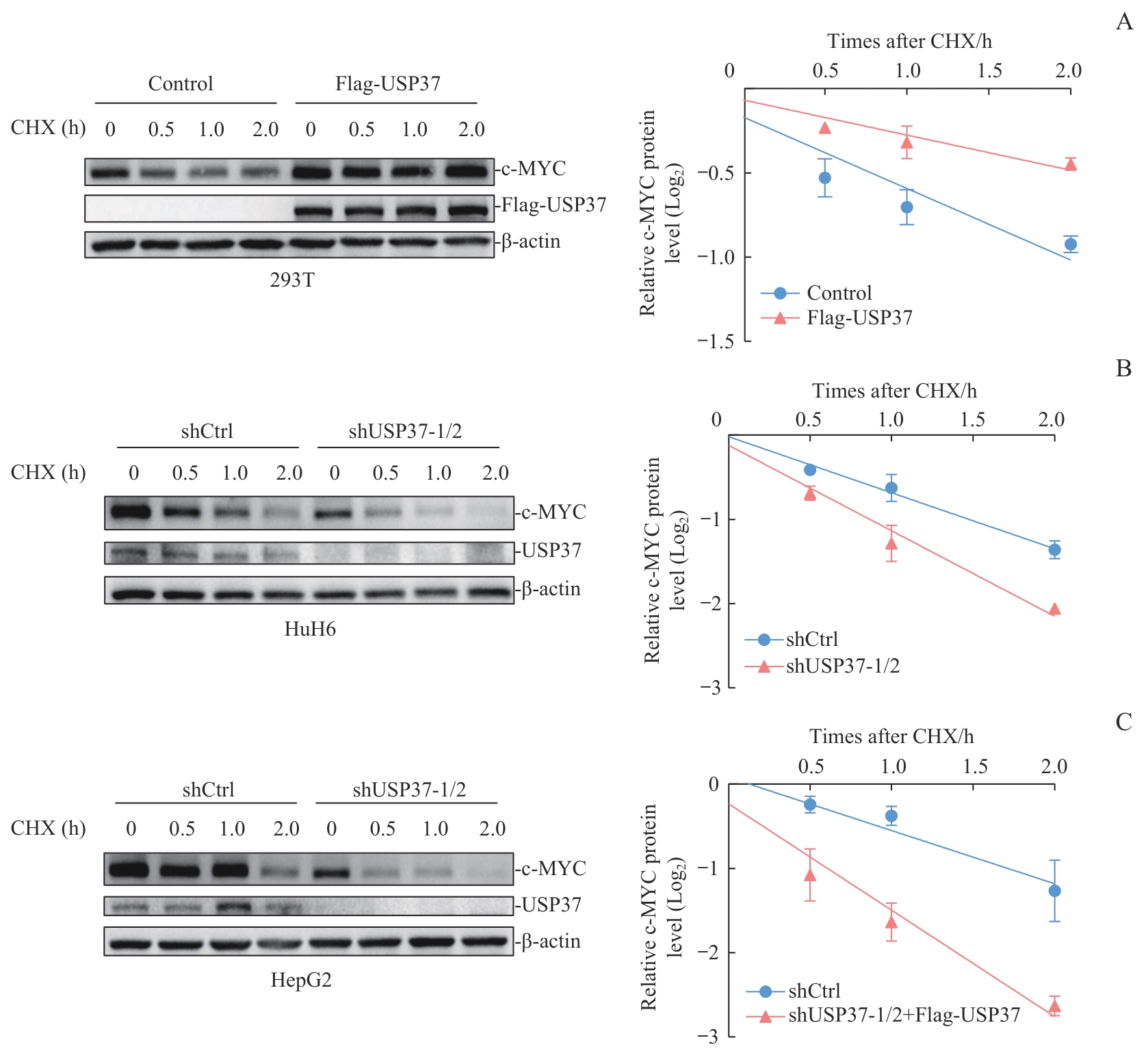
Fig 5 Analysis of c-MYC protein stability following modulation of USP37 expression
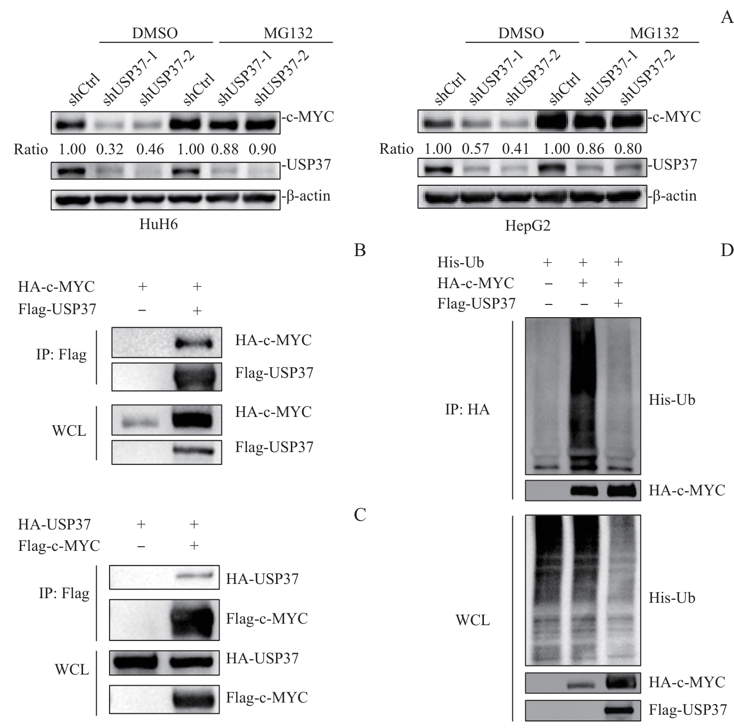
Fig 6 Mechanistic analysis of USP37-mediated deubiquitination and stabilization of c-MYC
| [1] | Hager J, Sergi C M. Liver Cancer[M]. Brisbane (AU): Exon Publications, 2021:145-164. |
| [2] | Pio L, O'Neill A F, Woodley H, et al. Hepatoblastoma[J]. Nat Rev Dis Primers, 2025, 11: 36. |
| [3] | Tang M J, Ma X L, He X L, et al. A multicenter prospective study on the management of hepatoblastoma in children: a report from the Chinese Children's Cancer Group[J]. World J Pediatr, 2024, 20(6): 590-601. |
| [4] | Zhou S M, Malvar J, Chi Y Y, et al. Independent assessment of the children's hepatic tumors international collaboration risk stratification for hepatoblastoma and the association of tumor histological characteristics with prognosis[J]. JAMA Netw Open, 2022, 5(2): e2148013. |
| [5] | Stine Z E, Walton Z E, Altman B J, et al. MYC, metabolism, and cancer[J]. Cancer Discov, 2015, 5(10): 1024-1039. |
| [6] | Dang C V. MYC on the path to cancer[J]. Cell, 2012, 149(1): 22-35. |
| [7] | Venkatraman S, Balasubramanian B, Thuwajit C, et al. Targeting MYC at the intersection between cancer metabolism and oncoimmunology[J]. Front Immunol, 2024, 15: 1324045. |
| [8] | Cairo S, Armengol C, de Reyniès A, et al. Hepatic stem-like phenotype and interplay of Wnt/beta-catenin and Myc signaling in aggressive childhood liver cancer[J]. Cancer Cell, 2008, 14(6): 471-484. |
| [9] | Wang D L, Tian J M, Yan Z Y, et al. Mitochondrial fragmentation is crucial for c-Myc-driven hepatoblastoma-like liver tumors[J]. Mol Ther, 2022, 30(4): 1645-1660. |
| [10] | Farrell A S, Sears R C. MYC degradation[J]. Cold Spring Harb Perspect Med, 2014, 4(3): a014365. |
| [11] | Lee J M, Hammarén H M, Savitski M M, et al. Control of protein stability by post-translational modifications[J]. Nat Commun, 2023, 14(1): 201. |
| [12] | Wang C, Zhang J W, Yin J, et al. Alternative approaches to target Myc for cancer treatment[J]. Signal Transduct Target Ther, 2021, 6(1): 117. |
| [13] | Nguyen L H, Robinton D A, Seligson M T, et al. Lin28b is sufficient to drive liver cancer and necessary for its maintenance in murine models[J]. Cancer Cell, 2014, 26(2): 248-261. |
| [14] | Sumazin P, Chen Y D, Treviño L R, et al. Genomic analysis of hepatoblastoma identifies distinct molecular and prognostic subgroups[J]. Hepatology, 2017, 65(1): 104-121. |
| [15] | Kwon Y T, Ciechanover A. The ubiquitin code in the ubiquitin-proteasome system and autophagy[J]. Trends Biochem Sci, 2017, 42(11): 873-886. |
| [16] | Mevissen T E T, Komander D. Mechanisms of deubiquitinase specificity and regulation[J]. Annu Rev Biochem, 2017, 86: 159-192. |
| [17] | Liu F F, Chen J Y, Li K, et al. Ubiquitination and deubiquitination in cancer: from mechanisms to novel therapeutic approaches[J]. Mol Cancer, 2024, 23(1): 148. |
| [18] | Sun T S, Liu Z N, Yang Q. The role of ubiquitination and deubiquitination in cancer metabolism[J]. Mol Cancer, 2020, 19(1): 146. |
| [19] | Hong K, Hu L X, Liu X J, et al. USP37 promotes deubiquitination of HIF2α in kidney cancer[J]. Proc Natl Acad Sci U S A, 2020, 117(23): 13023-13032. |
| [20] | Yang W C, Shih H M. The deubiquitinating enzyme USP37 regulates the oncogenic fusion protein PLZF/RARA stability[J]. Oncogene, 2013, 32(43): 5167-5175. |
| [21] | Pan J, Deng Q, Jiang C, et al. USP37 directly deubiquitinates and stabilizes c-Myc in lung cancer[J]. Oncogene, 2015, 34(30): 3957-3967. |
| [22] | Zhang X, Peng P, Bao L W, et al. Ubiquitin-specific protease 1 promotes bladder cancer progression by stabilizing c-MYC[J]. Cells, 2024, 13(21): 1798. |
| [23] | Xiao Z L, Wang Y, Pan D H, et al. USP3 promotes clear cell renal cell carcinoma progression by stabilizing MYC and enhancing glycolysis[J]. Biochim Biophys Acta Gen Subj, 2025, 1869(6): 130801. |
| [24] | Xia P, Zhang H, Lu H F, et al. METTL5 stabilizes c-Myc by facilitating USP5 translation to reprogram glucose metabolism and promote hepatocellular carcinoma progression[J]. Cancer Commun (Lond), 2023, 43(3): 338-364. |
| [25] | Gu J C, Xiao X, Zou C F, et al. Ubiquitin-specific protease 7 maintains c-Myc stability to support pancreatic cancer glycolysis and tumor growth[J]. J Transl Med, 2024, 22(1): 1135. |
| [26] | Li A N, Wang S J, Li F, et al. PCMTD1-127aa suppresses osteosarcoma progression by competitively binding to USP10 to promote c-MYC degradation[J]. iScience, 2026, 29(3): 114765. |
| [27] | Pornour M, Jeon H Y, Ryu H, et al. USP11 promotes prostate cancer progression by up-regulating AR and c-Myc activity[J]. Proc Natl Acad Sci U S A, 2024, 121(31): e2403331121. |
| [28] | Fang X G, Zhou W C, Wu Q L, et al. Deubiquitinase USP13 maintains glioblastoma stem cells by antagonizing FBXL14-mediated Myc ubiquitination[J]. J Exp Med, 2017, 214(1): 245-267. |
| [29] | Ge J C, Yu W D, Li J H, et al. USP16 regulates castration-resistant prostate cancer cell proliferation by deubiquitinating and stablizing c-Myc[J]. J Exp Clin Cancer Res, 2021, 40(1): 59. |
| [30] | Zhang D, Zhang X W, Chang S, et al. E2F1 activates USP19 to affect the stability of c-Myc to facilitate the progression of hepatocellular carcinoma[J]. Mutat Res, 2025, 830: 111902. |
| [31] | Kim D, Hong A, Park H I, et al. Deubiquitinating enzyme USP22 positively regulates c-Myc stability and tumorigenic activity in mammalian and breast cancer cells[J]. J Cell Physiol, 2017, 232(12): 3664-3676. |
| [32] | Ren D D, Cai Z Q, Zhang R, et al. USP26 promotes cell proliferation of gastric cancer by stabilizing c-myc[J]. DNA Cell Biol, 2025, 44(12): 662-670. |
| [33] | Han X, Ren C E, Lu C, et al. Phosphorylation of USP27X by PIM2 promotes glycolysis and breast cancer progression via deubiquitylation of MYC[J]. Oncogene, 2024, 43(33): 2493-2503. |
| [34] | Popov N, Wanzel M, Madiredjo M, et al. The ubiquitin-specific protease USP28 is required for MYC stability[J]. Nat Cell Biol, 2007, 9(7): 765-774. |
| [35] | Diefenbacher M E, Chakraborty A, Blake S M, et al. Usp28 counteracts Fbw7 in intestinal homeostasis and cancer[J]. Cancer Res, 2015, 75(7): 1181-1186. |
| [36] | Tu R F, Kang W Q, Yang M J, et al. USP29 coordinates MYC and HIF1α stabilization to promote tumor metabolism and progression[J]. Oncogene, 2021, 40(46): 6417-6429. |
| [37] | Zhang X Y, Han Y, Liu S L, et al. MF-094 nanodelivery inhibits oral squamous cell carcinoma by targeting USP30[J]. Cell Mol Biol Lett, 2022, 27(1): 107. |
| [38] | Ding T, Zhu Y J, Jin H M, et al. Circular RNA circ_0057558 controls prostate cancer cell proliferation through regulating miR-206/USP33/c-Myc axis[J]. Front Cell Dev Biol, 2021, 9: 644397. |
| [39] | Chen D J, Zhao Y, Zhang X B, et al. USP33-mediated stabilization of c-Myc drives glycolytic reprogramming and promotes ovarian cancer progression[J]. Biochim Biophys Acta Gen Subj, 2025, 1869(9): 130830. |
| [40] | Liu H L, Wei X L, Nie Y, et al. The knockdown of USP34 inhibits the progression of hepatocellular carcinoma by accelerating c-Myc degradation[J]. Turk J Gastroenterol, 2025, 36(9): 573-582. |
| [41] | Sun X X, He X, Yin L, et al. The nucleolar ubiquitin-specific protease USP36 deubiquitinates and stabilizes c-Myc[J]. Proc Natl Acad Sci U S A, 2015, 112(12): 3734-3739. |
| [42] | Xu Z J, Hu H, Fang D B, et al. The deubiquitinase USP38 promotes cell proliferation through stabilizing c-Myc[J]. Int J Biochem Cell Biol, 2021, 137: 106023. |
| [43] | Fu W, Wu Q L, Yao Y R, et al. Ubiquitin-specific peptidase 40 promotes cellular proliferation and inhibits apoptosis through deubiquitination-mediated stabilization of c-MYC in acute myeloid leukemia[J]. Biochim Biophys Acta Mol Basis Dis, 2026, 1872(3): 168138. |
| [44] | Li M X, Yu J T, Ju L G, et al. USP43 stabilizes c-Myc to promote glycolysis and metastasis in bladder cancer[J]. Cell Death Dis, 2024, 15(1): 44. |
| [45] | Cheng H X, Hua L Y, Tang H L, et al. CBX7 reprograms metabolic flux to protect against meningioma progression by modulating the USP44/c-MYC/LDHA axis[J]. J Mol Cell Biol, 2024, 15(10): mjad057. |
| [46] | Tu X, Li C C, Sun W, et al. Suppression of cancer cell stemness and drug resistance via MYC destabilization by deubiquitinase USP45 inhibition with a natural small molecule[J]. Cancers, 2023, 15(3): 930. |
| [47] | Shin H, Hwang S, Jeong J H, et al. Targeting USP47 enhances the efficacy of KRAS inhibitor in KRASG12C mutated non-small cell lung cancer by controlling deubiquitination of c-Myc[J]. Pharmacol Res, 2025, 215: 107722. |
| [48] | Ye M J, He J J, Zhang J J, et al. USP7 promotes hepatoblastoma progression through activation of PI3K/AKT signaling pathway[J]. Cancer Biomark, 2021, 31(2): 107-117. |
| [49] | Wu L, Zhao N, Zhou Z L, et al. PLAGL2 promotes the proliferation and migration of gastric cancer cells via USP37-mediated deubiquitination of Snail1[J]. Theranostics, 2021, 11(2): 700-714. |
| [50] | Das C M, Taylor P, Gireud M, et al. The deubiquitylase USP37 links REST to the control of p27 stability and cell proliferation[J]. Oncogene, 2013, 32(13): 1691-1701. |
| [51] | Dobson T H W, Hatcher R J, Swaminathan J, et al. Regulation of USP37 expression by REST-associated G9a-dependent histone methylation[J]. Mol Cancer Res, 2017, 15(8): 1073-1084. |
| [1] | ZHU Zhi-xing1, 2, JI Wei3, GU Jian-lei1, 4, LÜ Hui1, 2, 4, TIAN Guo-li3. Analysis of four mutations and protein structure of glucose-6-phosphate dehydrogenase gene [J]. JOURNAL OF SHANGHAI JIAOTONG UNIVERSITY (MEDICAL SCIENCE), 2020, 40(12): 1571-1578. |
| [2] | QU Guo-jun1, LU Yuan-feng2, LI Yu1. Mechanism of CCT2, a new downstream substrate of PDGFRα, on proliferation of tumor cells [J]. , 2019, 39(1): 28-. |
| [3] | LI Ze, REN Zun, TU Hong-hu, JIAN Zheng, YU Jian-xiu, DENG Rong . Research advance on BAP1 and its function in tumor suppression [J]. , 2017, 37(5): 689-. |
| [4] | SHENG Qi, YUAN Xiao-jun, ZHANG Qin, et al. Diagnosis and treatment of 12 children with hepatoblastoma [J]. , 2011, 31(4): 517-. |
| Viewed | ||||||
|
Full text |
|
|||||
|
Abstract |
|
|||||
