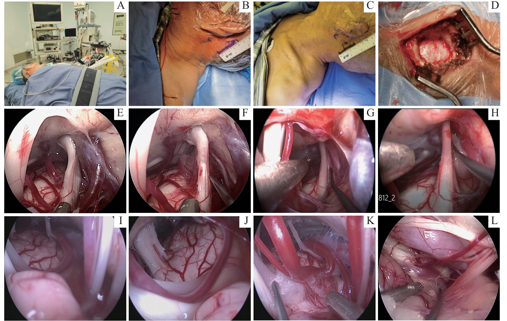
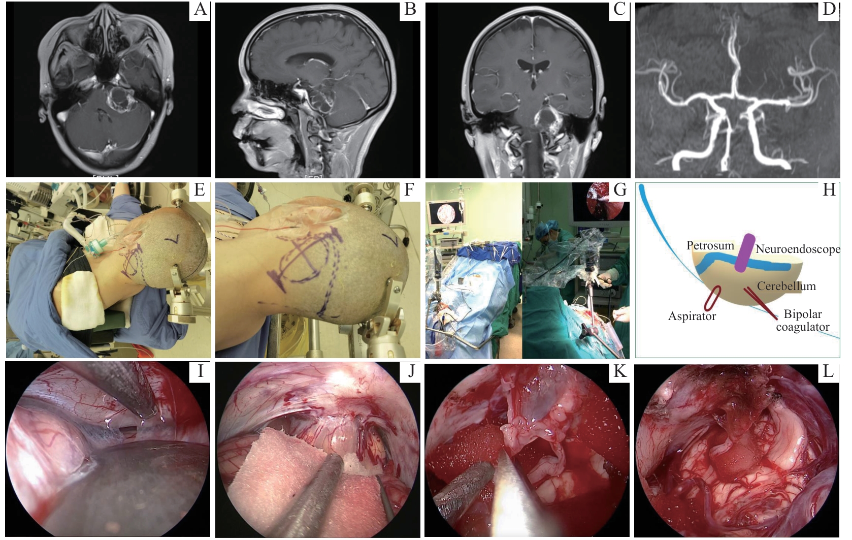
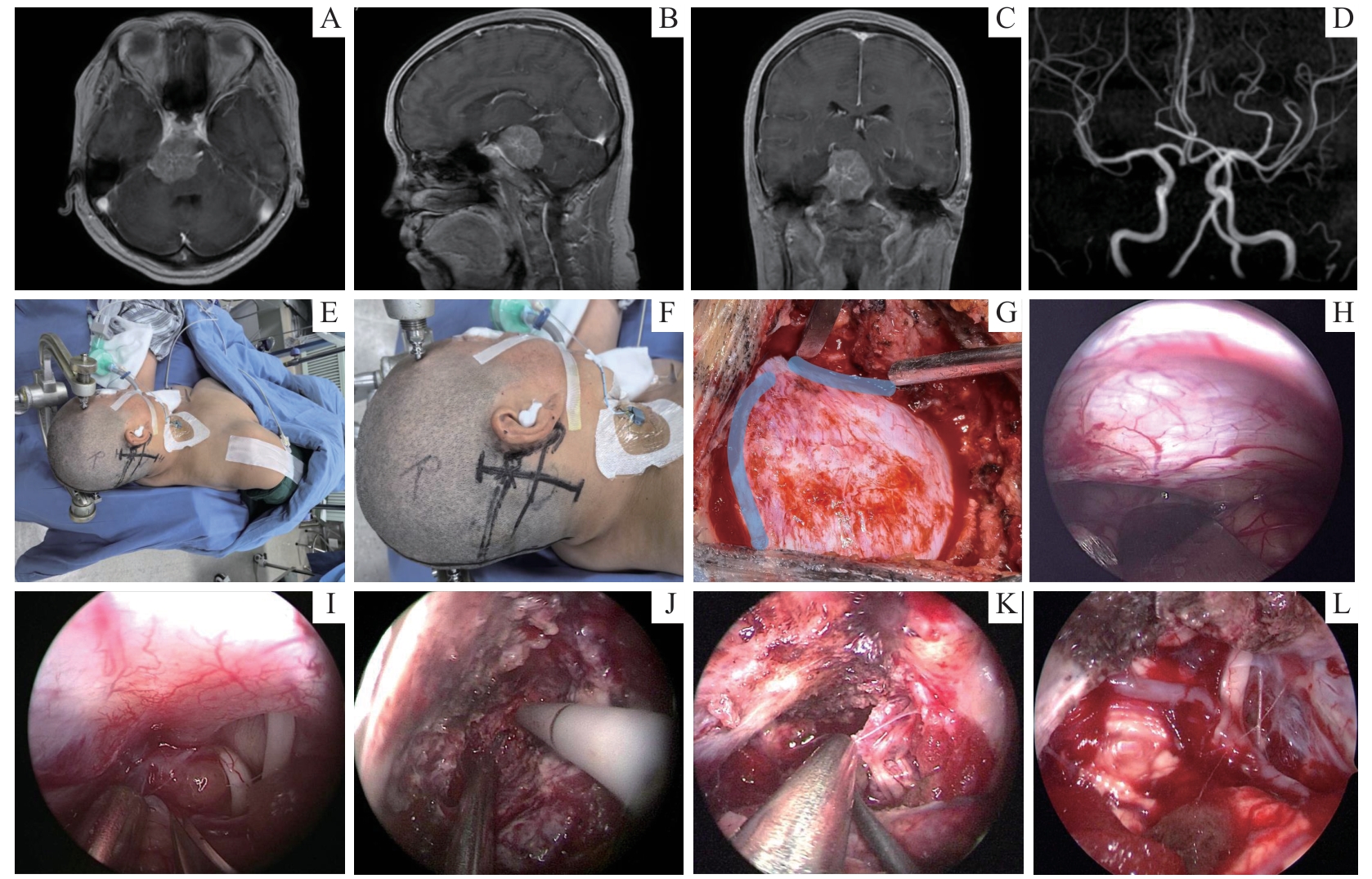
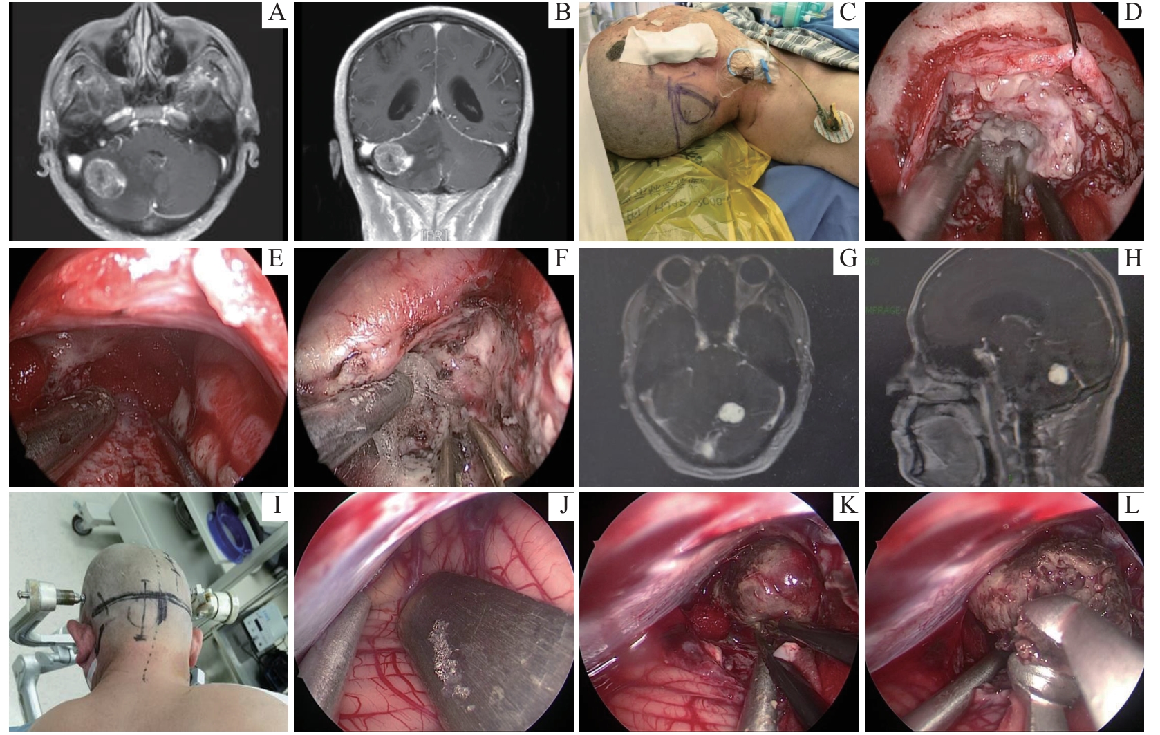
| 1 |
BRAY H N, SAPPINGTON J M. A review of posterior fossa lesions[J]. Mo Med, 2022, 119(6): 553-558.
|
| 2 |
ROLDAN-VALADEZ E, GONZALEZ-HERMOSILLO L M, MENDOZA-LOPEZ A C. Brain death diagnosis in primary posterior fossa lesions[J]. Neurol India, 2023, 71(1): 164-165.
|
| 3 |
TAMURA R, KATAYAMA M, YAMAMOTO K, et al. Suboccipital transhorizontal fissure approach for posterior cranial fossa lesions: a cadaveric study and first clinical experience[J]. Oper Neurosurg (Hagerstown), 2021, 21(6): E479-E487.
|
| 4 |
GAO H, LIU C H, ZHANG Y Z. Neuro-endoscope for skull base tumors[J]. Clin Neurol Neurosurg, 2018, 170: 102-105.
|
| 5 |
LI C Z, ZHU H B, ZONG X Y, et al. History, current situation, and future development of endoscopic neurosurgery in China[J]. World Neurosurg, 2018, 110: 270-275.
|
| 6 |
HUA W, XU H, ZHANG X, et al. Pure endoscopic resection of pineal region tumors through supracerebellar infratentorial approach with ‘head-up’ park-bench position[J]. Neurol Res, 2023, 45(4): 354-362.
|
| 7 |
BOSE A, PRASAD U, KUMAR A, et al. Characterizing various posterior fossa tumors in children and adults with diffusion-weighted imaging and spectroscopy[J]. Cureus, 2023, 15(5): e39144.
|
| 8 |
ARNAOUT M M, LUZZI S, GALZIO R, et al. Supraorbital keyhole approach: pure endoscopic and endoscope-assisted perspective[J]. Clin Neurol Neurosurg, 2020, 189: 105623.
|
| 9 |
CHOO J, TAKEUCHI K, NAGATA Y, et al. Neuroendoscopic cylinder surgery and 5-aminolevulinic acid photodynamic diagnosis of deep-seated intracranial lesions[J]. World Neurosurg, 2018, 116: e35-e41.
|
| 10 |
SCHROEDER H W S, SGOUROS S. Neuroendoscopy: history, endoscopes, and instrumentation[J]. Childs Nerv Syst, 2023, 39(10): 2729-2735.
|
| 11 |
TOSI U, GUADIX S W, SOUWEIDANE M M. Neuroendoscopy: the state of the art[J]. World Neurosurg, 2023, 178: 305-310.
|
| 12 |
TOSI U, GUADIX S W, COHEN A R, et al. Neuroendoscopy: how we got here[J]. World Neurosurg, 2023, 178: 298-304.
|
| 13 |
BEER-FURLAN A, VELLUTINI E A S, BALSALOBRE L, et al. Endoscopic endonasal approach to ventral posterior fossa meningiomas: from case selection to surgical management[J]. Neurosurg Clin N Am, 2015, 26(3): 413-426.
|
| 14 |
IDRIS Z, TAN Y C, KANDASAMY R, et al. Transfrontal transaqueductal, transtrigonal, and suboccipital infratentorial supracerebellar endoscopic fenestration of posterior fossa arachnoid cysts: three surgical cases[J]. J Neurol Surg A Cent Eur Neurosurg, 2017, 78(2): 210-215.
|
| 15 |
李炯, 钟东, 吕东, 等. 神经内镜辅助后颅窝显微神经外科手术治疗脑肿瘤[J]. 中华医学杂志, 2018, 98(17): 1311-1316.
|
|
LI J, ZHONG D, LÜ D, et al. Neuroendoscopy assisted microneurosurgery for posterior cranial fossa lesion[J]. National Medical Journal of China, 2018, 98(17): 1311-1316.
|
 ), ZHAO Hao2, MIAO Yifeng1(