Journal of Shanghai Jiao Tong University (Medical Science) ›› 2023, Vol. 43 ›› Issue (7): 923-930.doi: 10.3969/j.issn.1674-8115.2023.07.015
• Review • Previous Articles
MA Ben( ), ZHAO Cheng, SHU Yijun, DONG Ping()
Received:2022-10-18
Accepted:2023-06-20
Online:2023-07-28
Published:2023-07-28
Contact:
DONG Ping
E-mail:jsxzmaben@163.com;dongping1050@163.com
Supported by:CLC Number:
MA Ben, ZHAO Cheng, SHU Yijun, DONG Ping. Application progress of CT radiomics in gastrointestinal stromal tumor[J]. Journal of Shanghai Jiao Tong University (Medical Science), 2023, 43(7): 923-930.
Add to citation manager EndNote|Ris|BibTeX
URL: https://xuebao.shsmu.edu.cn/EN/10.3969/j.issn.1674-8115.2023.07.015
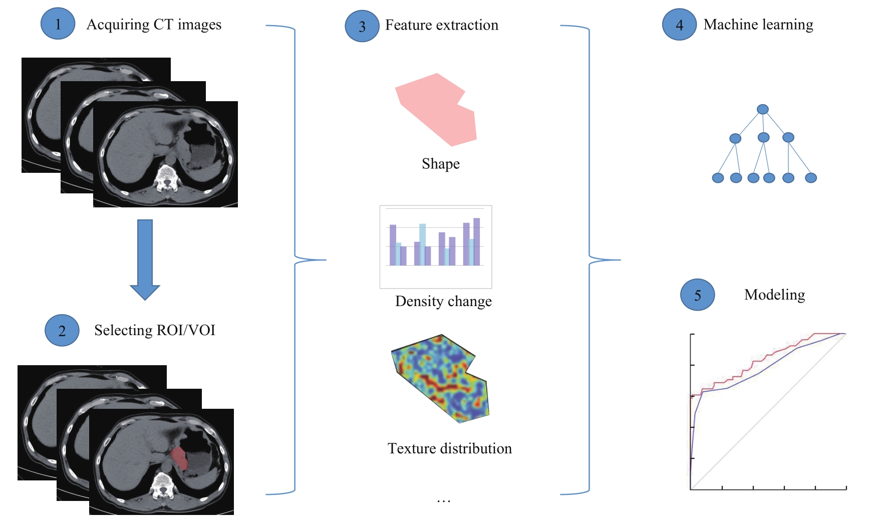
Fig 1 Basic flow of CT radiomics mode
| Disease | Group | Sample size/n | Result | Reference |
|---|---|---|---|---|
| GIST/gastric adenocarcinoma/gastric lymphoma | Turkey | 26/125/12 | GIST vs gastric lymphoma: Sensitivity=98%, Specificity=75% GIST vs gastric adenocarcinoma: Sensitivity=91%, Specificity=77% | [ |
| GIST/gastric cancer | China | 40/60 | Subjective CT signs model: AUC=0.806 (0.696‒0.917), Accuracy=75% Radiomic signature model: AUC=0.886 (0.809‒0.963), Accuracy=81% Combined model: AUC=0.903 (0.831‒0.975), Accuracy=86% | [ |
| GIST/gastric adenocarcinoma/gastric lymphoma | Austria | Arterial phase: 15/31/5 Venous phase: 17/23/8 | Arterial phase misclassification rate: Gastric adenocarcinoma vs gastric lymphoma=3.1% GIST vs gastric lymphoma=0 Venous phase misclassification rate: Gastric adenocarcinoma vs gastric lymphoma=9.7% GIST vs gastric lymphoma=8.0% GIST vs gastric adenocarcinoma=10.0% | [ |
Tab 1 Studies on differential diagnosis of GIST from gastric cancer and gastric lymphoma by CT radiomics
| Disease | Group | Sample size/n | Result | Reference |
|---|---|---|---|---|
| GIST/gastric adenocarcinoma/gastric lymphoma | Turkey | 26/125/12 | GIST vs gastric lymphoma: Sensitivity=98%, Specificity=75% GIST vs gastric adenocarcinoma: Sensitivity=91%, Specificity=77% | [ |
| GIST/gastric cancer | China | 40/60 | Subjective CT signs model: AUC=0.806 (0.696‒0.917), Accuracy=75% Radiomic signature model: AUC=0.886 (0.809‒0.963), Accuracy=81% Combined model: AUC=0.903 (0.831‒0.975), Accuracy=86% | [ |
| GIST/gastric adenocarcinoma/gastric lymphoma | Austria | Arterial phase: 15/31/5 Venous phase: 17/23/8 | Arterial phase misclassification rate: Gastric adenocarcinoma vs gastric lymphoma=3.1% GIST vs gastric lymphoma=0 Venous phase misclassification rate: Gastric adenocarcinoma vs gastric lymphoma=9.7% GIST vs gastric lymphoma=8.0% GIST vs gastric adenocarcinoma=10.0% | [ |
| Group | Sample size/n | Cutoff value of Ki-67/% | Result | Reference |
|---|---|---|---|---|
| China | 339 | 10 | Radiomic signature AUC: Training cohort: 0.787 (95%CI 0.632‒0.801) Internal validation cohort: 0.765 (95%CI 0.683‒0.847) External validation cohort: 0.754 (95%CI 0.666‒0.842) Radiomic nomogram AUC: Training cohort: 0.801 (95%CI 0.726‒0.876) Internal validation cohort: 0.828 (95%CI 0.681‒0.974) External validation cohort: 0.784 (95%CI 0.701‒0.868) | [ |
| China | 344 | 8 | Generated radiomic model AUC: Training cohort: 0.835 (95%CI 0.761‒0.908) External validation cohort: 0.784 (95%CI 0.691‒0.874) | [ |
| Japan | 64 | 5 | Fractal dimension: Sensitivity=66.7%, Specificity=69.8% | [ |
Tab 2 Studies on immunohistochemical analysis of GIST by CT radiomics
| Group | Sample size/n | Cutoff value of Ki-67/% | Result | Reference |
|---|---|---|---|---|
| China | 339 | 10 | Radiomic signature AUC: Training cohort: 0.787 (95%CI 0.632‒0.801) Internal validation cohort: 0.765 (95%CI 0.683‒0.847) External validation cohort: 0.754 (95%CI 0.666‒0.842) Radiomic nomogram AUC: Training cohort: 0.801 (95%CI 0.726‒0.876) Internal validation cohort: 0.828 (95%CI 0.681‒0.974) External validation cohort: 0.784 (95%CI 0.701‒0.868) | [ |
| China | 344 | 8 | Generated radiomic model AUC: Training cohort: 0.835 (95%CI 0.761‒0.908) External validation cohort: 0.784 (95%CI 0.691‒0.874) | [ |
| Japan | 64 | 5 | Fractal dimension: Sensitivity=66.7%, Specificity=69.8% | [ |
| 1 | JEMAL A, BRAY F, CENTER M M, et al. Global cancer statistics[J]. CA Cancer J Clin, 2011, 61(2): 69-90. |
| 2 | LEE I S, PARK Y S, LEE J H, et al. Pathologic discordance of differentiation between endoscopic biopsy and postoperative specimen in mucosal gastric adenocarcinomas[J]. Ann Surg Oncol, 2013, 20(13): 4231-4237. |
| 3 | JOENSUU H. Risk stratification of patients diagnosed with gastrointestinal stromal tumor[J]. Hum Pathol, 2008, 39(10): 1411-1419. |
| 4 | JOENSUU H, HOHENBERGER P, CORLESS C L. Gastrointestinal stromal tumour[J]. Lancet, 2013, 382(9896): 973-983. |
| 5 | DEMETRI G D, VON MEHREN M, BLANKE C D, et al. Efficacy and safety of imatinib mesylate in advanced gastrointestinal stromal tumors[J]. N Engl J Med, 2002, 347(7): 472-480. |
| 6 | VERWEIJ J, CASALI P G, ZALCBERG J, et al. Progression-free survival in gastrointestinal stromal tumours with high-dose imatinib: randomised trial[J]. Lancet, 2004, 364(9440): 1127-1134. |
| 7 | MIETTINEN M, SOBIN L H, LASOTA J. Gastrointestinal stromal tumors of the stomach: a clinicopathologic, immunohistochemical, and molecular genetic study of 1 765 cases with long-term follow-up[J]. Am J Surg Pathol, 2005, 29(1): 52-68. |
| 8 | GILLIES R J, KINAHAN P E, HRICAK H. Radiomics: images are more than pictures, they are data[J]. Radiology, 2016, 278(2): 563-577. |
| 9 | YIP S S F, AERTS H J W L. Applications and limitations of radiomics[J]. Phys Med Biol, 2016, 61(13): R150-R166. |
| 10 | ZHANG L J, KANG L Q, LI G C, et al. Computed tomography-based radiomics model for discriminating the risk stratification of gastrointestinal stromal tumors[J]. Radiol Med, 2020, 125(5): 465-473. |
| 11 | LAMBIN P, RIOS-VELAZQUEZ E, LEIJENAAR R, et al. Radiomics: extracting more information from medical images using advanced feature analysis[J]. Eur J Cancer, 2012, 48(4): 441-446. |
| 12 | CHEN T, LIU S Q, LI Y, et al. Developed and validated a prognostic nomogram for recurrence-free survival after complete surgical resection of local primary gastrointestinal stromal tumors based on deep learning[J]. EBioMedicine, 2019, 39: 272-279. |
| 13 | SUN Z Q, HU S D, LI J, et al. Radiomics study for differentiating gastric cancer from gastric stromal tumor based on contrast-enhanced CT images[J]. J Xray Sci Technol, 2019, 27(6): 1021-1031. |
| 14 | ZHENG J, XIA Y, XU A Q, et al. Combined model based on enhanced CT texture features in liver metastasis prediction of high-risk gastrointestinal stromal tumors[J]. Abdom Radiol (NY), 2022, 47(1): 85-93. |
| 15 | STARMANS M P A, TIMBERGEN M J M, VOS M, et al. Differential diagnosis and molecular stratification of gastrointestinal stromal tumors on CT images using a radiomics approach[J]. J Digit Imaging, 2022, 35(2): 127-136. |
| 16 | 李定杰, 吴慧, 刘如, 等. 基于诊断CT影像组学对食管癌放疗疗效早期评估[J]. 中华放射肿瘤学杂志, 2019, 28(10): 731-734. |
| LI D J, WU H, LIU R, et al. Early evaluation of radiotherapy effect of esophageal cancer based on diagnostic CT imaging histology[J]. Chinese Journal of Radiation Oncology, 2019, 28(10): 731-734. | |
| 17 | 李华秀, 李振辉, 李鹍, 等. CT影像组学预测局部进展期直肠癌新辅助治疗的效果[J]. 中国医学影像学杂志, 2020, 28(1): 44-50. |
| LI H X, LI Z H, LI K, et al. Efficacy of CT radiomics in predicting neoadjuvant therapy of locally advanced rectal cancer[J]. Chinese Journal of Medical Imaging, 2020, 28(1): 44-50. | |
| 18 | ZHOU Y, HE L, HUANG Y Q, et al. CT-based radiomics signature: a potential biomarker for preoperative prediction of early recurrence in hepatocellular carcinoma[J]. Abdom Radiol, 2017, 42(6): 1695-1704. |
| 19 | GAO X J, MA T T, CUI J L, et al. A radiomics-based model for prediction of lymph node metastasis in gastric cancer[J]. Eur J Radiol, 2020, 129: 109069. |
| 20 | BA-SSALAMAH A, MUIN D, SCHERNTHANER R, et al. Texture-based classification of different gastric tumors at contrast-enhanced CT[J]. Eur J Radiol, 2013, 82(10): e537-e543. |
| 21 | ZEYDANLI T, KILIC H K. Performance of quantitative CT texture analysis in differentiation of gastric tumors[J]. Jpn J Radiol, 2022, 40(1): 56-65. |
| 22 | CASTELLANO G, BONILHA L, LI L M, et al. Texture analysis of medical images[J]. Clin Radiol, 2004, 59(12): 1061-1069. |
| 23 | BASHIR U, SIDDIQUE M M, MCLEAN E, et al. Imaging heterogeneity in lung cancer: techniques, applications, and challenges[J]. AJR Am J Roentgenol, 2016, 207(3): 534-543. |
| 24 | YE H, XIN H, ZHENG Q, et al. Prognostic role of the primary tumour site in patients with operable small intestine and gastrointestinal stromal tumours: a large population-based analysis[J]. Oncotarget, 2017, 9(8): 8147-8154. |
| 25 | MIETTINEN M, LASOTA J. Gastrointestinal stromal tumors: review on morphology, molecular pathology, prognosis, and differential diagnosis[J]. Arch Pathol Lab Med, 2006, 130(10): 1466-1478. |
| 26 | MIETTINEN M, LASOTA J. Gastrointestinal stromal tumors: pathology and prognosis at different sites[J]. Semin Diagn Pathol, 2006, 23(2): 70-83. |
| 27 | FLETCHER C D M, BRIDGE J A, HOGENDOORN P C W, et al. WHO classification of tumours of soft tissue and bone[M]. 4th ed. Lyon: IARC Press, 2013. |
| 28 | 中国临床肿瘤学会胃肠间质瘤专家委员会. 中国胃肠间质瘤诊断治疗共识(2017年版)[J]. 肿瘤综合治疗电子杂志, 2018, 4(1): 31-43. |
| Expert Committee on Gastrointestinal Stromal Tumor, Chinese Society of Clinical Oncology. Chinese consensus on diagnosis and treatment of gastrointestinal stromal tumor (2017 edition)[J]. Journal of Multidisciplinary Cancer Management (Electronic Version), 2018, 4(1): 31-43. | |
| 29 | CHOI I Y, YEOM S K, CHA J, et al. Feasibility of using computed tomography texture analysis parameters as imaging biomarkers for predicting risk grade of gastrointestinal stromal tumors: comparison with visual inspection[J]. Abdom Radiol (NY), 2019, 44(7): 2346-2356. |
| 30 | SONG Y C, LI J, WANG H X, et al. Radiomics nomogram based on contrast-enhanced CT to predict the malignant potential of gastrointestinal stromal tumor: a two-center study[J]. Acad Radiol, 2022, 29(6): 806-816. |
| 31 | DEMETRI G D, VON MEHREN M, ANTONESCU C R, et al. NCCN Task Force report: update on the management of patients with gastrointestinal stromal tumors[J]. J Natl Compr Canc Netw, 2010, 8(Suppl 2): S1-S44. |
| 32 | RUTKOWSKI P, PRZYBYŁ J, ZDZIENICKI M. Extended adjuvant therapy with imatinib in patients with gastrointestinal stromal tumors[J]. Mol Diagn Ther, 2013, 17(1): 9-19. |
| 33 | ECKARDT A J, ADLER A, GOMES E M, et al. Endosonographic large-bore biopsy of gastric subepithelial tumors: a prospective multicenter study[J]. Eur J Gastroenterol Hepatol, 2012, 24(10): 1135-1144. |
| 34 | ZHAO Y L, FENG M B, WANG M H, et al. CT radiomics for the preoperative prediction of Ki67 index in gastrointestinal stromal tumors: a multi-center study[J]. Front Oncol, 2021, 11: 689136. |
| 35 | WANG C, LI H L, JIAERKEN Y, et al. Building CT radiomics-based models for preoperatively predicting malignant potential and mitotic count of gastrointestinal stromal tumors[J]. Transl Oncol, 2019, 12(9): 1229-1236. |
| 36 | BASILIO-DE-OLIVEIRA R P, PANNAIN V L N. Prognostic angiogenic markers (endoglin, VEGF, CD31) and tumor cell proliferation (Ki67) for gastrointestinal stromal tumors[J]. World J Gastroenterol, 2015, 21(22): 6924-6930. |
| 37 | ZHANG Q W, GAO Y J, ZHANG R Y, et al. Personalized CT-based radiomics nomogram preoperative predicting Ki-67 expression in gastrointestinal stromal tumors: a multicenter development and validation cohort[J]. Clin Transl Med, 2020, 9(1): 12. |
| 38 | KURATA Y, HAYANO K, OHIRA G, et al. Fractal analysis of contrast-enhanced CT images for preoperative prediction of malignant potential of gastrointestinal stromal tumor[J]. Abdom Radiol, 2018, 43(10): 2659-2664. |
| 39 | 朱从波, 廖国庆, 赵丁民. Ki-67对胃肠道间质瘤预后的评估价值[J]. 临床与病理杂志, 2018, 38(8): 1632-1639. |
| ZHU C B, LIAO G Q, ZHAO D M. Prognostic value of Ki-67 index in gastrointestinal stromal tumor[J]. Journal of Clinical and Pathological Research, 2018, 38(8): 1632-1639. | |
| 40 | XU F, MA X H, WANG Y C, et al. CT texture analysis can be a potential tool to differentiate gastrointestinal stromal tumors without KIT exon 11 mutation[J]. Eur J Radiol, 2018, 107: 90-97. |
| 41 | LIU X J, YIN Y, WANG X Z, et al. Gastrointestinal stromal tumors: associations between contrast-enhanced CT images and KIT exon 11 gene mutation[J]. Ann Transl Med, 2021, 9(19): 1496. |
| 42 | PALATRESI D, FEDELI F, DANTI G, et al. Correlation of CT radiomic features for GISTs with pathological classification and molecular subtypes: preliminary and monocentric experience[J]. Radiol Med, 2022, 127(2): 117-128. |
| 43 | CHEN T, NING Z Y, XU L L, et al. Radiomics nomogram for predicting the malignant potential of gastrointestinal stromal tumours preoperatively[J]. Eur Radiol, 2019, 29(3): 1074-1082. |
| 44 | CHU H R, PANG P P, HE J, et al. Value of radiomics model based on enhanced computed tomography in risk grade prediction of gastrointestinal stromal tumors[J]. Sci Rep, 2021, 11(1): 12009. |
| 45 | WANG M H, FENG Z, ZHOU L X, et al. Computed-tomography-based radiomics model for predicting the malignant potential of gastrointestinal stromal tumors preoperatively: a multi-classifier and multicenter study[J]. Front Oncol, 2021, 11: 582847. |
| 46 | REN C Y, WANG S P, ZHANG S J. Development and validation of a nomogram based on CT images and 3D texture analysis for preoperative prediction of the malignant potential in gastrointestinal stromal tumors[J]. Cancer Imaging, 2020, 20(1): 5. |
| 47 | ZHANG Q W, ZHOU X X, ZHANG R Y, et al. Comparison of malignancy-prediction efficiency between contrast and non-contract CT-based radiomics features in gastrointestinal stromal tumors: a multicenter study[J]. Clin Transl Med, 2020, 10(3): e291. |
| 48 | COLLEWET G, STRZELECKI M, MARIETTE F. Influence of MRI acquisition protocols and image intensity normalization methods on texture classification[J]. Magn Reson Imaging, 2004, 22(1): 81-91. |
| 49 | LUBNER M G, STABO N, LUBNER S J, et al. CT textural analysis of hepatic metastatic colorectal cancer: pre-treatment tumor heterogeneity correlates with pathology and clinical outcomes[J]. Abdom Imaging, 2015, 40(7): 2331-2337. |
| 50 | HUANG Y Q, LIANG C H, HE L, et al. Development and validation of a radiomics nomogram for preoperative prediction of lymph node metastasis in colorectal cancer[J]. J Clin Oncol, 2016, 34(18): 2157-2164. |
| [1] | Xiaofeng WANG, Lu ZHOU, Leyu YAO, Fan HE, Haixia PENG, Daming YANG, Xiaolin HUANG. Effect of DCNN model-assisted colorectal polyp detection system on the detection of colorectal polyps by junior physicians [J]. JOURNAL OF SHANGHAI JIAOTONG UNIVERSITY (MEDICAL SCIENCE), 2022, 42(2): 205-210. |
| [2] | Xin LI, Qing FAN. Application progress of machine learning in the study of facial features of patients with depression [J]. JOURNAL OF SHANGHAI JIAOTONG UNIVERSITY (MEDICAL SCIENCE), 2022, 42(1): 124-129. |
| [3] | RONG Wen-wen1, WANG Gang1, ZHU Qi-li2. Discussion on value of medical records-structured specialized disease database based on artificial intelligence in clinical research [J]. JOURNAL OF SHANGHAI JIAOTONG UNIVERSITY (MEDICAL SCIENCE), 2020, 40(7): 995-1000. |
| [4] | LU Yan-qiao, SHEN Lan, HE Ben. Application of artificial intelligence in assisted diagnosis and treatment of cardiovascular disease [J]. , 2020, 40(2): 259-. |
| [5] | JIA Zhi-ying1, 2, DONG Min-ye1, 2, SHI Zhen-su2, 3, JIN Chun-lin4, LI Guo-hong1, 2. Study of a screening system for mild cognitive impairment based on machine learning model [J]. , 2019, 39(8): 908-. |
| [6] | XU Yu-peng1*, DU Yu-chen1, 2*, CHEN Feng-e1. Automatic layer segmentation of optical coherence tomography images in retinal vascular diseases [J]. , 2019, 39(6): 613-. |
| [7] | SANG Chao1, XIE Guo-xiang2, LIANG Dan-dan1, ZHAO Ai-hua1, JIA Wei1, 2, CHEN Tian-lu1. Improvement of liver fibrosis diagnostic models based on Youden index [J]. , 2019, 39(10): 1156-. |
| [8] | ZHANG Xin-yu1*, ZHANG Jing2*, ZHU Xiao-qiang1, CAO Ying-ying1, CHEN Hao-yan1. Bacterial signatures for diagnosis of colorectal cancerfecal metagenomics analysis [J]. , 2018, 38(9): 1019-. |
| Viewed | ||||||
|
Full text |
|
|||||
|
Abstract |
|
|||||