Journal of Shanghai Jiao Tong University (Medical Science) ›› 2025, Vol. 45 ›› Issue (11): 1507-1514.doi: 10.3969/j.issn.1674-8115.2025.11.010
• Clinical research • Previous Articles
LI Lulu, WU Jianyong( )
)
Received:2025-02-19
Accepted:2025-06-30
Online:2025-11-28
Published:2025-12-03
Contact:
WU Jianyong
E-mail:wujianyong@xinhuamed.com.cn
CLC Number:
LI Lulu, WU Jianyong. A cone-beam computed tomographic study comparing characteristics of maxillary anterior regional alveolar bone remodeling under two reference systems[J]. Journal of Shanghai Jiao Tong University (Medical Science), 2025, 45(11): 1507-1514.
Add to citation manager EndNote|Ris|BibTeX
URL: https://xuebao.shsmu.edu.cn/EN/10.3969/j.issn.1674-8115.2025.11.010
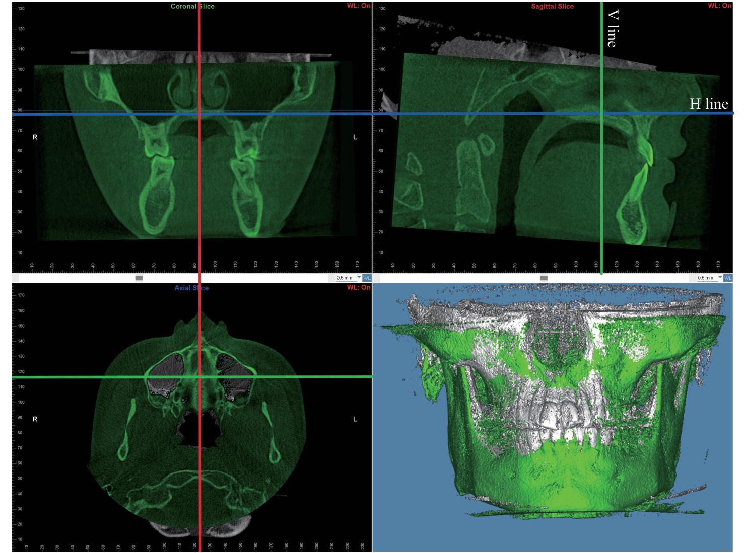
Fig 1 Positioning of the three-dimensional reconstructed dental and maxillofacial image
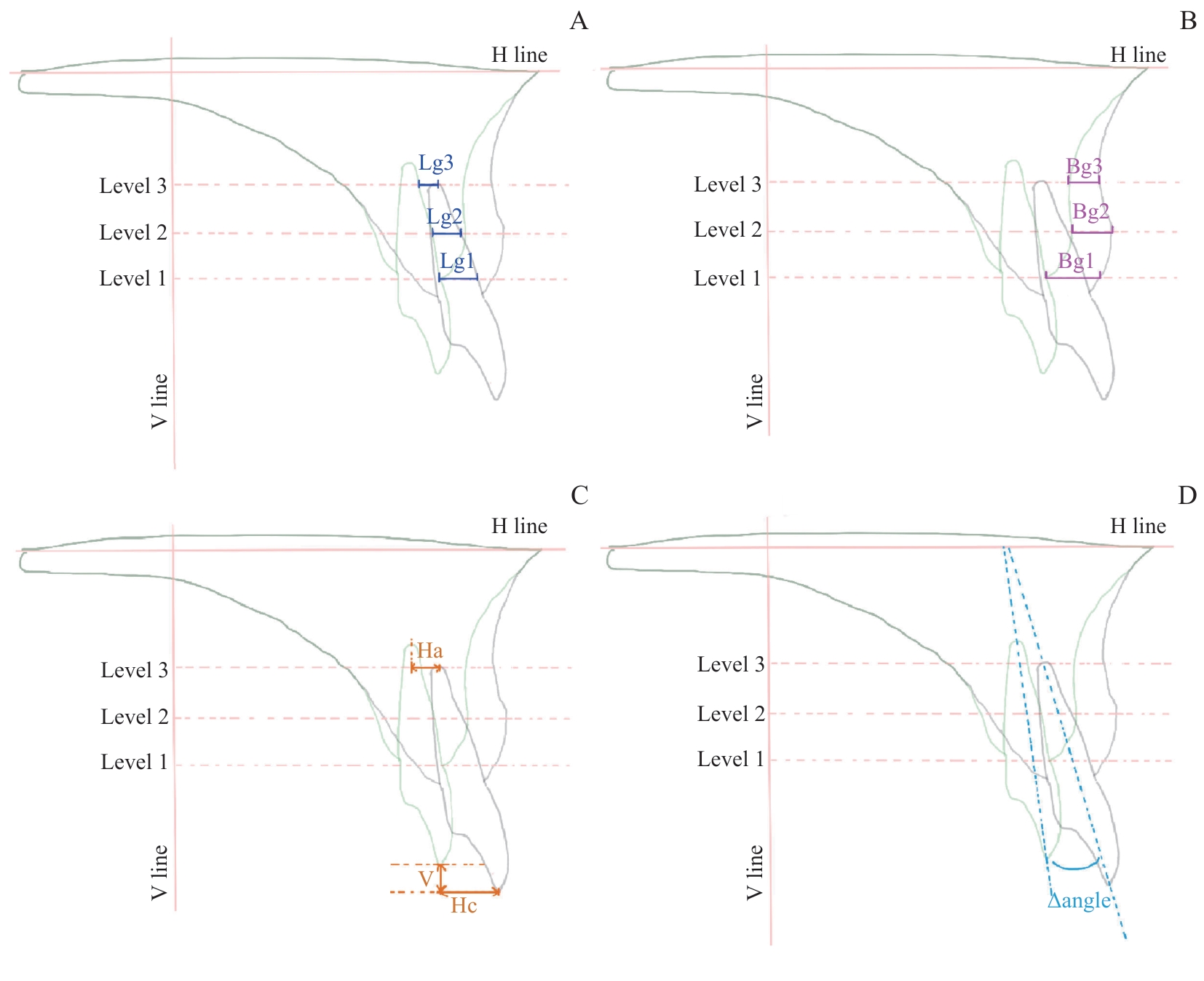
Fig 2 Diagram of measurement baselines and parameters in CBCT images
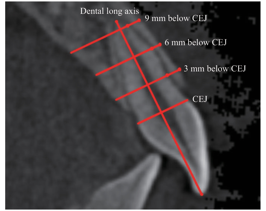
Fig 3 Schematic diagram of the section planes in the dental long- axis reference system
| Alveolar bone level | Tooth movement/mm | Alveolar bone remodeling/mm | B/T Ratio |
|---|---|---|---|
| T11 | |||
| Level 1 | 3.49±2.21 | 2.83±1.78 | 0.84(0.76, 1.03) |
| Level 2 | 3.05±2.29 | 1.96±1.46 | 0.75(0.59, 0.90) |
| Level 3 | 2.09±2.51 | 1.09±1.27 | 0.59(0.47, 0.81) |
| T21 | |||
| Level 1 | 3.55±2.06 | 2.83±1.65 | 0.74(0.67, 0.98) |
| Level 2 | 3.18±2.18 | 2.00±1.42 | 0.63(0.54, 0.76) |
| Level 3 | 2.81±2.54 | 1.29±1.25 | 0.55(0.44, 0.61) |
Tab 1 Tooth movement and alveolar bone remodeling in group G based on the skeletal stable structure reference system
| Alveolar bone level | Tooth movement/mm | Alveolar bone remodeling/mm | B/T Ratio |
|---|---|---|---|
| T11 | |||
| Level 1 | 3.49±2.21 | 2.83±1.78 | 0.84(0.76, 1.03) |
| Level 2 | 3.05±2.29 | 1.96±1.46 | 0.75(0.59, 0.90) |
| Level 3 | 2.09±2.51 | 1.09±1.27 | 0.59(0.47, 0.81) |
| T21 | |||
| Level 1 | 3.55±2.06 | 2.83±1.65 | 0.74(0.67, 0.98) |
| Level 2 | 3.18±2.18 | 2.00±1.42 | 0.63(0.54, 0.76) |
| Level 3 | 2.81±2.54 | 1.29±1.25 | 0.55(0.44, 0.61) |
| Alveolar bone level | Tooth movement/mm | Alveolar bone remodeling/mm | B/T Ratio |
|---|---|---|---|
| T11 | |||
| Level 1 | 4.93±1.87 | 4.28±1.27 | 0.88(0.79, 1.00) |
| Level 2 | 4.71±1.98 | 3.77±1.30 | 0.84(0.73, 0.92) |
| Level 3 | 4.54±2.15 | 2.92±1.29 | 0.72(0.49, 0.81) |
| T21 | |||
| Level 1 | 5.08±1.60 | 4.40±1.23 | 0.89(0.82, 0.97) |
| Level 2 | 4.94±1.69 | 3.79±1.03 | 0.81(0.65, 0.93) |
| Level 3 | 5.00±1.90 | 3.01±1.11 | 0.64(0.50, 0.73) |
Tab 2 Tooth movement and alveolar bone remodeling in group Y based on the dental long-axis reference system
| Alveolar bone level | Tooth movement/mm | Alveolar bone remodeling/mm | B/T Ratio |
|---|---|---|---|
| T11 | |||
| Level 1 | 4.93±1.87 | 4.28±1.27 | 0.88(0.79, 1.00) |
| Level 2 | 4.71±1.98 | 3.77±1.30 | 0.84(0.73, 0.92) |
| Level 3 | 4.54±2.15 | 2.92±1.29 | 0.72(0.49, 0.81) |
| T21 | |||
| Level 1 | 5.08±1.60 | 4.40±1.23 | 0.89(0.82, 0.97) |
| Level 2 | 4.94±1.69 | 3.79±1.03 | 0.81(0.65, 0.93) |
| Level 3 | 5.00±1.90 | 3.01±1.11 | 0.64(0.50, 0.73) |
| Group | M(Q1, Q3) | Z value | P value | |
|---|---|---|---|---|
| T11 B/T Ratio | ||||
| Level 1 | Group G | 0.58(0.47, 0.86) | -2.798 | 0.005 |
| Group Y | 0.35(0.19, 0.56) | |||
| Level 2 | Group G | 0.56(0.46, 0.62) | -4.010 | 0.000 |
| Group Y | 0.28(0.21, 0.33) | |||
| Level 3 | Group G | 0.81(0.70, 0.90) | -3.498 | 0.000 |
| Group Y | 0.40(0.25, 0.46) | |||
| T21 B/T Ratio | ||||
| Level 1 | Group G | 0.43(0.28, 0.86) | -0.016 | 0.987 |
| Group Y | 0.54(0.41, 0.72) | |||
| Level 2 | Group G | 0.37(0.27, 0.51) | -1.977 | 0.048 |
| Group Y | 0.57(0.22, 0.83) | |||
| Level 3 | Group G | 0.65(0.53, 0.70) | -2.419 | 0.016 |
| Group Y | 0.51(0.34, 0.62) | |||
Tab 3 Comparison of normalized B/T Ratio between group G and group Y
| Group | M(Q1, Q3) | Z value | P value | |
|---|---|---|---|---|
| T11 B/T Ratio | ||||
| Level 1 | Group G | 0.58(0.47, 0.86) | -2.798 | 0.005 |
| Group Y | 0.35(0.19, 0.56) | |||
| Level 2 | Group G | 0.56(0.46, 0.62) | -4.010 | 0.000 |
| Group Y | 0.28(0.21, 0.33) | |||
| Level 3 | Group G | 0.81(0.70, 0.90) | -3.498 | 0.000 |
| Group Y | 0.40(0.25, 0.46) | |||
| T21 B/T Ratio | ||||
| Level 1 | Group G | 0.43(0.28, 0.86) | -0.016 | 0.987 |
| Group Y | 0.54(0.41, 0.72) | |||
| Level 2 | Group G | 0.37(0.27, 0.51) | -1.977 | 0.048 |
| Group Y | 0.57(0.22, 0.83) | |||
| Level 3 | Group G | 0.65(0.53, 0.70) | -2.419 | 0.016 |
| Group Y | 0.51(0.34, 0.62) | |||
| [1] | DEANGELIS V. Observations on the response of alveolar bone to orthodontic force[J]. Am J Orthod, 1970, 58(3): 284-294. |
| [2] | VARDIMON A D, OREN E, BEN-BASSAT Y. Cortical bone remodeling/tooth movement ratio during maxillary incisor retraction with tip versus torque movements[J]. Am J Orthod Dentofac Orthop, 1998, 114(5): 520-529. |
| [3] | 连旭丽, 魏玢. 锥形线束CT测量对拔牙矫治上颌前突的患者牙根吸收及牙槽骨改建的影响[J]. 中国医疗器械信息, 2023, 29(13):65-67. |
| LIAN X L, WEI F. Effect of CBCT measurement on root resorption and alveolar bone remodeling in patients with maxillary protrusion[J]. China Medical Device Information, 2023, 29(13): 65-67. | |
| [4] | 林祥祥, 余飞, 宋艺蔚, 等.内收上颌切牙时唇腭侧牙槽骨改建影响因素的研究进展[J]. 口腔医学, 2023, 43(11): 1053-1056. |
| LIN X X, YU F, SONG Y W, et al. Progress of research on influencing factors of alveolar bone remodeling at lip and palate side during maxillary incisor retraction[J]. Stomatology, 2023, 43(11):1053-1056 | |
| [5] | 贾向敏. 不同年龄上颌前突患者内收前后上切牙牙根及牙槽骨变化的CBCT研究[D]. 大连: 大连医科大学, 2021. |
| JIA X M. A CBCT study of changes in roots of maxillary incisors and alveolar bone before and after retraction in patients with maxillary protrusion at different ages[D]. Dalian: Dalian Medical University, 2021. | |
| [6] | CHANMANEE P, CHAROEMRATROTE C. Maxillary anterior gingiva and dentoalveolar changes after en-masse retraction between thick and thin gingival biotypes[J]. Am J Orthod Dentofac Orthop, 2022, 161(6): 838-848. |
| [7] | 李玲凤. 正畸患者拔牙矫治后上下颌前牙区牙槽骨改建的回顾性研究[D]. 重庆: 重庆医科大学, 2023. |
| LI L F. Changes in alveolar bone of maxillary and mandibular anterior teeth after orthodontic extraction: a retrospective study[D]. Chongqing: Chongqing Medical University, 2023. | |
| [8] | XU B, YANG K. Changes in alveolar bone structure during orthodontic tooth movement in adolescent and adult rats: a microcomputed tomography study[J]. Orthod Craniofac Res, 2023, 26(4): 568-575. |
| [9] | ZHENG Y, ZHU C, ZHU M, et al. Difference in the alveolar bone remodeling between the adolescents and adults during upper incisor retraction: a retrospective study[J]. Sci Rep, 2022, 12(1): 9161. |
| [10] | LINJAWI A. Predictive factors affecting the maxillary alveolar bone thickness: a cone-beam computed tomography study[J]. Clin Cosmet Investig Dent, 2020, 12: 359-365. |
| [11] | 林雪芬. 成人患者拔牙与非拔牙矫治前后第一磨牙牙槽骨形态学研究[D]. 济南: 山东大学, 2022. |
| LIN X F. Morphological study on alveolar bone of the first molars in adult patients before and after extraction and non-extraction orthodontic treatment[D]. Jinan: Shandong University, 2022. | |
| [12] | GUO Z, ZHANG R, GUO C, et al. A retrospective study of alveolar bone remodelling after anterior retraction in orthodontic tooth extraction cases with clear aligners and fixed appliances[J]. Orthod Craniofac Res, 2024, 27(2): 220-227. |
| [13] | SON E J, KIM S J, HONG C, et al. A study on the morphologic change of palatal alveolar bone shape after intrusion and retraction of maxillary incisors[J]. Sci Rep, 2020, 10(1): 14454. |
| [14] | YANG C Y M, ATSAWASUWAN P, VIANA G, et al. Cone-beam computed tomography assessment of maxillary anterior alveolar bone remodelling in extraction and non-extraction orthodontic cases using stable extra-alveolar reference[J]. Orthod Craniofac Res, 2023, 26(2): 265-276. |
| [15] | SUN Q, LU W, ZHANG Y, et al. Morphological changes of the anterior alveolar bone due to retraction of anterior teeth: a retrospective study[J]. Head Face Med, 2021, 17(1): 30. |
| [16] | 林薇怡. 不同唇倾度的下切牙内收不同程度与牙槽骨改建的研究及病例报告[D]. 福州: 福建医科大学, 2022. |
| LIN W Y. Study on different angle change of different long axis of lower incisors and the reconstruction of alveolar bone & case report[D]. Fuzhou: Fujian Medical University, 2022. | |
| [17] | LUO N, CHEN Y, LI L, et al. Multivariate analysis of alveolar bone dehiscence and fenestration in anterior teeth after orthodontic treatment: a retrospective study[J]. Orthod Craniofac Res, 2024, 27(2): 287-296. |
| [18] | KALINA E, GRZEBYTA A, ZADURSKA M. Bone remodeling during orthodontic movement of lower incisors-narrative review[J]. Int J Environ Res Public Health, 2022, 19(22): 15002. |
| [19] | ZHANG F, LEE S C, LEE J B, et al. Geometric analysis of alveolar bone around the incisors after anterior retraction following premolar extraction[J]. Angle Orthod, 2020, 90(2): 173-180. |
| [20] | WANG S, LIU D W, GUO R Z, et al. Maxillary cortical bone remodeling characteristics in extraction patients: a cone-beam computed tomography study[J]. Am J Orthod Dentofac Orthop, 2023, 164(2): 160-171. |
| [21] | CHAIMONGKOL P, THONGUDOMPORN U, LINDAUER S J. Alveolar bone response to light-force tipping and bodily movement in maxillary incisor advancement: a prospective randomized clinical trial[J]. Angle Orthod, 2018, 88(1): 58-66. |
| [22] | EKSRIWONG T, THONGUDOMPORN U. Alveolar bone response to maxillary incisor retraction using stable skeletal structures as a reference[J]. Angle Orthod, 2021, 91(1): 30-35. |
| [23] | GUO R Z, LI L W, LIN Y F, et al. Long-term bone remodeling of maxillary anterior teeth with post-treatment alveolar bone defect in adult patients with maxillary protrusion: a prospective follow-up study[J]. Prog Orthod, 2023, 24(1): 36. |
| [24] | MAO H, YANG A, PAN Y, et al. Displacement in root apex and changes in incisor inclination affect alveolar bone remodeling in adult bimaxillary protrusion patients: a retrospective study[J]. Head Face Med, 2020, 16(1): 29. |
| [25] | SHEERAN S, HARTSFIELD J, OMAMI G, et al. Comparison of two 3-dimensional user-friendly voxel-based maxillary and 2-dimensional superimposition methods[J]. Am J Orthod Dentofacial Orthop, 2023, 163(1): 117-125. |
| [26] | 吴晓玫, 郭宏铭, 曾东, 等. 双颌前突患者拔牙内收后前牙区牙槽骨改建的CBCT研究[J]. 北京口腔医学, 2023, 31(6): 410-413. |
| WU X M, GUO H M, ZENG D, et al. Evaluation of alveolar bone remodeling after anterior teeth retraction in patients with bimaxillary protrusion using cone beam computed tomography[J]. Beijing Journal of Stomatology, 2023, 31(6): 410-413. | |
| [27] | AHN H W, MOON S C, BAEK S H. Morphometric evaluation of changes in the alveolar bone and roots of the maxillary anterior teeth before and after en masse retraction using cone-beam computed tomography[J]. Angle Orthod, 2013, 83(2): 212-221. |
| [1] | LIU Jingyi, XU Hongyuan, DAI Qinggang, JIANG Lingyong. Progress in the regulatory mechanisms of mandibular condylar development and deformity [J]. Journal of Shanghai Jiao Tong University (Medical Science), 2024, 44(8): 951-958. |
| [2] | GUO Liqiang, ZHAO Shitian, SHU Bing. Research progress in the roles of Notch signaling pathway during fracture healing [J]. Journal of Shanghai Jiao Tong University (Medical Science), 2023, 43(2): 222-229. |
| [3] | WANG Xijun, JIN Anting, HUANG Xiangru, XU Hongyuan, GAO Xin, YANG Yiling, DAI Qinggang, JIANG Lingyong. Study on Prrx1+ periodontal ligament stem cells during orthodontic tooth movement by lineage tracing [J]. Journal of Shanghai Jiao Tong University (Medical Science), 2022, 42(8): 1008-1015. |
| [4] | Yan-qing LU, Xing ZHOU, Jiao LI, Jian-ping PENG, Chuan-dong WANG, Xiao-ling ZHANG. Promotive effect of antitumor drug etoposide on osteogenic differentiation of mesenchymal stem cells [J]. JOURNAL OF SHANGHAI JIAOTONG UNIVERSITY (MEDICAL SCIENCE), 2021, 41(7): 849-857. |
| [5] | LUO Hong1, 2, WU Hong-yan1, 3, TAN Xi1, 3, DAI Hong-wei1, 2, 3, HUANG Lan1, 2, 3. Study on the difference of orthodontic tooth movement rate and pressure side bone remodeling between obese and obesity-resistant rats [J]. JOURNAL OF SHANGHAI JIAOTONG UNIVERSITY (MEDICAL SCIENCE), 2020, 40(8): 1055-1062. |
| [6] | YU Jin-feng, HU Yun, HUANG Ming-na, CHEN Jun, MING Ye, ZHENG Lei-lei. Three-dimensional study on symmetry of the first molar and its peripheric alveolar bone in skeletal class Ⅲ patients with mandibular deviation [J]. , 2018, 38(3): 288-. |
| [7] | ZHOU Di, WU Yan, WANG Yun-ji, FAN Xiao-ping. Comparison of alveolar bone changes in maxillary anterior area secondary to different kinds of retraction method of anterior teeth: a cone-beam computed tomography study [J]. , 2018, 38(11): 1375-. |
| [8] | CHEN Peng, YANG Feng-xue, ZHOU Jian-ping, DAI Hong-wei . Micro-structure changes in rat tooth movement process through micro-computed tomography dynamic observation [J]. , 2017, 37(2): 193-. |
| Viewed | ||||||
|
Full text |
|
|||||
|
Abstract |
|
|||||