Journal of Shanghai Jiao Tong University (Medical Science) ›› 2025, Vol. 45 ›› Issue (3): 253-260.doi: 10.3969/j.issn.1674-8115.2025.03.001
• Basic research • Next Articles
MAO Chenzhou1( ), ZHANG Ruiyun2, CHEN Haige2(), YIN Fangfei1(), ZUO Xiaolei1()
Received:2024-10-24
Accepted:2025-01-21
Online:2025-03-28
Published:2025-03-28
Contact:
CHEN Haige, YIN Fangfei, ZUO Xiaolei
E-mail:mcz1588_dpz@sjtu.edu.cn;chenhage@renji.com;yinfangfei@sjtu.edu.cn;zuoxiaolei@sjtu.edu.cn
Supported by:CLC Number:
MAO Chenzhou, ZHANG Ruiyun, CHEN Haige, YIN Fangfei, ZUO Xiaolei. Framework nucleic acid-based linear amplification platform for sensitive detection of bladder cancer-related miRNAs[J]. Journal of Shanghai Jiao Tong University (Medical Science), 2025, 45(3): 253-260.
Add to citation manager EndNote|Ris|BibTeX
URL: https://xuebao.shsmu.edu.cn/EN/10.3969/j.issn.1674-8115.2025.03.001
| DNA | Sequence (5′→3′) |
|---|---|
| A37-1 | CCCTGTACTGGCTAGGAATTCACGTTTTAATCTGGGCTTTGGGTTAAGAAACTCCCCG |
| A37-2 | CGCTGGAGGCGCATCACCGTTTGCGTATGTGTTCTGTGCGGCCTGCCGTCCCGTGTGGG |
| B37-1 | CGGTGATGCGCCTCCAGCGCGGGGAGTTTCTTAACCCTTTCCGACTTACAAGAGCCGG |
| B37-2 | GCGAGACTCAGGTGGTGCCTTTGGCATTCGACCAGGAGATATCGCGTTCAGCTATGCCC |
| C37-1 | CCCATGAGAATAATACCGCCGATTTACGTCAGTCCGGTTTCCCACACGGGACGGCAGGC |
| C37-2 | CGCACAGAACACATACGCTTTGGGCATAGCTGAACGCGATATCTCCTGGTCGAATGCC |
| D37-1 | GCCCAGATTAAAACGTGAATTCCTAGCCAGTACAGGGTTTCCGGACTGACGTAAATCGG |
| D37-2 | CGGTATTATTCTCATGGGTTTGGCACCACCTGAGTCTCGCCCGGCTCTTGTAAGTCGG |
Tab 1 DNA sequence for DTN
| DNA | Sequence (5′→3′) |
|---|---|
| A37-1 | CCCTGTACTGGCTAGGAATTCACGTTTTAATCTGGGCTTTGGGTTAAGAAACTCCCCG |
| A37-2 | CGCTGGAGGCGCATCACCGTTTGCGTATGTGTTCTGTGCGGCCTGCCGTCCCGTGTGGG |
| B37-1 | CGGTGATGCGCCTCCAGCGCGGGGAGTTTCTTAACCCTTTCCGACTTACAAGAGCCGG |
| B37-2 | GCGAGACTCAGGTGGTGCCTTTGGCATTCGACCAGGAGATATCGCGTTCAGCTATGCCC |
| C37-1 | CCCATGAGAATAATACCGCCGATTTACGTCAGTCCGGTTTCCCACACGGGACGGCAGGC |
| C37-2 | CGCACAGAACACATACGCTTTGGGCATAGCTGAACGCGATATCTCCTGGTCGAATGCC |
| D37-1 | GCCCAGATTAAAACGTGAATTCCTAGCCAGTACAGGGTTTCCGGACTGACGTAAATCGG |
| D37-2 | CGGTATTATTCTCATGGGTTTGGCACCACCTGAGTCTCGCCCGGCTCTTGTAAGTCGG |
| miRNA | Sequence (5′→3′) |
|---|---|
| miRNA-10a-5p | UACCCUGUAGAUCCGAAUUUGUG |
| miRNA-182-5p | UUUGGCAAUGGUAGAACUCACACU |
| miRNA-1-3p | UGGAAUGUAAAGAAGUAUGUAU |
| miRNA-30a-5p | UGUAAACAUCCUCGACUGGAAG |
| miRNA-100-5p | AACCCGUAGAUCCGAACUUGUG |
Tab 2 miRNA sequence for DTN
| miRNA | Sequence (5′→3′) |
|---|---|
| miRNA-10a-5p | UACCCUGUAGAUCCGAAUUUGUG |
| miRNA-182-5p | UUUGGCAAUGGUAGAACUCACACU |
| miRNA-1-3p | UGGAAUGUAAAGAAGUAUGUAU |
| miRNA-30a-5p | UGUAAACAUCCUCGACUGGAAG |
| miRNA-100-5p | AACCCGUAGAUCCGAACUUGUG |
| DNA | Sequence (5′→3′) |
|---|---|
| Signal-10a-Cy5 | Cy5-CACAAATTCGGATCTACTAA |
| Signal-182-Cy5 | Cy5-AGTGTGAGTTCTACCATTGA |
| Signal-1-Cy5 | Cy5-ATACATACTTCTTTACATGA |
| Signal-30a-Cy5 | Cy5-CTTCCAGTCGAGGATGTTGA |
| Signal-100-Cy5 | Cy5-CACAAGTTCGGATCTACTGA |
Tab 3 DNA sequence for specificity
| DNA | Sequence (5′→3′) |
|---|---|
| Signal-10a-Cy5 | Cy5-CACAAATTCGGATCTACTAA |
| Signal-182-Cy5 | Cy5-AGTGTGAGTTCTACCATTGA |
| Signal-1-Cy5 | Cy5-ATACATACTTCTTTACATGA |
| Signal-30a-Cy5 | Cy5-CTTCCAGTCGAGGATGTTGA |
| Signal-100-Cy5 | Cy5-CACAAGTTCGGATCTACTGA |
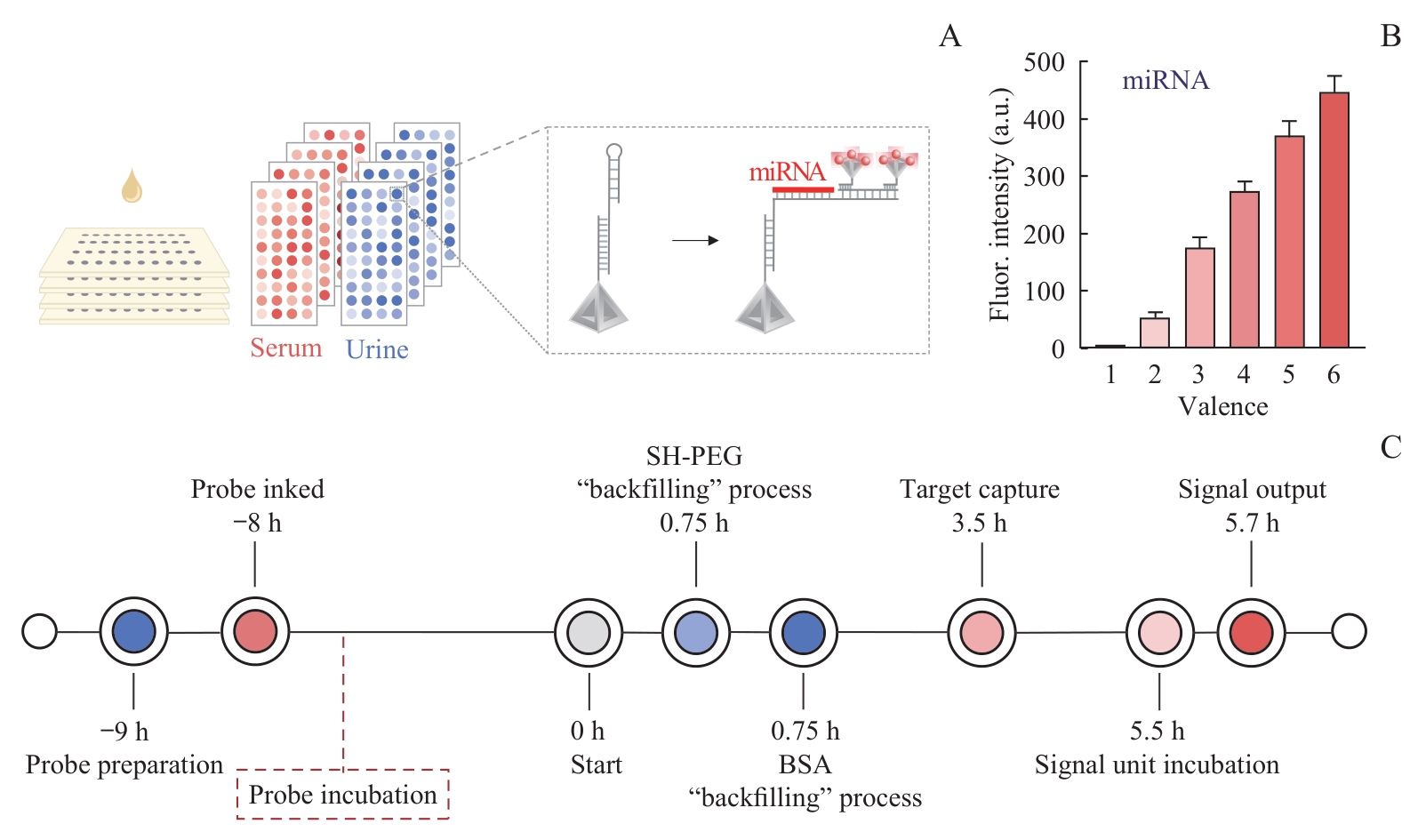
Fig 1 Schematic illustration of the design of a high-throughput multi-target detection platform
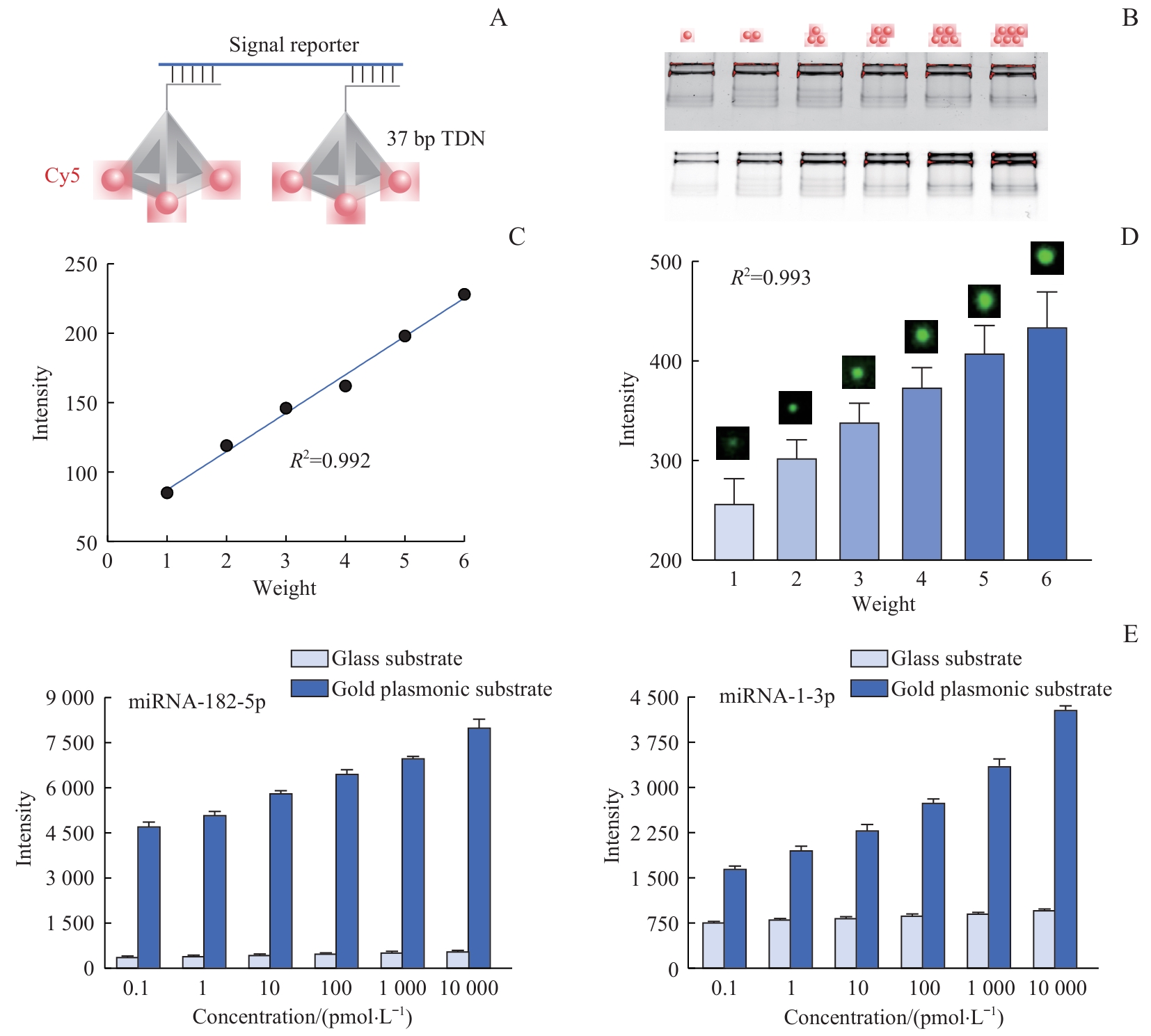
Fig 2 Performance comparison of the high-throughput multi-target joint detection platform
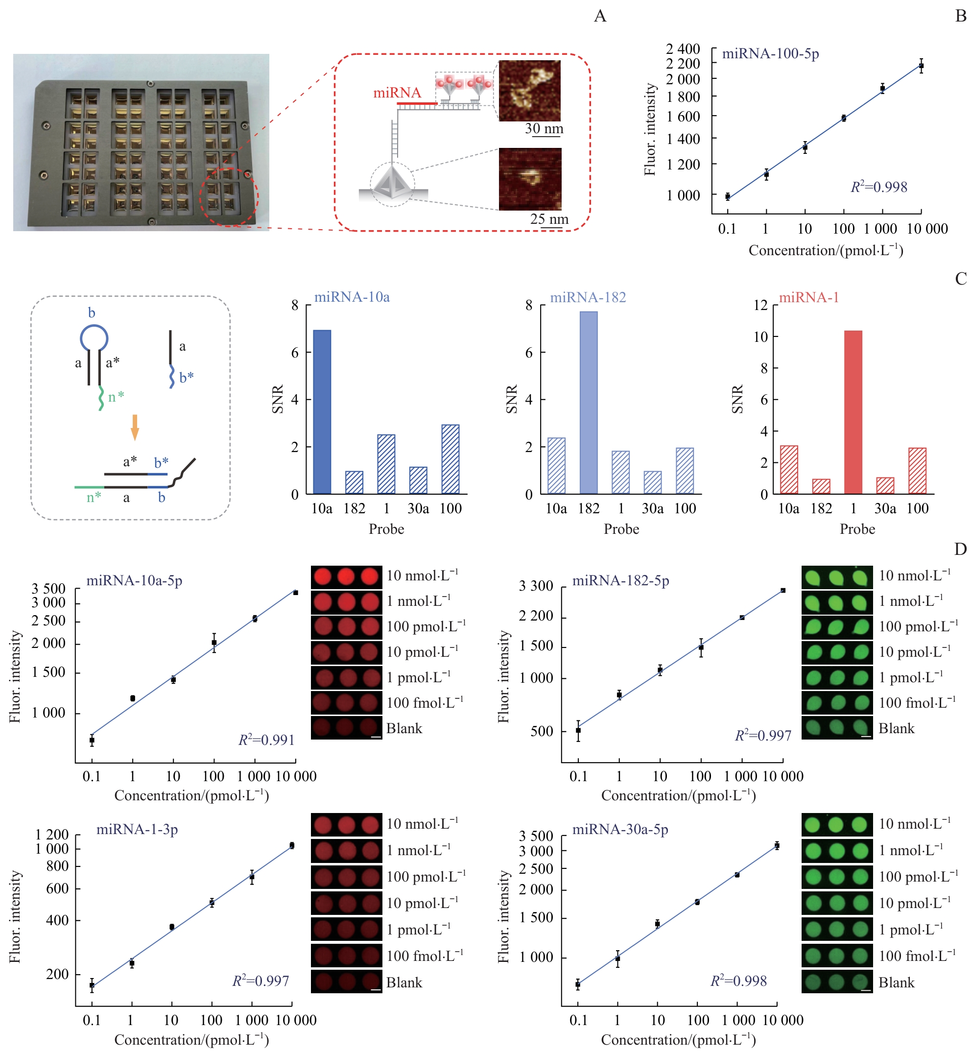
Fig 3 Performance of the high-throughput multi-target detection platform
| 1 | VAN HOOGSTRATEN L M C, VRIELING A, VAN DER HEIJDEN A G, et al. Global trends in the epidemiology of bladder cancer: challenges for public health and clinical practice[J]. Nat Rev Clin Oncol, 2023, 20(5): 287-304. |
| 2 | BRAY F, LAVERSANNE M, SUNG H, et al. Global cancer statistics 2022: globocan estimates of incidence and mortality worldwide for 36 cancers in 185 countries[J]. CA Cancer J Clin, 2024, 74(3): 229-263. |
| 3 | JASSIM A, RAHRMANN E P, SIMONS B D, et al. Cancers make their own luck: theories of cancer origins[J]. Nat Rev Cancer, 2023, 23(10): 710-724. |
| 4 | YUAN S P, ALMAGRO J, FUCHS E. Beyond genetics: driving cancer with the tumour microenvironment behind the wheel[J]. Nat Rev Cancer, 2024, 24(4): 274-286. |
| 5 | TRAN L, XIAO J F, AGARWAL N, et al. Advances in bladder cancer biology and therapy[J]. Nat Rev Cancer, 2021, 21(2): 104-121. |
| 6 | WAHIDA A, BUSCHHORN L, FRÖHLING S, et al. The coming decade in precision oncology: six riddles[J]. Nat Rev Cancer, 2023, 23(1): 43-54. |
| 7 | KON E, AD-EL N, HAZAN-HALEVY I, et al. Targeting cancer with mRNA-lipid nanoparticles: key considerations and future prospects[J]. Nat Rev Clin Oncol, 2023, 20(11): 739-754. |
| 8 | HIAM-GALVEZ K J, ALLEN B M, SPITZER M H. Systemic immunity in cancer[J]. Nat Rev Cancer, 2021, 21(6): 345-359. |
| 9 | LU Z H, CHEN Y, LIU D, et al. The landscape of cancer research and cancer care in China[J]. Nat Med, 2023, 29(12): 3022-3032. |
| 10 | DYRSKJØT L, HANSEL D E, EFSTATHIOU J A, et al. Bladder cancer[J]. Nat Rev Dis Primers, 2023, 9(1): 58. |
| 11 | LENIS A T, LEC P M, CHAMIE K, et al. Bladder cancer: a review[J]. JAMA, 2020, 324(19): 1980-1991. |
| 12 | SANLI O, DOBRUCH J, KNOWLES M A, et al. Bladder cancer[J]. Nat Rev Dis Primers, 2017, 3: 17022. |
| 13 | LOPEZ-BELTRAN A, COOKSON M S, GUERCIO B J, et al. Advances in diagnosis and treatment of bladder cancer[J]. BMJ, 2024, 384: e076743. |
| 14 | MATULEWICZ R S, DELANCEY J O, MEEKS J J. Cystoscopy[J]. JAMA, 2017, 317(11): 1187. |
| 15 | MAAS M, TODENHÖFER T, BLACK P C. Urine biomarkers in bladder cancer: current status and future perspectives[J]. Nat Rev Urol, 2023, 20: 597-614. |
| 16 | ROSE K M, HUELSTER H L, MEEKS J J, et al. Circulating and urinary tumour DNA in urothelial carcinoma: upper tract, lower tract and metastatic disease[J]. Nat Rev Urol, 2023, 20: 406-419. |
| 17 | DONG H F, LEI J P, DING L, et al. MicroRNA: function, detection, and bioanalysis[J]. Chem Rev, 2013, 113(8): 6207-6233. |
| 18 | LIU Z L, WANG H, LIU J, et al. MicroRNA-21 (miR-21) expression promotes growth, metastasis, and chemo- or radioresistance in non-small cell lung cancer cells by targeting PTEN[J]. Mol Cell Biochem, 2013, 372(1/2): 35-45. |
| 19 | CAUSA F, ALIBERTI A, CUSANO A M, et al. Supramolecular spectrally encoded microgels with double strand probes for absolute and direct miRNA fluorescence detection at high sensitivity[J]. J Am Chem Soc, 2015, 137(5): 1758-1761. |
| 20 | GE Z L, LIN M H, WANG P, et al. Hybridization chain reaction amplification of microRNA detection with a tetrahedral DNA nanostructure-based electrochemical biosensor[J]. Anal Chem, 2014, 86(4): 2124-2130. |
| 21 | YIN F F, LIU H Q, LI Q, et al. Trace microRNA quantification by means of plasmon-enhanced hybridization chain reaction[J]. Anal Chem, 2016, 88(9): 4600-4604. |
| 22 | DUAN R X, ZUO X L, WANG S T, et al. Lab in a tube: ultrasensitive detection of microRNAs at the single-cell level and in breast cancer patients using quadratic isothermal amplification[J]. J Am Chem Soc, 2013, 135(12): 4604-4607. |
| 23 | LIN M H, WANG J J, ZHOU G B, et al. Programmable engineering of a biosensing interface with tetrahedral DNA nanostructures for ultrasensitive DNA detection[J]. Angew Chem Int Ed, 2015, 54(7): 2151-2155. |
| 24 | SONG P, SHEN J W, YE D K, et al. Programming bulk enzyme heterojunctions for biosensor development with tetrahedral DNA framework[J]. Nat Commun, 2020, 11(1): 838. |
| 25 | LI F Q, MAO X H, LI F, et al. Ultrafast DNA sensors with DNA framework-bridged hybridization reactions[J]. J Am Chem Soc, 2020, 142(22): 9975-9981. |
| 26 | SQUIRES T M, MESSINGER R J, MANALIS S R. Making it stick: convection, reaction and diffusion in surface-based biosensors[J]. Nat Biotechnol, 2008, 26(4): 417-426. |
| 27 | LIU Q, GE Z L, MAO X H, et al. Valency-controlled framework nucleic acid signal amplifiers[J]. Angew Chem Int Ed, 2018, 57(24): 7131-7135. |
| 28 | YIN F F, ZHAO H P, LU S S, et al. DNA-framework-based multidimensional molecular classifiers for cancer diagnosis[J]. Nat Nanotechnol, 2023, 18(6): 677-686. |
| 29 | ZAIDI N, SIDDIQUI Z, SANKHWAR S N, et al. Urinary microRNA-10a levels in diagnosis and prognosis of urinary bladder cancer[J]. J Cancer Res Ther, 2023, 19(5): 1324-1329. |
| 30 | GU C H, ZHAO K Y, ZHOU N C, et al. UBAC2 promotes bladder cancer proliferation through BCRC-3/miRNA-182-5p/p27 axis[J]. Cell Death Dis, 2020, 11(9): 733. |
| 31 | YAMASAKI T, YOSHINO H, ENOKIDA H, et al. Novel molecular targets regulated by tumor suppressors microRNA-1 and microRNA-133a in bladder cancer[J]. Int J Oncol, 2012, 40(6): 1821-1830. |
| 32 | ZHANG C, MA X, DU J, et al. MicroRNA-30a as a prognostic factor in urothelial carcinoma of bladder inhibits cellular malignancy by antagonising Notch1[J]. BJU Int, 2016, 118(4): 578-589. |
| 33 | XU C L, ZENG Q S, XU W D, et al. miRNA-100 inhibits human bladder urothelial carcinogenesis by directly targeting mTOR[J]. Mol Cancer Ther, 2013, 12(2): 207-219. |
| 34 | BLANCA A, SANCHEZ-GONZALEZ A, REQUENA M J, et al. Expression of miR-100 and miR-138 as prognostic biomarkers in non-muscle-invasive bladder cancer[J]. APMIS, 2019, 127(8): 545-553. |
| [1] | YIN Ziming, WANG Rongqin, YANG Ziyi, LIU Yingbin, CHEN Tao, SHU Yijun, GONG Wei. Graph neural network-based auxiliary diagnostic model for gallbladder cancer on CT imaging [J]. Journal of Shanghai Jiao Tong University (Medical Science), 2025, 45(9): 1221-1231. |
| [2] | YANG Jingxiao, JIA Ziyao, WU Wenguang, WU Xiangsong, ZHANG Fei, LI Huaifeng, ZHU Yidi, LI Maolan. Effect of BRCA1 R1325K mutation on proliferation and apoptosis of gallbladder cancer cells [J]. Journal of Shanghai Jiao Tong University (Medical Science), 2023, 43(9): 1071-1079. |
| [3] | XIE Shasha, LÜ Yehui, LIN Jian. Application and research progress of tetrahedral framework nucleic acids in the field of medicine [J]. Journal of Shanghai Jiao Tong University (Medical Science), 2023, 43(3): 380-384. |
| [4] | YANG Chenkai, LI Wei, CAO Xiangqian, HE Lei, LI Shengzhou, SHEN Bing. Research progress in the treatment of bladder cancer based on nanotechnology [J]. Journal of Shanghai Jiao Tong University (Medical Science), 2023, 43(12): 1562-1568. |
| [5] | QI Yangyang, XIONG Ying. Phenotype, function and clinical significance of galectin-9 positive tumor-associated macrophages in muscle-invasive bladder cancer [J]. Journal of Shanghai Jiao Tong University (Medical Science), 2022, 42(12): 1666-1676. |
| [6] | Xiao-han WANG, Yun-lin YE, Kang-hua XIAO, Zhi-xin ZHAO, Jing-ya WU. Effects of knockdown of immunoregulatory protein B7-H3 gene on malignant proliferation, invasion and stem cell-like characteristics of bladder cancer cells [J]. JOURNAL OF SHANGHAI JIAOTONG UNIVERSITY (MEDICAL SCIENCE), 2021, 41(11): 1454-1460. |
| [7] | Kun ZHOU, Yu-mei CHEN, Jian-jun LIU, Gang HUANG. Prognostic value of delayed diuretic 18F-FDG PET/CT before radical cystectomy for bladder cancer [J]. JOURNAL OF SHANGHAI JIAOTONG UNIVERSITY (MEDICAL SCIENCE), 2021, 41(10): 1336-1343. |
| [8] | WU Ke1*, YAO Zhi-xian1*, ZHENG Zhong1*, CHENG Lei-lei2, LIU Zhi-hong1. Cisplatin promotes heart failure in bladder cancer miceimpairing granulocytic myeloid-derived suppressor cells [J]. , 2019, 39(9): 1011-. |
| [9] | QIN Yi-yu, ZHOU Xue-ping, LIN Pei-yi, et al. Inhibitory effects of combination of gemcitabine with sorafenib on proliferation and invasion of gallbladder cancer cells [J]. , 2014, 34(3): 318-. |
| [10] | HUANG Sheng-song, ZHU Zi-qi, SUN Li, et al. Expression of JARID1B in bladder cancer tissues and construction and function of lentiviral expression vector [J]. , 2010, 30(6): 693-. |
| Viewed | ||||||
|
Full text |
|
|||||
|
Abstract |
|
|||||