Journal of Shanghai Jiao Tong University (Medical Science) ›› 2025, Vol. 45 ›› Issue (9): 1221-1231.doi: 10.3969/j.issn.1674-8115.2025.09.014
• Techniques and methods • Previous Articles Next Articles
YIN Ziming1, WANG Rongqin1, YANG Ziyi2, LIU Yingbin3, CHEN Tao3, SHU Yijun2, GONG Wei2( )
)
Received:2025-06-09
Accepted:2025-08-25
Online:2025-09-28
Published:2025-09-30
Contact:
GONG Wei
E-mail:gongwei@xinhuamed.com.cn
Supported by:CLC Number:
YIN Ziming, WANG Rongqin, YANG Ziyi, LIU Yingbin, CHEN Tao, SHU Yijun, GONG Wei. Graph neural network-based auxiliary diagnostic model for gallbladder cancer on CT imaging[J]. Journal of Shanghai Jiao Tong University (Medical Science), 2025, 45(9): 1221-1231.
Add to citation manager EndNote|Ris|BibTeX
URL: https://xuebao.shsmu.edu.cn/EN/10.3969/j.issn.1674-8115.2025.09.014
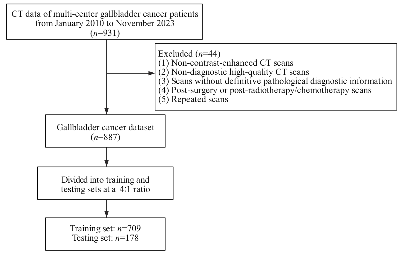
Fig 1 Process of data inclusion
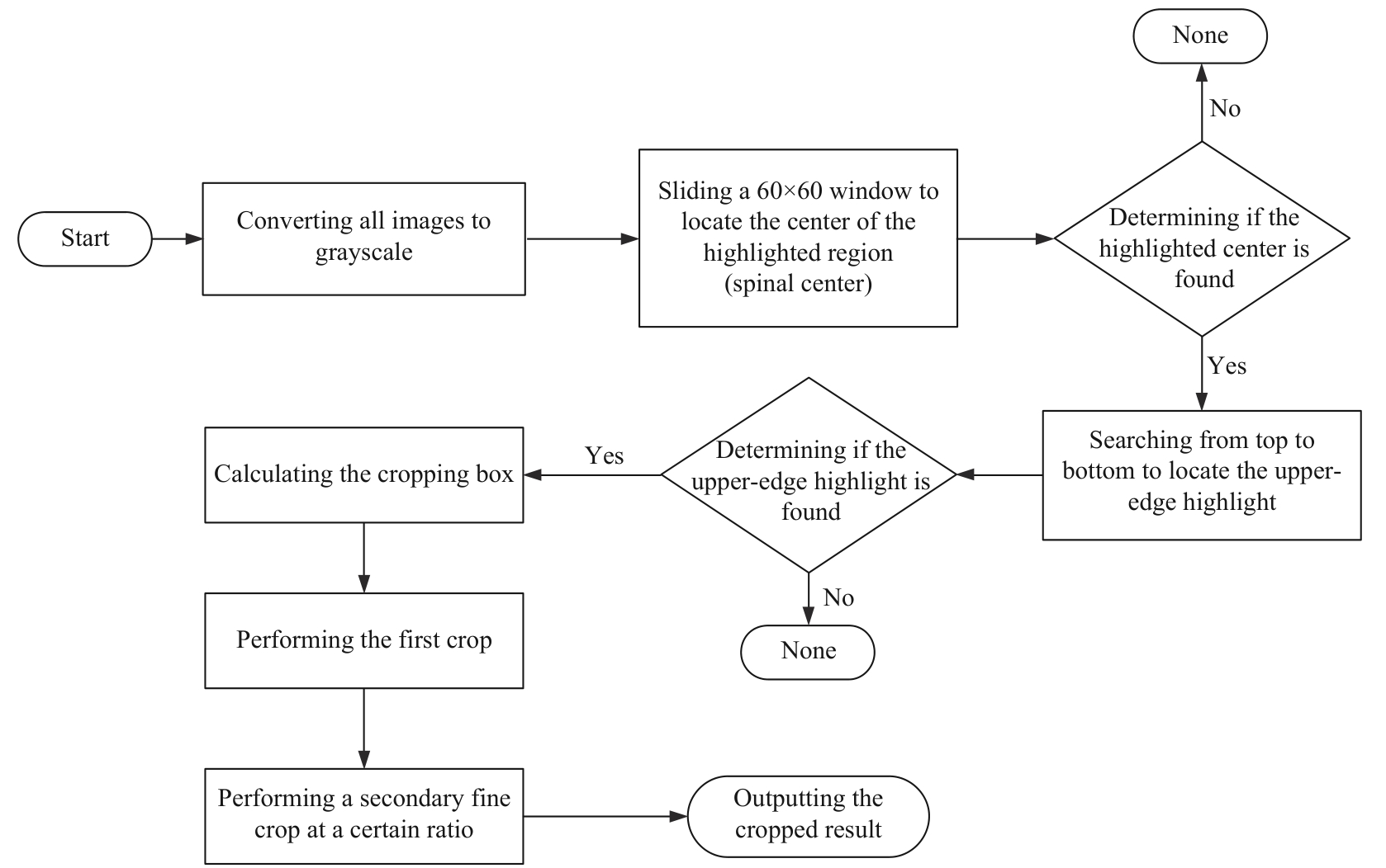
Fig 2 Flow chart of the fully automatic gallbladder region recognition and clipping algorithm
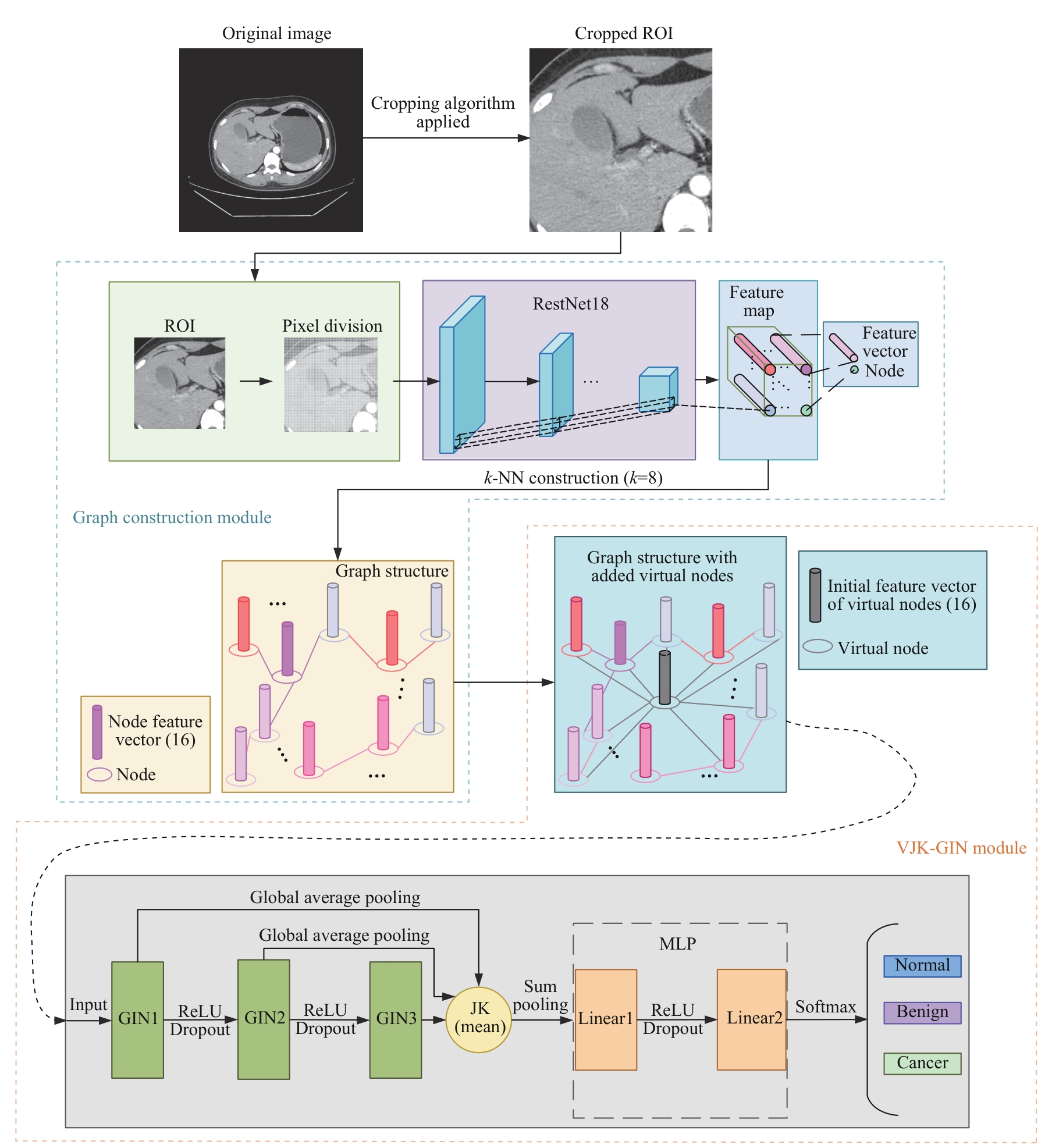
Fig 3 Overall schematic diagram of the model for gallbladder cancer diagnosis
| Item | Total | Training set | Testing set | P value |
|---|---|---|---|---|
| Number of patients/n | 887 | 709 | 178 | ‒ |
| Number of CT slices/n | 1 774 | 1419 | 355 | ‒ |
| Male/n (%) | 447 (50.4) | 360 (50.8) | 87 (49.2) | 0.712 |
| Age/year | 64.6±10.4 | 65.0±10.6 | 63.0±9.6 | 0.331 |
| BMI/(kg·m-2) | 24.3±3.1 | 24.4±3.5 | 23.9±0.18 | 0.383 |
| Disease type/n (%) | >0.999 | |||
| Cancer | 266 (30) | 213 (30) | 53 (30) | |
| Benign | 266 (30) | 213 (30) | 53 (30) | |
| Normal | 355 (40) | 283 (40) | 72 (40) |
Tab 1 Comparison of baseline characteristics between the training and test sets (one fold of 5-fold cross-validation)
| Item | Total | Training set | Testing set | P value |
|---|---|---|---|---|
| Number of patients/n | 887 | 709 | 178 | ‒ |
| Number of CT slices/n | 1 774 | 1419 | 355 | ‒ |
| Male/n (%) | 447 (50.4) | 360 (50.8) | 87 (49.2) | 0.712 |
| Age/year | 64.6±10.4 | 65.0±10.6 | 63.0±9.6 | 0.331 |
| BMI/(kg·m-2) | 24.3±3.1 | 24.4±3.5 | 23.9±0.18 | 0.383 |
| Disease type/n (%) | >0.999 | |||
| Cancer | 266 (30) | 213 (30) | 53 (30) | |
| Benign | 266 (30) | 213 (30) | 53 (30) | |
| Normal | 355 (40) | 283 (40) | 72 (40) |
| Index | GNN | CNN | ||||||
|---|---|---|---|---|---|---|---|---|
| VJK-GIN | GCN | GraphSAGE | GAT | GIN | ConvNeXt | ViT | EfficientNetV2 | |
Recall (95%CI) | 0.795 (0.773‒0.817) | 0.668 (0.641‒0.695) | 0.713 (0.689‒0.737) | 0.749 (0.729‒0.769) | 0.760 (0.744‒0.776) | 0.777 (0.760‒0.795) | 0.752 (0.729‒0.776) | 0.620 (0.594‒0.645) |
Precision (95%CI) | 0.799 (0.775‒0.823) | 0.667 (0.638‒0.696) | 0.715 (0.695‒0.735) | 0.742 (0.721‒0.764) | 0.758 (0.743‒0.776) | 0.778 (0.759‒0.798) | 0.754 (0.730‒0.778) | 0.621 (0.593‒0.648) |
F1-score (95%CI) | 0.799 (0.775‒0.823) | 0.667 (0.638‒0.696) | 0.714 (0.694‒0.734) | 0.742 (0.721‒0.764) | 0.758 (0.743‒0.776) | 0.778 (0.758‒0.797) | 0.753 (0.729‒0.776) | 0.619 (0.592‒0.646) |
Accuracy (95%CI) | 0.773 (0.748‒0.798) | 0.668 (0.644‒0.692) | 0.713 (0.695‒0.731) | 0.728 (0.708‒0.748) | 0.740 (0.718‒0.762) | 0.777 (0.758‒0.797) | 0.752 (0.731‒0.774) | 0.620 (0.592‒0.647) |
AUC (95%CI) | 0.812 (0.792‒0.832) | 0.697 (0.672‒0.722) | 0.713 (0.691‒0.735) | 0.762 (0.742‒0.782) | 0.775 (0.755‒0.795) | 0.757 (0.735‒0.778) | 0.751 (0.727‒0.774) | 0.703 (0.677‒0.728) |
Tab 2 Comparison of main indicators of each model
| Index | GNN | CNN | ||||||
|---|---|---|---|---|---|---|---|---|
| VJK-GIN | GCN | GraphSAGE | GAT | GIN | ConvNeXt | ViT | EfficientNetV2 | |
Recall (95%CI) | 0.795 (0.773‒0.817) | 0.668 (0.641‒0.695) | 0.713 (0.689‒0.737) | 0.749 (0.729‒0.769) | 0.760 (0.744‒0.776) | 0.777 (0.760‒0.795) | 0.752 (0.729‒0.776) | 0.620 (0.594‒0.645) |
Precision (95%CI) | 0.799 (0.775‒0.823) | 0.667 (0.638‒0.696) | 0.715 (0.695‒0.735) | 0.742 (0.721‒0.764) | 0.758 (0.743‒0.776) | 0.778 (0.759‒0.798) | 0.754 (0.730‒0.778) | 0.621 (0.593‒0.648) |
F1-score (95%CI) | 0.799 (0.775‒0.823) | 0.667 (0.638‒0.696) | 0.714 (0.694‒0.734) | 0.742 (0.721‒0.764) | 0.758 (0.743‒0.776) | 0.778 (0.758‒0.797) | 0.753 (0.729‒0.776) | 0.619 (0.592‒0.646) |
Accuracy (95%CI) | 0.773 (0.748‒0.798) | 0.668 (0.644‒0.692) | 0.713 (0.695‒0.731) | 0.728 (0.708‒0.748) | 0.740 (0.718‒0.762) | 0.777 (0.758‒0.797) | 0.752 (0.731‒0.774) | 0.620 (0.592‒0.647) |
AUC (95%CI) | 0.812 (0.792‒0.832) | 0.697 (0.672‒0.722) | 0.713 (0.691‒0.735) | 0.762 (0.742‒0.782) | 0.775 (0.755‒0.795) | 0.757 (0.735‒0.778) | 0.751 (0.727‒0.774) | 0.703 (0.677‒0.728) |
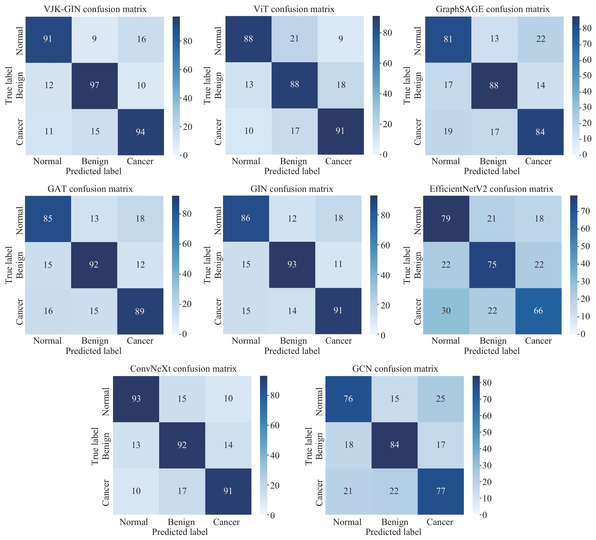
Fig 4 Confusion matrix heatmaps of each model in the test set
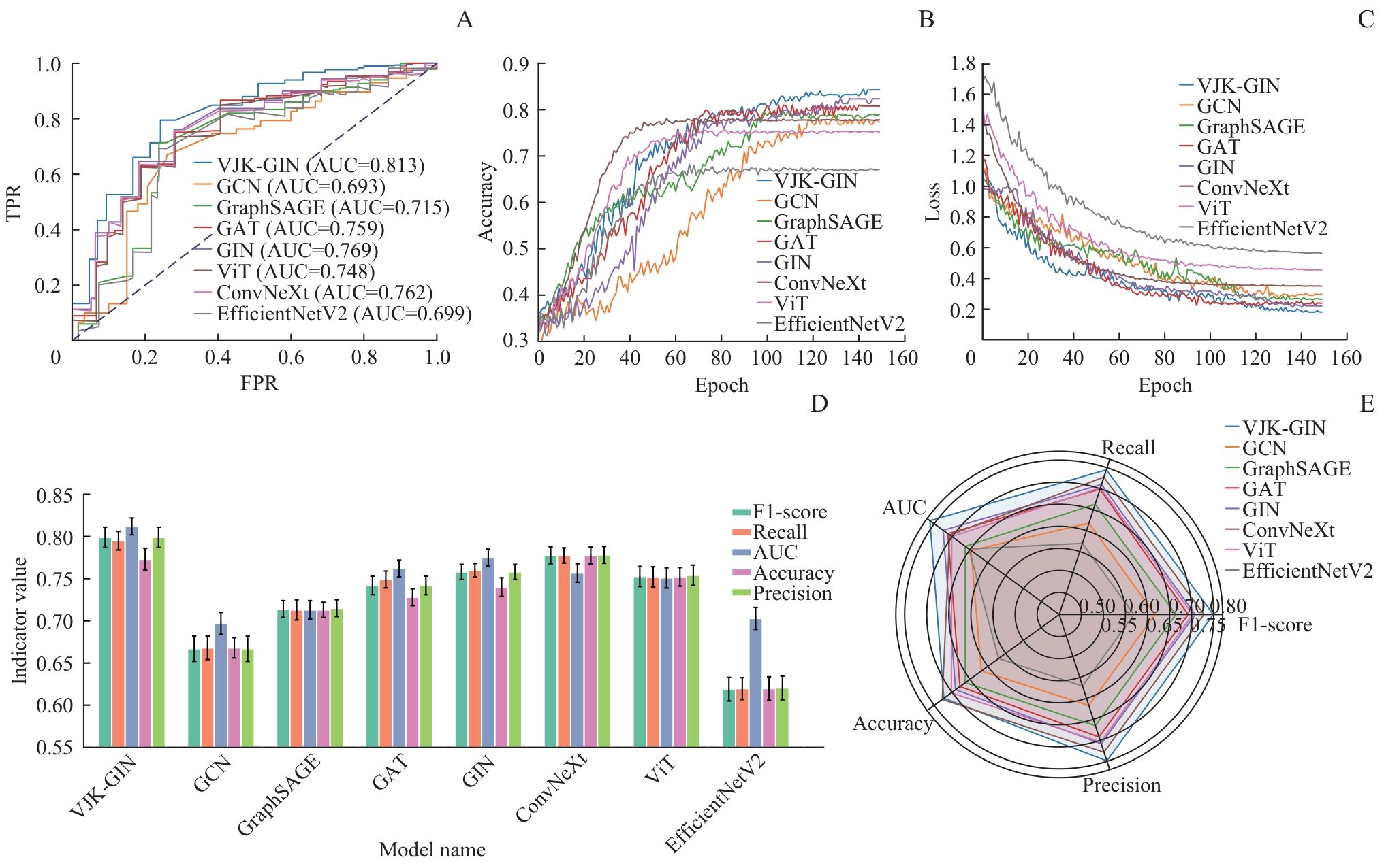
Fig 5 Visualization of the comprehensive performance of each model
| [1] | 刘颖斌, 陈炜. 重视胆囊癌的临床和基础研究[J]. 中华消化外科杂志, 2025, 24(1): 64-71. |
| LIU Y B, CHEN W. Attach importance to the clinical and basic research on gallbladder carcinoma[J]. Chinese Journal of Digestive Surgery, 2025, 24(1): 64-71. | |
| [2] | 刘珊山, 李初谊, 郑英, 等. 1990—2019年中国胆囊癌疾病负担研究及未来趋势分析[J]. 中国全科医学, 2024, 27(14): 1742-1749. |
| LIU S S, LI C Y, ZHENG Y, et al. Disease burden of gallbladder cancer in China from 1990 to 2019 and the analysis of its future trends[J]. Chinese General Practice, 2024, 27(14): 1742-1749. | |
| [3] | 吴玉梅, 郝金钢. 影像组学在胆囊癌诊疗中的研究进展[J]. 国际医学放射学杂志, 2024, 47(5): 594-598. |
| WU Y M, HAO J G. Research progress of radiomics in the diagnosis and treatment of gallbladder cancer[J]. International Journal of Medical Radiology, 2024, 47(5): 594-598. | |
| [4] | 罗明伟, 张译中, 周田, 等. 意外胆囊癌规范化诊疗策略的研究进展[J]. 腹部外科, 2022, 35(6): 457-461. |
| LUO M W, ZHANG Y Z, ZHOU T, et al. Research advances on standardized diagnosis and treatment strategies for unexpected gallbladder cancer[J]. Journal of Abdominal Surgery, 2022, 35(6): 457-461. | |
| [5] | 马振威, 朱博, 刘赋斌, 等. 血小板和淋巴细胞比值联合CA19-9在胆囊癌术后患者预后评估中的价值[J]. 中华肝脏外科手术学电子杂志, 2024, 13(2): 163-168. |
| MA Z W, ZHU B, LIU F B, et al. Evaluation value of platelet-to-lymphocyte ratio combined with CA19-9 for postoperative prognosis of patients with gallbladder cancer[J]. Chinese Journal of Hepatic Surgery (Electronic Edition), 2024, 13(2): 163-168. | |
| [6] | 杨志新, 赵丽珠, 邓玥, 等. 基于卷积神经网络在卵巢肿瘤良恶性鉴别诊断中的研究[J]. 昆明医科大学学报, 2023, 44(10): 134-139. |
| YANG Z X, ZHAO L Z, DENG Y, et al. Differential diagnosis of benign and malignant ovarian tumors based on convolutional neural network[J]. Journal of Kunming Medical University, 2023, 44(10): 134-139. | |
| [7] | 沈天乐, 杜向慧. 人工智能在恶性肿瘤放疗领域中的应用与前景[J]. 浙江医学, 2018, 40(8): 783-785, 795. |
| SHEN T L, DU X H. Application and prospect of artificial intelligence in radiotherapy for malignant tumors[J]. Zhejiang Medicine, 2018, 40(8): 783-785, 795. | |
| [8] | 蒋西然, 蒋韬, 孙嘉瑶, 等. 深度学习人工智能技术在医学影像辅助分析中的应用[J]. 中国医疗设备, 2021, 36(6): 164-171. |
| JIANG X R, JIANG T, SUN J Y, et al. Deep learning in computer aided analyses of medical images[J]. China Medical Devices, 2021, 36(6): 164-171. | |
| [9] | 张礼, 王嘉瑞. 基于图同构网络的自闭症功能磁共振影像诊断算法[J]. 南京大学学报(自然科学), 2021, 57(5): 801-809. |
| ZHANG L, WANG J R. Diagnosing autism spectrum disorder from functional MRI using graph isomorphic network[J]. Journal of Nanjing University (Natural Sciences), 2021, 57(5): 801-809. | |
| [10] | 覃智威, 刘钊, 陆允敏, 等. 基于广义卷积神经网络的阿尔茨海默病多模态磁共振图像分类方法研究[J]. 生物医学工程学杂志, 2023, 40(2): 217-225. |
| QIN Z W, LIU Z, LU Y M, et al. Research on classification method of multimodal magnetic resonance images of Alzheimer's disease based on generalized convolutional neural networks[J]. Journal of Biomedical Engineering, 2023, 40(2): 217-225. | |
| [11] | JEONG Y, KIM J H, CHAE H D, et al. Deep learning-based decision support system for the diagnosis of neoplastic gallbladder polyps on ultrasonography: preliminary results[J]. Sci Rep, 2020, 10(1): 7700. |
| [12] | ZHAO H Y, MIAO C, ZHU Y D, et al. An end-to-end interpretable machine-learning-based framework for early-stage diagnosis of gallbladder cancer using multi-modality medical data[J]. BMC Cancer, 2025, 25(1): 1178. |
| [13] | YANG M, ZHAO Y H, LI C, et al. Multimodal integration of liquid biopsy and radiology for the noninvasive diagnosis of gallbladder cancer and benign disorders[J]. Cancer Cell, 2025, 43(3): 398-412.e4. |
| [14] | YIN Y C, YAKAR D, SLANGEN J J G, et al. The value of deep learning in gallbladder lesion characterization[J]. Diagnostics (Basel), 2023, 13(4): 704. |
| [15] | GUPTA P, BASU S, RANA P, et al. Deep-learning enabled ultrasound based detection of gallbladder cancer in northern India: a prospective diagnostic study[J]. Lancet Reg Health Southeast Asia, 2024, 24: 100279. |
| [16] | OBAID A M, TURKI A, BELLAAJ H, et al. Detection of gallbladder disease types using deep learning: an informative medical method[J]. Diagnostics (Basel), 2023, 13(10): 1744. |
| [17] | WANG H X, HUANG G, ZHAO Z, et al. CCF-GNN: a unified model aggregating appearance, microenvironment, and topology for pathology image classification[J]. IEEE Trans Med Imaging, 2023, 42(11): 3179-3193. |
| [18] | ZHANG L, ZHAO Y, CHE T T, et al. Graph neural networks for image-guided disease diagnosis: a review[J]. iRADIOLOGY, 2023, 1(2): 151-166. |
| [19] | ADDOUNE S E, OUAHBI M I. A hybrid UNet-GNN architecture for enhanced medical image segmentation[D]. Ouargla, Algeria: Kasdi Merbah University, 2024. |
| [20] | DUAN J W, XIONG J Q, LI Y H, et al. Deep learning based multimodal biomedical data fusion: an overview and comparative review[J]. Inf Fusion, 2024, 112: 102536. |
| [21] | LING R H, CEN X X, WU S Y, et al. Explainable graph neural network based on metabolic brain imaging for differential diagnosis of Parkinsonism[J]. Front Aging Neurosci, 2025, 17: 1580910. |
| [22] | ZHENG K Z, YU S J, CHEN L J, et al. BPI-GNN: interpretable brain network-based psychiatric diagnosis and subtyping[J]. Neuroimage, 2024, 292: 120594. |
| [23] | XU K, HU W, LESKOVEC J, et al. How powerful are graph neural networks?[DB/OL]. (2019-02-22)[2025-08-03]. https://arxiv.org/abs/1810.00826. |
| [24] | WANG H, GAO K, ZHANG X, et al. Pixel- and patch-wise context-aware learning with CNN and GCN collaboration for hyperspectral image classification[C]//IGARSS 2023‒2023 IEEE International Geoscience and Remote Sensing Symposium, Pasadena, CA, USA. New York: IEEE, 2023: 7555-7558. |
| [25] | WANG Y F, YU X D, DONG H B, et al. Graph convolutional network with local and global feature fusion for hyperspectral image classification[J]. IEEE Trans Geosci Remote Sens, 2024, 62: 5536915. |
| [26] | HE K M, ZHANG X Y, REN S Q, et al. Deep residual learning for image recognition[C]//2016 IEEE Conference on Computer Vision and Pattern Recognition (CVPR), Las Vegas, NV, USA. New York: IEEE, 2016: 770-778. |
| [27] | DOSOVITSKIY A, BEYER L, KOLESNIKOV A, et al. An image is worth 16×16 words: transformers for image recognition at scale[DB/OL]. (2021-06-03)[2025-08-03]. https://arxiv.org/abs/2010.11929. |
| [28] | TAN M, LE Q V. EfficientNetV2: smaller models and faster training[DB/OL]. (2021-06-23)[2025-08-03]. https://arxiv.org/abs/2104.00298. |
| [29] | FENG J W, TAN H L, LI W W, et al. Conv2NeXt: reconsidering ConvNeXt network design for image recognition[C]//2022 International Conference on Computers and Artificial Intelligence Technologies (CAIT), Quzhou, China. New York: IEEE, 2022: 53-60. |
| [30] | KIPF T N, WELLING M. Semi-supervised classification with graph convolutional networks[DB/OL]. (2017-02-22)[2025-08-03]. https://arxiv.org/abs/1609.02907. |
| [31] | HAMILTON W L, YING R, LESKOVEC J. Inductive representation learning on large graphs[DB/OL]. (2018-09-10)[2025-08-03]. https://arxiv.org/abs/1706.02216. |
| [32] | VELIČKOVIĆ P, CUCURULL G, CASANOVA A, et al. Graph attention networks[DB/OL]. (2018-02-04)[2025-08-03]. https://arxiv.org/abs/1710.10903. |
| [1] | Zhu Menglin, Liu Xiao, Xu Xiaodan, Wang Ganhong, Xia Kaijian, Chen Jian. Prediction of delayed post-polypectomy bleeding using a multimodal model [J]. Journal of Shanghai Jiao Tong University (Medical Science), 2026, 46(4): 509-520. |
| [2] | CHEN Jian, WANG Zhenni, XIA Kaijian, WANG Ganhong, LIU Luojie, XU Xiaodan. Comparative study on methods for colon polyp endoscopic image segmentation and classification based on deep learning [J]. Journal of Shanghai Jiao Tong University (Medical Science), 2024, 44(6): 762-772. |
| [3] | LIU Yang, WU Mengyi, HU Yao, QI Kun, WANG Yubin, ZHAO Yue, SONG Jinlin. Preliminary application of a cervical vertebra segmentation method based on Transformer and diffusion model for lateral cephalometric radiographs in orthodontic clinical practice [J]. Journal of Shanghai Jiao Tong University (Medical Science), 2024, 44(12): 1579-1586. |
| [4] | YANG Jingxiao, JIA Ziyao, WU Wenguang, WU Xiangsong, ZHANG Fei, LI Huaifeng, ZHU Yidi, LI Maolan. Effect of BRCA1 R1325K mutation on proliferation and apoptosis of gallbladder cancer cells [J]. Journal of Shanghai Jiao Tong University (Medical Science), 2023, 43(9): 1071-1079. |
| [5] | ZHAO Keke, JIANG Beibei, ZHANG Lu, WANG Lingyun, ZHANG Yaping, XIE Xueqian. Feasibility of ultra-low-dose noncontrast CT based on deep learning image reconstruction to evaluate chest lesions [J]. Journal of Shanghai Jiao Tong University (Medical Science), 2022, 42(8): 1062-1069. |
| [6] | LIN Jiaxi, WANG Shengjia, ZHAO Xin, GAO Xin, YIN Minyue, ZHU Jinzhou. Development of endoscopic image classification models of Barrett's esophagus based on deep convolutional neural networks [J]. Journal of Shanghai Jiao Tong University (Medical Science), 2022, 42(5): 653-659. |
| [7] | CHEN Liqi, XUE Zhuowei, WU Qingkai. Review of MRI-based three-dimensional digital model reconstruction of female pelvic floor organs [J]. Journal of Shanghai Jiao Tong University (Medical Science), 2022, 42(3): 381-386. |
| [8] | LU Yan-qiao, SHEN Lan, HE Ben. Application of artificial intelligence in assisted diagnosis and treatment of cardiovascular disease [J]. , 2020, 40(2): 259-. |
| [9] | QIN Yi-yu, ZHOU Xue-ping, LIN Pei-yi, et al. Inhibitory effects of combination of gemcitabine with sorafenib on proliferation and invasion of gallbladder cancer cells [J]. , 2014, 34(3): 318-. |
| Viewed | ||||||
|
Full text |
|
|||||
|
Abstract |
|
|||||