Journal of Shanghai Jiao Tong University (Medical Science) ›› 2026, Vol. 46 ›› Issue (1): 15-24.doi: 10.3969/j.issn.1674-8115.2026.01.002
• Basic research • Previous Articles Next Articles
Zhou Peng1,2, Liu Yanna1, Yan Jingbin1,3( )
)
Received:2025-03-25
Accepted:2025-05-13
Online:2026-01-28
Published:2026-01-30
Contact:
Yan Jingbin
E-mail:m18917128323@163.com
About author:First author contact:Zhou Peng, Liu Yanna, and Yan Jingbin contributed to the design of the experiments. Zhou Peng and Yan Jingbin were responsible for drafting the manuscript and conducting critical revisions. All authors have reviewed the final version of the manuscript and consented to its submission.
Supported by:CLC Number:
Zhou Peng, Liu Yanna, Yan Jingbin. Centromere proteomic analysis of Down syndrome-derived amniocytes[J]. Journal of Shanghai Jiao Tong University (Medical Science), 2026, 46(1): 15-24.
Add to citation manager EndNote|Ris|BibTeX
URL: https://xuebao.shsmu.edu.cn/EN/10.3969/j.issn.1674-8115.2026.01.002
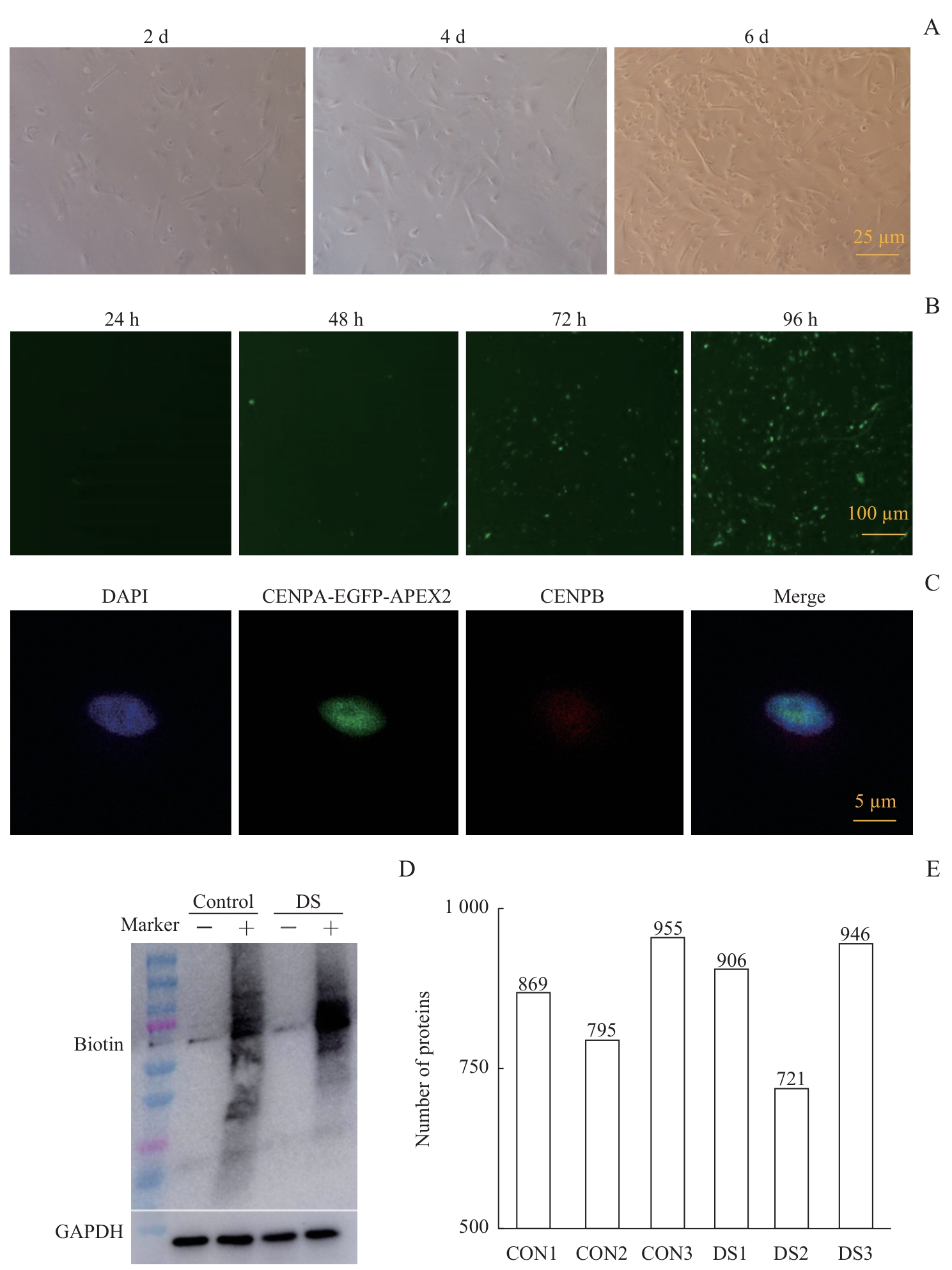
Fig 1 Establishment of DS-derived amniocytes models and identification of CENPA proximity-labeled proteins
| Case | Maternal age/year | Gestation age/week | Fetal sex | Indication for testing | Chromosomal karyotype |
|---|---|---|---|---|---|
| 1 | 40 | 16+3 | Male | High-risk NIPT result | 47, XY, +21 |
| 2 | 30 | 16+5 | Male | High-risk NIPT result | 47, XY, +21 |
| 3 | 44 | 18+2 | Female | High-risk NIPT result | 47, XX, +21 |
| 4 | 35 | 18+2 | Male | High-risk NIPT result | 47, XY, +21 |
| 5 | 41 | 19+3 | Female | High-risk NIPT result | 47, XX, +21 |
| 6 | 31 | 24+4 | Female | High-risk NIPT result | 47, XX, +21 |
| 7 | 32 | 13 | Female | High-risk NIPT result | 47, XX, +21 |
| 8 | 31 | 18+3 | Male | High-risk NIPT result | 47, XY, +21 |
| 9 | 25 | 16+1 | Male | High-risk NIPT result | 47, XY, +21 |
| 10 | 38 | 18 | Female | Advanced maternal age | 47, XX, +21 |
| 11 | 46 | 24+5 | Male | Positive screening for trisomy 21 | 46, XY |
| 12 | 34 | 19+4 | Male | Positive screening for trisomy 21 | 46, XY |
| 13 | 33 | 21+6 | Male | Positive screening for trisomy 21 | 46, XY |
| 14 | 27 | 16+5 | Male | High-risk serum markers | 46, XY |
| 15 | 30 | 17+5 | Female | High-risk serum markers | 46, XX |
| 16 | 42 | 23+3 | Male | Advanced maternal age | 46, XY |
Tab 1 Overview of amniocytes
| Case | Maternal age/year | Gestation age/week | Fetal sex | Indication for testing | Chromosomal karyotype |
|---|---|---|---|---|---|
| 1 | 40 | 16+3 | Male | High-risk NIPT result | 47, XY, +21 |
| 2 | 30 | 16+5 | Male | High-risk NIPT result | 47, XY, +21 |
| 3 | 44 | 18+2 | Female | High-risk NIPT result | 47, XX, +21 |
| 4 | 35 | 18+2 | Male | High-risk NIPT result | 47, XY, +21 |
| 5 | 41 | 19+3 | Female | High-risk NIPT result | 47, XX, +21 |
| 6 | 31 | 24+4 | Female | High-risk NIPT result | 47, XX, +21 |
| 7 | 32 | 13 | Female | High-risk NIPT result | 47, XX, +21 |
| 8 | 31 | 18+3 | Male | High-risk NIPT result | 47, XY, +21 |
| 9 | 25 | 16+1 | Male | High-risk NIPT result | 47, XY, +21 |
| 10 | 38 | 18 | Female | Advanced maternal age | 47, XX, +21 |
| 11 | 46 | 24+5 | Male | Positive screening for trisomy 21 | 46, XY |
| 12 | 34 | 19+4 | Male | Positive screening for trisomy 21 | 46, XY |
| 13 | 33 | 21+6 | Male | Positive screening for trisomy 21 | 46, XY |
| 14 | 27 | 16+5 | Male | High-risk serum markers | 46, XY |
| 15 | 30 | 17+5 | Female | High-risk serum markers | 46, XX |
| 16 | 42 | 23+3 | Male | Advanced maternal age | 46, XY |
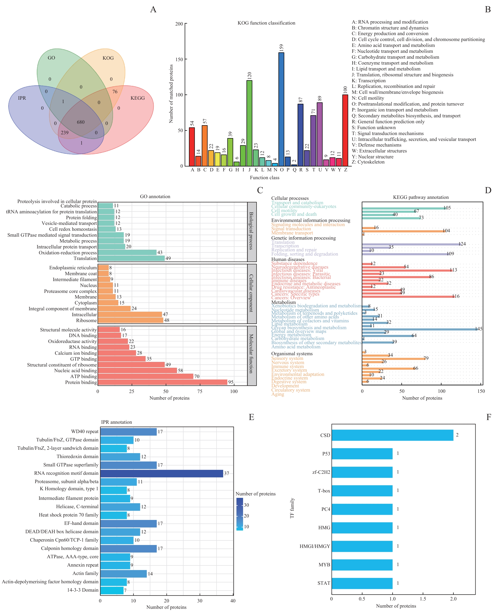
Fig 2 Functional annotation of proximity-labeled proteins of CENPA
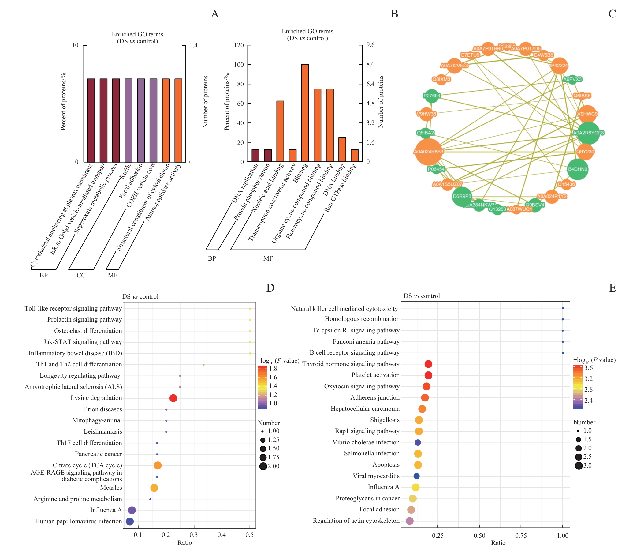
Fig 3 DEPs enrichment analysis of CENPA proximity-labeling proteins in amniocytes between the DS group and the normal group
| [1] | Antonarakis S E, Lyle R, Dermitzakis E T, et al. Chromosome 21 and Down syndrome: from genomics to pathophysiology[J]. Nat Rev Genet, 2004, 5(10): 725-738. |
| [2] | Lamb N E, Feingold E, Savage A, et al. Characterization of susceptible chiasma configurations that increase the risk for maternal nondisjunction of chromosome 21[J]. Hum Mol Genet, 1997, 6(9): 1391-1399. |
| [3] | Lanzillotta C, Di Domenico F. Stress responses in Down syndrome neurodegeneration: state of the art and therapeutic molecules[J]. Biomolecules, 2021, 11(2): 266. |
| [4] | Shapiro B L. Down syndrome: a disruption of homeostasis[J]. Am J Med Genet, 1983, 14(2): 241-269. |
| [5] | Pritchard M A, Kola I. The “gene dosage effect” hypothesis versus the “amplified developmental instability” hypothesis in Down syndrome[J]. J Neural Transm Suppl, 1999, 57: 293-303. |
| [6] | Krivega M, Stiefel C M, Storchova Z. Consequences of chromosome gain: a new view on trisomy syndromes[J]. Am J Hum Genet, 2022, 109(12): 2126-2140. |
| [7] | Verdaasdonk J S, Bloom K. Centromeres: unique chromatin structures that drive chromosome segregation[J]. Nat Rev Mol Cell Biol, 2011, 12(5): 320-332. |
| [8] | James S J, Pogribna M, Pogribny I P, et al. Abnormal folate metabolism and mutation in the methylenetetrahydrofolate reductase gene may be maternal risk factors for Down syndrome[J]. Am J Clin Nutr, 1999, 70(4): 495-501. |
| [9] | Oliver T R, Middlebrooks C D, Tinker S W, et al. An examination of the relationship between hotspots and recombination associated with chromosome 21 nondisjunction[J]. PLoS One, 2014, 9(6): e99560. |
| [10] | Coppedè F, Bosco P, Tannorella P, et al. DNMT3B promoter polymorphisms and maternal risk of birth of a child with Down syndrome[J]. Hum Reprod, 2013, 28(2): 545-550. |
| [11] | Jaiswal S K, Sukla K K, Kumari N, et al. Maternal risk for Down syndrome and polymorphisms in the promoter region of the DNMT3B gene: a case-control study[J]. Birth Defects Res A Clin Mol Teratol, 2015, 103(4): 299-305. |
| [12] | Contreras-Galindo R, Fischer S, Saha A K, et al. Rapid molecular assays to study human centromere genomics[J]. Genome Res, 2017, 27(12): 2040-2049. |
| [13] | Sullivan K D, Evans D, Pandey A, et al. Trisomy 21 causes changes in the circulating proteome indicative of chronic autoinflammation[J]. Sci Rep, 2017, 7(1): 14818. |
| [14] | Gao L, Zhang J, Ran X J, et al. Urinary proteomics for noninvasive prenatal screening of trisomy 21: new biomarker candidates[J]. OMICS, 2021, 25(11): 738-744. |
| [15] | Sobol M, Klar J, Laan L, et al. Transcriptome and proteome profiling of neural induced pluripotent stem cells from individuals with Down syndrome disclose dynamic dysregulations of key pathways and cellular functions[J]. Mol Neurobiol, 2019, 56(10): 7113-7127. |
| [16] | Hwang S, Cavaliere P, Li R, et al. Consequences of aneuploidy in human fibroblasts with trisomy 21[J]. Proc Natl Acad Sci USA, 2021, 118(6): e2014723118. |
| [17] | Liu Y S, Borel C, Li L, et al. Systematic proteome and proteostasis profiling in human trisomy 21 fibroblast cells[J]. Nat Commun, 2017, 8(1): 1212. |
| [18] | Lam S S, Martell J D, Kamer K J, et al. Directed evolution of APEX2 for electron microscopy and proximity labeling[J]. Nat Methods, 2015, 12(1): 51-54. |
| [19] | Paek J, Kalocsay M, Staus D P, et al. Multidimensional tracking of GPCR signaling via peroxidase-catalyzed proximity labeling[J]. Cell, 2017, 169(2): 338-349.e11. |
| [20] | Gao X D, Tu L C, Mir A, et al. C-BERST: defining subnuclear proteomic landscapes at genomic elements with dCas9-APEX2[J]. Nat Methods, 2018, 15(6): 433-436. |
| [21] | Myers S A, Wright J, Peckner R, et al. Discovery of proteins associated with a predefined genomic locus via dCas9-APEX-mediated proximity labeling[J]. Nat Methods, 2018, 15(6): 437-439. |
| [22] | Rueda N, FlóRez J, Dierssen M, et al. Translational validity and implications of pharmacotherapies in preclinical models of Down syndrome[J]. Prog Brain Res, 2020, 251: 245-268. |
| [23] | Younesi S, Taheri Amin M M, Hantoushzadeh S, et al. Karyotype analysis of amniotic fluid cells and report of chromosomal abnormalities in 15 401 cases of Iranian women[J]. Sci Rep, 2021, 11(1): 19402. |
| [24] | You S H, Lee Y S, Chang Y J, et al. Gene expression profiling of amniotic fluid mesenchymal stem cells of monozygotic twins discordant for trisomy 21[J]. Gene, 2020, 738: 144461. |
| [25] | Salvolini E, Orciani M, Lucarini G, et al. VEGF and nitric oxide synthase immunoexpression in Down′s syndrome amniotic fluid stem cells[J]. Eur J Clin Invest, 2011, 41(1): 23-29. |
| [26] | Guedj F, Siegel A E, Pennings J L A, et al. Apigenin as a candidate prenatal treatment for trisomy 21: effects in human amniocytes and the Ts1Cje mouse model[J]. Am J Hum Genet, 2020, 107(5): 911-931. |
| [27] | Liu Y Y, Zhang X, Zhang L L, et al. Sex differences in protein expression and their perturbations in amniotic fluid cells of Down syndrome fetuses[J]. ACS Omega, 2022, 7(40): 35981-35992. |
| [28] | Mowery C T, Reyes J M, Cabal-Hierro L, et al. Trisomy of a Down syndrome critical region globally amplifies transcription via HMGN1 overexpression[J]. Cell Rep, 2018, 25(7): 1898-1911.e5. |
| [29] | Zhou C J, Wang X Y, Dong Y H, et al. CENP-F-dependent DRP1 function regulates APC/C activity during oocyte meiosis I[J]. Nat Commun, 2022, 13: 7732. |
| [30] | Courtois A, Yoshida S, Takenouchi O, et al. Stable kinetochore-microtubule attachments restrict MTOC position and spindle elongation in oocytes[J]. EMBO Rep, 2021, 22(4): e51400. |
| [31] | Tazi-Ahnini R, Di Giovine F S, Mcdonagh A J, et al. Structure and polymorphism of the human gene for the interferon-induced P78 protein (MX1): evidence of association with alopecia areata in the Down syndrome region[J]. Hum Genet, 2000, 106(6): 639-645. |
| [32] | Hewitt C A, Ling K H, Merson T D, et al. Gene network disruptions and neurogenesis defects in the adult Ts1Cje mouse model of Down syndrome[J]. PLoS One, 2010, 5(7): e11561. |
| [33] | Fan G P, Martinowich K, Chin M H, et al. DNA methylation controls the timing of astrogliogenesis through regulation of JAK-STAT signaling[J]. Development, 2005, 132(15): 3345-3356. |
| [34] | PohóCzky K, Kun J, Szentes N, et al. Discovery of novel targets in a complex regional pain syndrome mouse model by transcriptomics: TNF and JAK-STAT pathways[J]. Pharmacol Res, 2022, 182: 106347. |
| [35] | Totten S P, Im Y K, Cañedo E C, et al. STAT1 potentiates oxidative stress revealing a targetable vulnerability that increases phenformin efficacy in breast cancer[J]. Nat Commun, 2021, 12(1): 3299. |
| [36] | Illner D, Scherthan H. Ionizing irradiation-induced radical stress stalls live meiotic chromosome movements by altering the actin cytoskeleton[J]. Proc Natl Acad Sci USA, 2013, 110(40): 16027-16032. |
| [37] | Paul S, Kaplan M H, Khanna D, et al. Centromere defects, chromosome instability, and cGAS-STING activation in systemic sclerosis[J]. Nat Commun, 2022, 13(1): 7074. |
| [38] | Mogessie B, Schuh M. Actin protects mammalian eggs against chromosome segregation errors[J]. Science, 2017, 357(6353): eaal1647. |
| [39] | So C, Menelaou K, Uraji J, et al. Mechanism of spindle pole organization and instability in human oocytes[J]. Science, 2022, 375(6581): eabj3944. |
| [40] | Liu J C Y, Kühbacher U, Larsen N B, et al. Mechanism and function of DNA replication-independent DNA-protein crosslink repair via the SUMO-RNF4 pathway[J]. EMBO J, 2021, 40(18): e107413. |
| [41] | Laurent A P, Siret A, Ignacimouttou C, et al. Constitutive activation of RAS/MAPK pathway cooperates with trisomy 21 and is therapeutically exploitable in down syndrome B-cell leukemia[J]. Clin Cancer Res, 2020, 26(13): 3307-3318. |
| [42] | Mcmillan E L, Kamps A L, Lake S S, et al. Gene expression changes in the MAPK pathway in both fragile X and Down syndrome human neural progenitor cells[J]. Am J Stem Cells, 2012, 1(2): 154-162. |
| [43] | Xie W, Zhu H C, Zhao M, et al. Crucial roles of different RNA-binding hnRNP proteins in stem cells[J]. Int J Biol Sci, 2021, 17(3): 807-817. |
| [44] | Li C Y, Ren J, Zhang M F, et al. The heterogeneity of microglial activation and its epigenetic and non-coding RNA regulations in the immunopathogenesis of neurodegenerative diseases[J]. Cell Mol Life Sci, 2022, 79(10): 511. |
| [1] | Zhou Peng, Liu Yanna, Yan Jingbin. Centromere proteomic analysis of Down syndrome-derived amniocytes [J]. Journal of Shanghai Jiao Tong University (Medical Science), 2026, (): 1-10. |
| [2] | CHEN Huaihuang, ZUO Wu, BIAN Qian. CTCF regulates lipid metabolism and gene expression in mouse AML12 liver cell line [J]. Journal of Shanghai Jiao Tong University (Medical Science), 2024, 44(9): 1069-1082. |
| [3] | LIU Yu, WANG Huanhuan, XIAO Bing, TANG Lifang. High-throughput sequencing analysis of deletion mutation of TRAPPC2 in an X-linked spondyloepiphyseal dysplasia tarda pedigree [J]. Journal of Shanghai Jiao Tong University (Medical Science), 2024, 44(3): 407-411. |
| [4] | WU Xiafei, FANG Jie, QI Hongbo, YU Xinyang. Neuropsychiatric effects of gestational diabetes mellitus in adult offspring in C57BL/6J mice [J]. Journal of Shanghai Jiao Tong University (Medical Science), 2022, 42(4): 422-432. |
| [5] | Peng GU, Xing SUN. Research advances in mechanisms of super-enhancers-driven oncogenesis [J]. JOURNAL OF SHANGHAI JIAOTONG UNIVERSITY (MEDICAL SCIENCE), 2021, 41(10): 1378-1383. |
| Viewed | ||||||
|
Full text |
|
|||||
|
Abstract |
|
|||||