Journal of Shanghai Jiao Tong University (Medical Science) ›› 2025, Vol. 45 ›› Issue (3): 342-348.doi: 10.3969/j.issn.1674-8115.2025.03.011
• Clinical research • Previous Articles Next Articles
SUN Yidan( ), YANG Xin()
Received:2024-10-28
Accepted:2024-11-19
Online:2025-03-28
Published:2025-03-28
Contact:
YANG Xin
E-mail:923030392@qq.com;yangxin@xinhuamed.com.cn
Supported by:CLC Number:
SUN Yidan, YANG Xin. Functional MRI study on anxiety-enhanced temporomandibular joint pain[J]. Journal of Shanghai Jiao Tong University (Medical Science), 2025, 45(3): 342-348.
Add to citation manager EndNote|Ris|BibTeX
URL: https://xuebao.shsmu.edu.cn/EN/10.3969/j.issn.1674-8115.2025.03.011
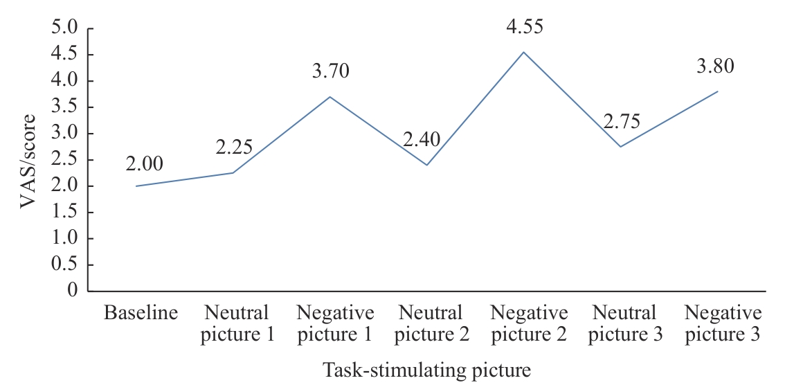
Fig 1 Effect of negative and neutral picture stimulation on temporomandibular joint pain in TMD patients
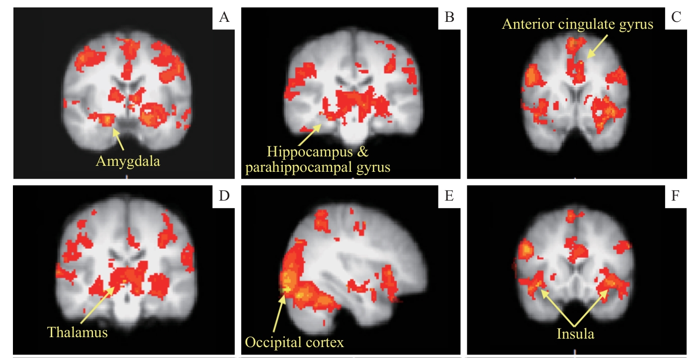
Fig 2 Active brain regions in TMD patients with temporomandibular joint pain under anxiety in the coronal and sagittal plane
| Active brain region | Cluster extreme point (MNI coordinate) | Cluster extremum( Z-max) | P value | ||
|---|---|---|---|---|---|
| X | Y | Z | |||
| Left anterior cingulate cortex | -6 | 12 | 42 | 4.71 | <0.001 |
| Right amygdala | 22 | -4 | -16 | 4.75 | <0.001 |
| Left amygdala | -20 | -6 | -18 | 4.74 | <0.001 |
| Left insula, temporal pole | -44 | 12 | -8 | 4.61 | <0.001 |
| Left orbital frontal cortex, temporal pole | -30 | 12 | -20 | 4.64 | <0.001 |
| Right insula, orbital frontal cortex | 40 | 16 | -8 | 4.68 | <0.001 |
| Right thalamus | 4 | -18 | 0 | 4.79 | <0.001 |
| Left thalamus | -10 | -22 | -2 | 4.81 | <0.001 |
| Left temporal pole, orbital frontal cortex, parahippocampal gyrus | -30 | 8 | -22 | 4.68 | <0.001 |
| Right hippocampus, parahippocampal gyrus | 24 | -22 | -16 | 4.02 | <0.001 |
Tab 1 Active brain regions in TMD patients with temporomandibular joint pain under anxiety
| Active brain region | Cluster extreme point (MNI coordinate) | Cluster extremum( Z-max) | P value | ||
|---|---|---|---|---|---|
| X | Y | Z | |||
| Left anterior cingulate cortex | -6 | 12 | 42 | 4.71 | <0.001 |
| Right amygdala | 22 | -4 | -16 | 4.75 | <0.001 |
| Left amygdala | -20 | -6 | -18 | 4.74 | <0.001 |
| Left insula, temporal pole | -44 | 12 | -8 | 4.61 | <0.001 |
| Left orbital frontal cortex, temporal pole | -30 | 12 | -20 | 4.64 | <0.001 |
| Right insula, orbital frontal cortex | 40 | 16 | -8 | 4.68 | <0.001 |
| Right thalamus | 4 | -18 | 0 | 4.79 | <0.001 |
| Left thalamus | -10 | -22 | -2 | 4.81 | <0.001 |
| Left temporal pole, orbital frontal cortex, parahippocampal gyrus | -30 | 8 | -22 | 4.68 | <0.001 |
| Right hippocampus, parahippocampal gyrus | 24 | -22 | -16 | 4.02 | <0.001 |
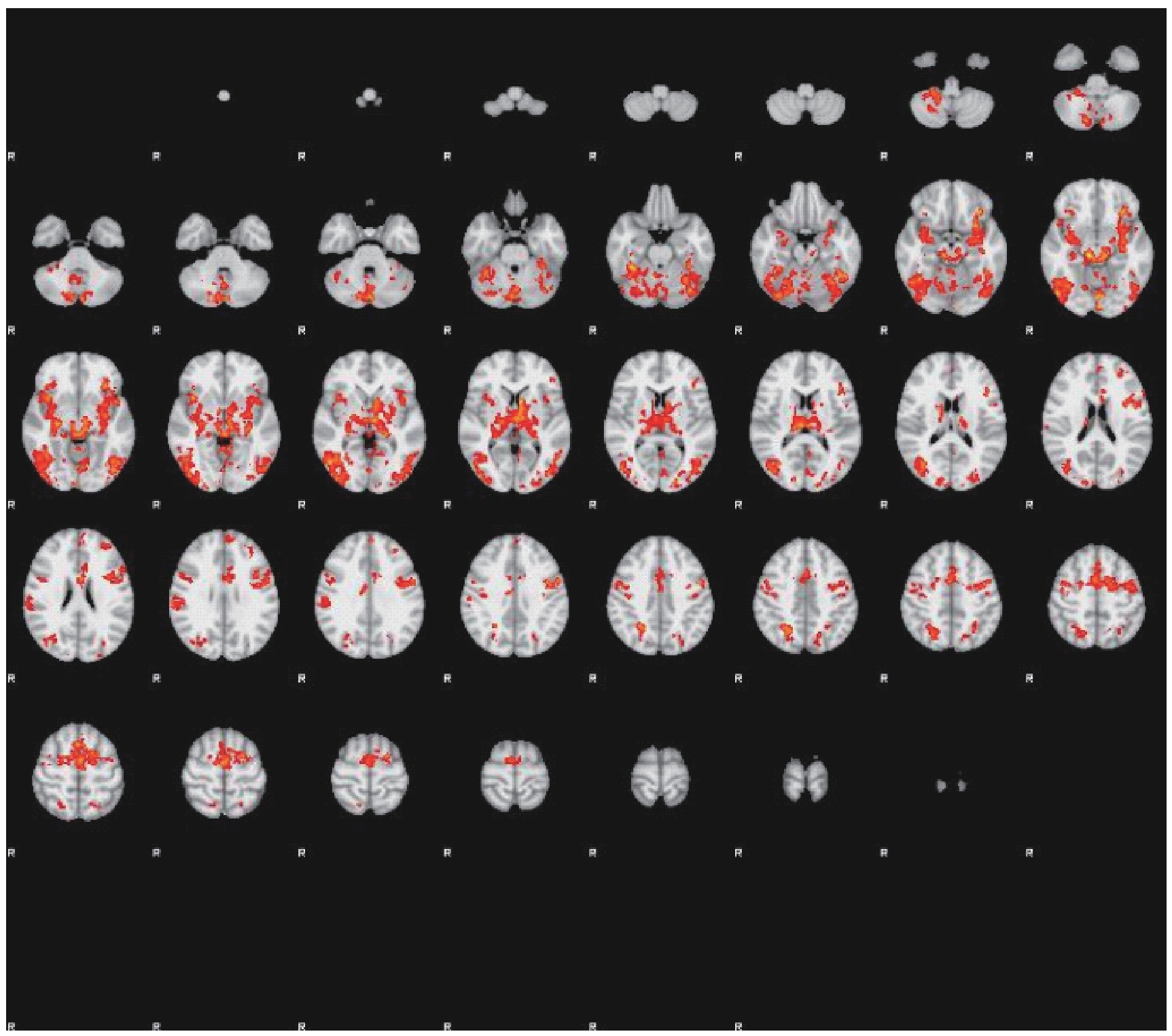
Fig 3 Active brain regions of TMD patients with temporomandibular joint pain in the coronal plane
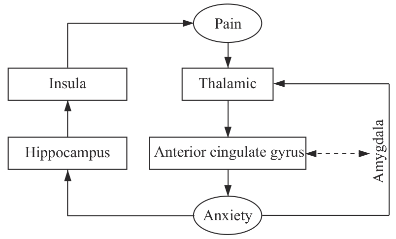
Fig 4 Comorbidity mechanism of TMD pain and anxiety
| 1 | RONGO R, MICHELOTTI A, PEDERSEN T K, et al. Management of temporomandibular joint arthritis in children and adolescents: an introduction for orthodontists[J]. Orthod Craniofac Res, 2023, 26(Suppl 1): 151-163. |
| 2 | ASSIRI K. Relationships between personality factors and DC/TMD Axis Ⅱ scores of psychosocial impairment among patients with pain related temporomandibular disorders[J]. Sci Rep, 2024, 14: 26869. |
| 3 | LIU S N, CHANG C H, LIN C J, et al. Modified dialectical behavior therapy-informed transdiagnostic intervention for emotional disorders: protocol for a randomized controlled trial[J]. BMC Psychiatry, 2024, 24(1): 771. |
| 4 | JIANG Y, OATHES D, HUSH J, et al. Perturbed connectivity of the amygdala and its subregions with the central executive and default mode networks in chronic pain[J]. Pain, 2016, 157(9): 1970-1978. |
| 5 | RESTREPO C, ORTIZ A M, HENAO A C, et al. Association between psychological factors and temporomandibular disorders in adolescents of rural and urban zones[J]. BMC Oral Health, 2021, 21(1): 140. |
| 6 | PASANTA D, HE J L, FORD T, et al. Functional MRS studies of GABA and glutamate/Glx: a systematic review and meta-analysis[J]. Neurosci Biobehav Rev, 2023, 144: 104940. |
| 7 | KOBAYASHI Y, KURATA J, SEKIGUCHI M, et al. Augmented cerebral activation by lumbar mechanical stimulus in chronic low back pain patients: an fMRI study[J]. Spine, 2009, 34(22): 2431-2436. |
| 8 | KLEPZIG K, DOMIN M, KORDASS B, et al. Pain catastrophizing and functional activation during occlusion in TMD patients: an interventional study[J]. Hum Brain Mapp, 2024, 45(15): e70051. |
| 9 | SCHIFFMAN E, OHRBACH R, TRUELOVE E, et al. Diagnostic Criteria for Temporomandibular Disorders (DC/TMD) for clinical and research applications: recommendations of the International RDC/TMD Consortium Network* and Orofacial Pain Special Interest Group[J]. J Oral Facial Pain Headache, 2014, 28(1): 6-27. |
| 10 | BRANCO D, GONÇALVES Ó F, BADIA S B I. A systematic review of International Affective Picture System (IAPS) around the world[J]. Sensors (Basel), 2023, 23(8): 3866. |
| 11 | KNOWLES K A, OLATUNJI B O. Specificity of trait anxiety in anxiety and depression: meta-analysis of the State-Trait Anxiety Inventory[J]. Clin Psychol Rev, 2020, 82: 101928. |
| 12 | BALIKI M N, VANIA APKARIAN A. Nociception, pain, negative moods, and behavior selection[J]. Neuron, 2015, 87(3): 474-491. |
| 13 | SEYMOUR B, CROOK R J, CHEN Z S. Post-injury pain and behaviour: a control theory perspective[J]. Nat Rev Neurosci, 2023, 24(6): 378-392. |
| 14 | LANÇON K, SÉGUÉLA P. Dysregulated neuromodulation in the anterior cingulate cortex in chronic pain[J]. Front Pharmacol, 2023, 14: 1289218. |
| 15 | SONG Y C, WANG X Z, SU Q, et al. Pain-discriminating information decoded from spatiotemporal patterns of hemodynamic responses measured by fMRI in the human brain[J]. Hum Brain Mapp, 2024, 45(16): e70065. |
| 16 | YOSHINO A, OKAMOTO Y, ONODA K, et al. Sadness enhances the experience of pain via neural activation in the anterior cingulate cortex and amygdala: an fMRI study[J]. Neuroimage, 2010, 50(3): 1194-1201. |
| 17 | LIU W Q, CHEN Q Y, LI X H, et al. Cortical tagged synaptic long-term depression in the anterior cingulate cortex of adult mice[J]. J Neurosci, 2024, 44(35): e0028242024. |
| 18 | SUENAGA S, NAGAYAMA K, NAGASAWA T, et al. The usefulness of diagnostic imaging for the assessment of pain symptoms in temporomandibular disorders[J]. Jpn Dent Sci Rev, 2016, 52(4): 93-106. |
| 19 | CHEN L Q, LV X J, GUO Q H, et al. Asymmetric activation of microglia in the hippocampus drives anxiodepressive consequences of trigeminal neuralgia in rodents[J]. Br J Pharmacol, 2023, 180(8): 1090-1113. |
| 20 | MCNAUGHTON N, GRAY J A. Anxiolytic action on the behavioural inhibition system implies multiple types of arousal contribute to anxiety[J]. J Affect Disord, 2000, 61(3): 161-176. |
| 21 | HE S S, LI F, GU T, et al. Reduced corticostriatal functional connectivity in temporomandibular disorders[J]. Hum Brain Mapp, 2018, 39(6): 2563-2572. |
| 22 | ICHESCO E, QUINTERO A, CLAUW D J, et al. Altered functional connectivity between the insula and the cingulate cortex in patients with temporomandibular disorder: a pilot study[J]. Headache, 2012, 52(3): 441-454. |
| 23 | CHEN Y R, TONG S Y, XU Y L, et al. Involvement of basolateral amygdala-rostral anterior cingulate cortex in mechanical allodynia and anxiety-like behaviors and potential mechanisms of electroacupuncture[J]. CNS Neurosci Ther, 2024, 30(9): e70035. |
| 24 | UEDA S, TAKEMOTO-KIMURA S. Exploring the molecular and neuronal bases involved in central amygdala-dependent control of emotional behaviors[J]. Nihon Yakurigaku Zasshi, 2024, 159(5): 316-320. |
| 25 | SESSLE B J. Modulatory processes in craniofacial pain states[J]. Adv Neurobiol, 2024, 35: 107-124. |
| [1] | Liu Li, Fan Haixia, Geng Haixia. Clinical research progress in multidisciplinary collaborative diagnosis and treatment of temporomandibular disorders [J]. Journal of Shanghai Jiao Tong University (Medical Science), 2026, 46(4): 529-536. |
| [2] | Li Jian, Wang Suping. Latent profile analysis of comorbidity of depression and anxiety symptoms in college students and its correlation with social networking service addiction [J]. Journal of Shanghai Jiao Tong University (Medical Science), 2026, 46(2): 213-219. |
| [3] | ZHOU Yining, YE Zhiyun, CHEN Huiwen, XIE Xinyi, ZHOU Wei, SONG Zhongchen. Effect of Th17-specific Stat3 knockout on anxiety- and depressive-like behaviors in periodontitis mice [J]. Journal of Shanghai Jiao Tong University (Medical Science), 2025, 45(7): 838-845. |
| [4] | SUN Lei, DAI Shifeng, CHEN Yuhua, XU Xinyi, JIANG Kele, LI Xiaowen, LI Chengjing, WU Tingting. Quantitative analysis of the distance between articular disc and condyle in patients with temporomandibular disorders [J]. Journal of Shanghai Jiao Tong University (Medical Science), 2025, 45(6): 684-692. |
| [5] | SU Shanshan, JIANG Wenhui, WANG Shuting, XU Mizhen, REN Xueqing, QIU Jianyin. Impact of anxiety levels and alexithymia degree on the quality of life in patients with anxiety disorders [J]. Journal of Shanghai Jiao Tong University (Medical Science), 2024, 44(5): 584-590. |
| [6] | LIAO Bingbing, WANG Zhen. Research progress of affective touch intervention on early life stress-related anxiety disorders [J]. Journal of Shanghai Jiao Tong University (Medical Science), 2024, 44(5): 647-652. |
| [7] | LI Huxiao, LI Xiaotian, ZHAO Xuri, ZHANG Huanyu, ZHOU Wei, SONG Zhongchen. Effects of gingipain extract on the biological characteristics of oral squamous cell carcinoma cell HN6 [J]. Journal of Shanghai Jiao Tong University (Medical Science), 2024, 44(2): 161-168. |
| [8] | LU Qifan, LIU Qiming, ZHOU Hongmei, CHAI Yezi, JIANG Meng, PU Jun. Effect of somatic symptoms, anxiety and depression on clinical prognosis in patients with chronic heart failure [J]. Journal of Shanghai Jiao Tong University (Medical Science), 2023, 43(9): 1153-1161. |
| [9] | WANG Xiaoyu, PENG Yinhui, MA Wenlin, YAO Boshuang, LI Yifan, ZHAO Li, YANG Chunxia. A longitudinal study on new onset anxiety among children and adolescents during the COVID-19 epidemic [J]. Journal of Shanghai Jiao Tong University (Medical Science), 2023, 43(8): 963-970. |
| [10] | GAO Nan, HAO Gem, MA Bingjie, JIN Tian, MA Ke, LIU Xiaoming. Translocator protein activates autophagy in diabetic neuropathic pain rats via regulation of the Keap1/Nrf2/HO-1 signaling [J]. Journal of Shanghai Jiao Tong University (Medical Science), 2023, 43(8): 988-996. |
| [11] | YANG Haixia, XU Lili, WANG Bocheng, CHEN Minjie. Evaluation of clinical effect of manipulation on masticatory muscle pain guided by MRI [J]. Journal of Shanghai Jiao Tong University (Medical Science), 2023, 43(5): 540-544. |
| [12] | MA CUI, YE Yujuan, YAN Xingke. Research progress on the neural circuit of pain emotion mediated by amygdala [J]. Journal of Shanghai Jiao Tong University (Medical Science), 2023, 43(10): 1304-1310. |
| [13] | WANG Yakun, XU Jiarui, WU Qianqian, ZHANG Xiaohua, ZHU Yingchun, BAI Shoujun. Effect of combination of medical care and nursing on the quality of life and mental state of elderly patients with chronic kidney disease in Shanghai suburbs [J]. Journal of Shanghai Jiao Tong University (Medical Science), 2022, 42(7): 904-910. |
| [14] | ZHANG Huanyu, JIANG Yiting, ZHU Xiaochen, HE Zhiyan, ZHOU Wei, SONG Zhongchen. Effects of gingipain extracts on brain neuroinflammation in mice [J]. Journal of Shanghai Jiao Tong University (Medical Science), 2022, 42(5): 570-577. |
| [15] | WU Xiafei, FANG Jie, QI Hongbo, YU Xinyang. Neuropsychiatric effects of gestational diabetes mellitus in adult offspring in C57BL/6J mice [J]. Journal of Shanghai Jiao Tong University (Medical Science), 2022, 42(4): 422-432. |
| Viewed | ||||||
|
Full text |
|
|||||
|
Abstract |
|
|||||