Journal of Shanghai Jiao Tong University (Medical Science) ›› 2026, Vol. 46 ›› Issue (3): 322-331.doi: 10.3969/j.issn.1674-8115.2026.03.006
• Clinical research • Previous Articles Next Articles
Quyang Danzeng, Xiao Huoyuan, Zhang Qingchen, Liu Yuting, Kang Sang, Feng Rui, Pan Jingwei( )
)
Received:2025-08-13
Accepted:2026-02-13
Online:2026-03-28
Published:2026-03-30
Contact:
Pan Jingwei
E-mail:jwpan@sjtu.edu.cn
Supported by:CLC Number:
Quyang Danzeng, Xiao Huoyuan, Zhang Qingchen, Liu Yuting, Kang Sang, Feng Rui, Pan Jingwei. Diagnostic value of cardiac magnetic resonance for myocardial injury in patients with mild COVID-19 infection[J]. Journal of Shanghai Jiao Tong University (Medical Science), 2026, 46(3): 322-331.
Add to citation manager EndNote|Ris|BibTeX
URL: https://xuebao.shsmu.edu.cn/EN/10.3969/j.issn.1674-8115.2026.03.006
| Variable | Overall (n=101) | Control group (n=37) | cTnI(+) group (n=26) | cTnI(-) group (n=38) | P value |
|---|---|---|---|---|---|
| Age/year | 32.00 (25.00, 41.00) | 32.00 (24.00, 42.00) | 29.50 (25.00, 38.75) | 36.50 (28.00, 40.75) | 0.273 |
| Male/n% | 49 (48.51) | 17 (45.95) | 19 (73.08) | 13 (34.21) | 0.009 |
| Time interval between symptom onset and CMR examination/n% | <0.001 | ||||
| 1 week | 32 (31.68) | 0 (0) | 18 (69.23) | 14 (36.84) | |
| 2 week | 12 (11.88) | 0 (0) | 5 (19.23) | 7 (18.42) | |
| 1 month | 16 (15.84) | 0 (0) | 3 (11.54) | 13 (34.21) | |
| 3 month | 2 (1.98) | 0 (0) | 0 (0) | 2 (5.26) | |
| 6 month | 2 (1.98) | 0 (0) | 0 (0) | 2 (5.26) | |
| Symptoms at presentation/n% | <0.001 | ||||
| Chest tightness | 11 (10.89) | 0 (0) | 5 (19.23) | 6 (15.79) | |
| Chest pain | 23 (22.77) | 0 (0) | 10 (38.46) | 13 (34.21) | |
| Palpitation | 14 (13.86) | 0 (0) | 2 (7.69) | 12 (31.58) | |
| Dizziness | 10 (9.90) | 0 (0) | 8 (30.77) | 2 (5.26) | |
| Fatigue | 4 (3.96) | 0 (0) | 1 (3.85) | 3 (7.89) | |
| Other symptoms | 2 (1.98) | 0 (0) | 0 (0) | 2 (5.26) | |
| Comorbidity/n% | <0.001 | ||||
| Positive | 5 (4.95) | 0 (0) | 3 (11.53) | 2 (5.26) | |
| Negative | 96 (95.05) | 37 (100.00) | 23 (88.46) | 36 (94.74) |
Tab 1 Comparison of baseline clinical characteristics among the three groups
| Variable | Overall (n=101) | Control group (n=37) | cTnI(+) group (n=26) | cTnI(-) group (n=38) | P value |
|---|---|---|---|---|---|
| Age/year | 32.00 (25.00, 41.00) | 32.00 (24.00, 42.00) | 29.50 (25.00, 38.75) | 36.50 (28.00, 40.75) | 0.273 |
| Male/n% | 49 (48.51) | 17 (45.95) | 19 (73.08) | 13 (34.21) | 0.009 |
| Time interval between symptom onset and CMR examination/n% | <0.001 | ||||
| 1 week | 32 (31.68) | 0 (0) | 18 (69.23) | 14 (36.84) | |
| 2 week | 12 (11.88) | 0 (0) | 5 (19.23) | 7 (18.42) | |
| 1 month | 16 (15.84) | 0 (0) | 3 (11.54) | 13 (34.21) | |
| 3 month | 2 (1.98) | 0 (0) | 0 (0) | 2 (5.26) | |
| 6 month | 2 (1.98) | 0 (0) | 0 (0) | 2 (5.26) | |
| Symptoms at presentation/n% | <0.001 | ||||
| Chest tightness | 11 (10.89) | 0 (0) | 5 (19.23) | 6 (15.79) | |
| Chest pain | 23 (22.77) | 0 (0) | 10 (38.46) | 13 (34.21) | |
| Palpitation | 14 (13.86) | 0 (0) | 2 (7.69) | 12 (31.58) | |
| Dizziness | 10 (9.90) | 0 (0) | 8 (30.77) | 2 (5.26) | |
| Fatigue | 4 (3.96) | 0 (0) | 1 (3.85) | 3 (7.89) | |
| Other symptoms | 2 (1.98) | 0 (0) | 0 (0) | 2 (5.26) | |
| Comorbidity/n% | <0.001 | ||||
| Positive | 5 (4.95) | 0 (0) | 3 (11.53) | 2 (5.26) | |
| Negative | 96 (95.05) | 37 (100.00) | 23 (88.46) | 36 (94.74) |
| Variable | Control group (n=37) | cTnI(+) group (n=26) | cTnI(-) group (n=38) | P value | |||
|---|---|---|---|---|---|---|---|
| All | Control vs cTnI(+) | Control vs cTnI(-) | cTnI(+) vs cTnI(-) | ||||
| LVEF/% | 63.60 | 62.24 | 63.61 | 0.672 | 0.269 | 0.996 | 0.447 |
| LVEDV/mL | 117.11 (89.39, 131.87) | 103.76 (87.43, 133.41) | 113.30 (93.08, 139.83) | 0.562 | 0.388 | 0.971 | 0.328 |
| LVESV/mL | 39.95 (34.87, 51.58) | 38.32 (35.14, 49.57) | 38.56 (31.85, 57.64) | 0.949 | 0.742 | 0.979 | 0.823 |
| LVSV/mL | 73.22 (59.30, 83.28) | 65.75 (53.35, 81.94) | 70.90 (59.03, 85.21) | 0.440 | 0.281 | 0.998 | 0.204 |
Tab 2 Comparison of left ventricular function and volumetric parameters among the three groups
| Variable | Control group (n=37) | cTnI(+) group (n=26) | cTnI(-) group (n=38) | P value | |||
|---|---|---|---|---|---|---|---|
| All | Control vs cTnI(+) | Control vs cTnI(-) | cTnI(+) vs cTnI(-) | ||||
| LVEF/% | 63.60 | 62.24 | 63.61 | 0.672 | 0.269 | 0.996 | 0.447 |
| LVEDV/mL | 117.11 (89.39, 131.87) | 103.76 (87.43, 133.41) | 113.30 (93.08, 139.83) | 0.562 | 0.388 | 0.971 | 0.328 |
| LVESV/mL | 39.95 (34.87, 51.58) | 38.32 (35.14, 49.57) | 38.56 (31.85, 57.64) | 0.949 | 0.742 | 0.979 | 0.823 |
| LVSV/mL | 73.22 (59.30, 83.28) | 65.75 (53.35, 81.94) | 70.90 (59.03, 85.21) | 0.440 | 0.281 | 0.998 | 0.204 |
| Variable | Control group (n=37) | cTnI(+) group (n=26) | cTnI(-) group (n=38) | P value | |||
|---|---|---|---|---|---|---|---|
| All | Control vs cTnI(+) | Control vs cTnI(-) | cTnI(+) vs cTnI(-) | ||||
| GRS/% | 30.20 (27.80, 39.01) | 30.24 (27.21, 43.19) | 32.62 (28.52, 37.93) | 0.990 | 0.933 | 0.344 | 0.880 |
| GCS/% | -20.87 | -17.38 | -20.27 | <0.001 | <0.001 | 0.352 | <0.001 |
| GLS/% | -14.68 | -13.48 | -13.86 | 0.288 | 0.153 | 0.911 | 0.621 |
| CSBasal/% | -18.86 | -15.18 | -17.46 | <0.001 | <0.001 | >0.999 | 0.008 |
| CSMid/% | -20.97 | -16.04 | -20.32 | <0.001 | <0.001 | 0.326 | <0.001 |
| CSApi/% | -23.14 (-24.56, -21.06) | -22.54 (-25.57, -19.88) | -24.81 (-27.47, -22.47) | 0.035 | 0.775 | 0.231 | 0.027 |
Tab 3 Comparison of global and segmental left ventricular strain parameters among the three groups
| Variable | Control group (n=37) | cTnI(+) group (n=26) | cTnI(-) group (n=38) | P value | |||
|---|---|---|---|---|---|---|---|
| All | Control vs cTnI(+) | Control vs cTnI(-) | cTnI(+) vs cTnI(-) | ||||
| GRS/% | 30.20 (27.80, 39.01) | 30.24 (27.21, 43.19) | 32.62 (28.52, 37.93) | 0.990 | 0.933 | 0.344 | 0.880 |
| GCS/% | -20.87 | -17.38 | -20.27 | <0.001 | <0.001 | 0.352 | <0.001 |
| GLS/% | -14.68 | -13.48 | -13.86 | 0.288 | 0.153 | 0.911 | 0.621 |
| CSBasal/% | -18.86 | -15.18 | -17.46 | <0.001 | <0.001 | >0.999 | 0.008 |
| CSMid/% | -20.97 | -16.04 | -20.32 | <0.001 | <0.001 | 0.326 | <0.001 |
| CSApi/% | -23.14 (-24.56, -21.06) | -22.54 (-25.57, -19.88) | -24.81 (-27.47, -22.47) | 0.035 | 0.775 | 0.231 | 0.027 |
| Variable | Control group (n=37) | cTnI(+) group (n=26) | cTnI(-) group (n=38) | P value | |||
|---|---|---|---|---|---|---|---|
| All | Control vs cTnI(+) | Control vs cTnI(-) | cTnI(+) vs cTnI(-) | ||||
| T2WI/n(%) | <0.001 | <0.001 | >0.999 | <0.001 | |||
| Negative | 37 (100.00) | 16 (61.54) | 37 (97.37) | ||||
| Positive | 0 (0) | 10 (38.46) | 1 (2.63) | ||||
| Native T1 mapping/ms | 1 216.90 (1 189.00, 1 238.69) | 1 268.90 (1 248.67, 1 317.00) | 1 235.49 (1 203.37, 1 264.05) | <0.001 | <0.001 | 0.007 | <0.001 |
| LGE/n(%) | <0.001 | <0.001 | <0.001 | <0.001 | |||
| Negative | 37 (100.00) | 2 (7.69) | 28 (73.68) | ||||
| Positive | 0 (0) | 24 (92.31) | 10 (26.32) | ||||
| LGE/% | / | 18.05 (14.43, 22.18) | 0.00 (0.00, 8.10) | / | / | / | <0.001 |
| LGE segment/n(%) | <0.001 | <0.001 | 0.004 | <0.001 | |||
| LAT | 0 (0) | 14 (58.33) | 3 (30.00) | ||||
| ANT | 0 (0) | 5 (20.83) | 5 (50.00) | ||||
| SEPT | 0 (0) | 4 (16.67) | 1 (10.00) | ||||
| Other segment | 0 (0) | 1 (4.17) | 1 (10.00) | ||||
| LGE location/n(%) | <0.001 | <0.001 | 0.003 | <0.001 | |||
| Mid-level | 37 (100.00) | 2 (8.33) | 4 (40.00) | ||||
| Basal-level | 0 (0) | 5 (20.83) | 3 (30.00) | ||||
| Apical-level | 0 (0) | 3 (12.50) | 0 (0) | ||||
| Basal-Mid | 0 (0) | 6 (25.00) | 3 (30.00) | ||||
| Mid-Apical | 0 (0) | 5 (20.83) | 0 (0) | ||||
| All-level | 0 (0) | 3 (12.50) | 0 (0) | ||||
| LGE pattern/n(%) | <0.001 | <0.001 | <0.001 | <0.001 | |||
| Mid-wall | 0 (0) | 5 (20.83) | 4 (40.00) | ||||
| Subepicardial | 0 (0) | 3 (12.50) | 1 (10.00) | ||||
| Mid-Subepi | 0 (0) | 15 (62.50) | 5 (50.00) | ||||
| Subendocardial | 0 (0) | 1 (4.17) | 0 (0) | ||||
Tab 4 Comparison of left ventricular myocardial tissue characterization parameters among the three groups
| Variable | Control group (n=37) | cTnI(+) group (n=26) | cTnI(-) group (n=38) | P value | |||
|---|---|---|---|---|---|---|---|
| All | Control vs cTnI(+) | Control vs cTnI(-) | cTnI(+) vs cTnI(-) | ||||
| T2WI/n(%) | <0.001 | <0.001 | >0.999 | <0.001 | |||
| Negative | 37 (100.00) | 16 (61.54) | 37 (97.37) | ||||
| Positive | 0 (0) | 10 (38.46) | 1 (2.63) | ||||
| Native T1 mapping/ms | 1 216.90 (1 189.00, 1 238.69) | 1 268.90 (1 248.67, 1 317.00) | 1 235.49 (1 203.37, 1 264.05) | <0.001 | <0.001 | 0.007 | <0.001 |
| LGE/n(%) | <0.001 | <0.001 | <0.001 | <0.001 | |||
| Negative | 37 (100.00) | 2 (7.69) | 28 (73.68) | ||||
| Positive | 0 (0) | 24 (92.31) | 10 (26.32) | ||||
| LGE/% | / | 18.05 (14.43, 22.18) | 0.00 (0.00, 8.10) | / | / | / | <0.001 |
| LGE segment/n(%) | <0.001 | <0.001 | 0.004 | <0.001 | |||
| LAT | 0 (0) | 14 (58.33) | 3 (30.00) | ||||
| ANT | 0 (0) | 5 (20.83) | 5 (50.00) | ||||
| SEPT | 0 (0) | 4 (16.67) | 1 (10.00) | ||||
| Other segment | 0 (0) | 1 (4.17) | 1 (10.00) | ||||
| LGE location/n(%) | <0.001 | <0.001 | 0.003 | <0.001 | |||
| Mid-level | 37 (100.00) | 2 (8.33) | 4 (40.00) | ||||
| Basal-level | 0 (0) | 5 (20.83) | 3 (30.00) | ||||
| Apical-level | 0 (0) | 3 (12.50) | 0 (0) | ||||
| Basal-Mid | 0 (0) | 6 (25.00) | 3 (30.00) | ||||
| Mid-Apical | 0 (0) | 5 (20.83) | 0 (0) | ||||
| All-level | 0 (0) | 3 (12.50) | 0 (0) | ||||
| LGE pattern/n(%) | <0.001 | <0.001 | <0.001 | <0.001 | |||
| Mid-wall | 0 (0) | 5 (20.83) | 4 (40.00) | ||||
| Subepicardial | 0 (0) | 3 (12.50) | 1 (10.00) | ||||
| Mid-Subepi | 0 (0) | 15 (62.50) | 5 (50.00) | ||||
| Subendocardial | 0 (0) | 1 (4.17) | 0 (0) | ||||
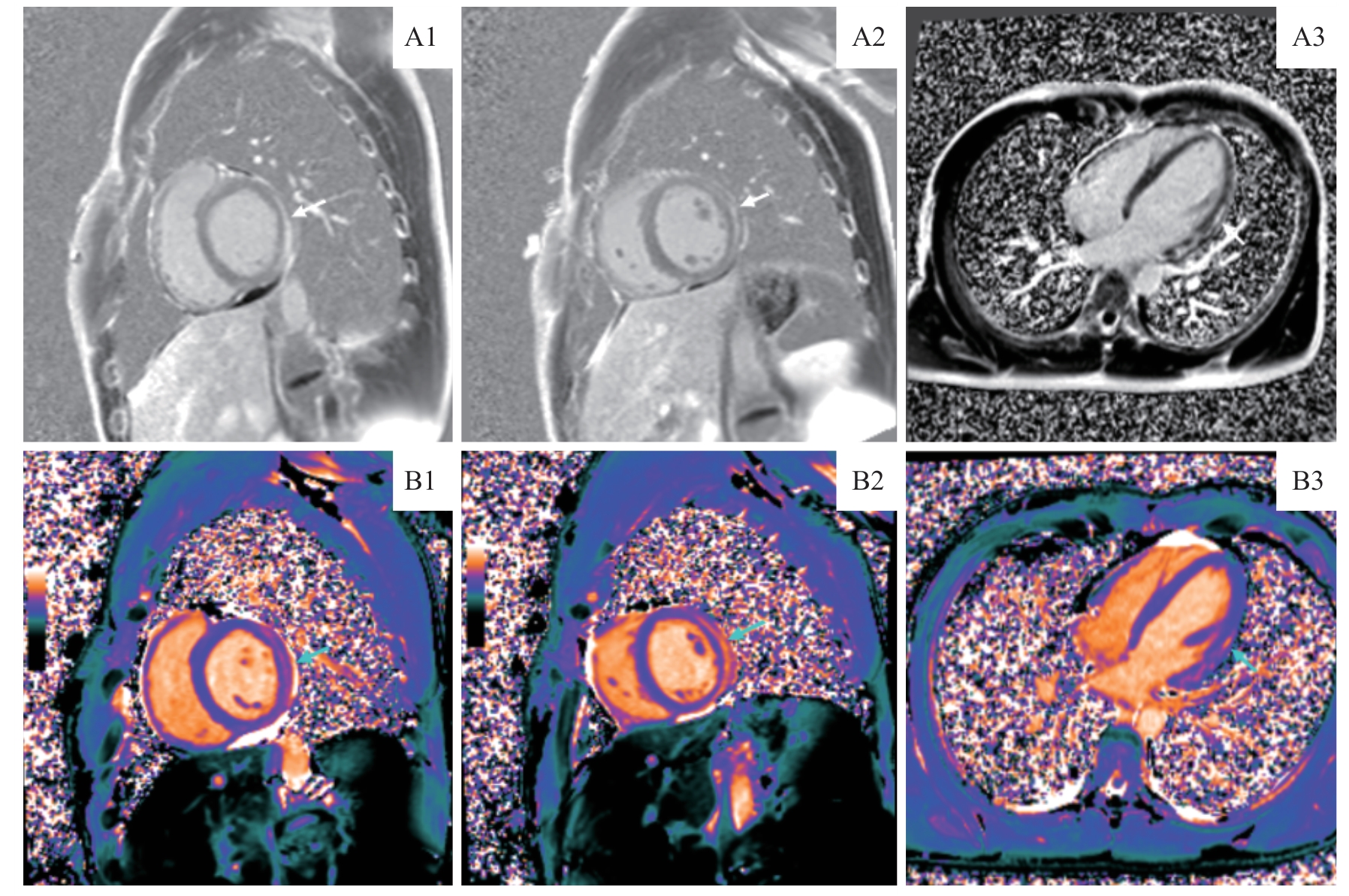
Fig 1 Representative LGE and Native T1 mapping images of a typical case
| Statistic | Gender | Time interval between symptom onset and CMR examination | T2WI | Native T1 mapping | GCS | CSBasal | CSMid |
|---|---|---|---|---|---|---|---|
| r | 0.383 | 0.370 | -0.466 | -0.422 | -0.462 | -0.366 | -0.600 |
| P | 0.003 | 0.003 | <0.001 | <0.001 | 0.001 | 0.003 | <0.001 |
Tab 5 Correlation analysis of cTnI with baseline clinical characteristics as well as key CMR parameters
| Statistic | Gender | Time interval between symptom onset and CMR examination | T2WI | Native T1 mapping | GCS | CSBasal | CSMid |
|---|---|---|---|---|---|---|---|
| r | 0.383 | 0.370 | -0.466 | -0.422 | -0.462 | -0.366 | -0.600 |
| P | 0.003 | 0.003 | <0.001 | <0.001 | 0.001 | 0.003 | <0.001 |
| Variable | Univariate Logistic regression analysis | Multivariate Logistic regression analysis | |||||||||
|---|---|---|---|---|---|---|---|---|---|---|---|
| β | S.E | Z | P | OR (95% CI) | β | S.E | Z | P | OR (95% CI) | ||
| Male | -0.27 | 0.42 | -0.65 | 0.515 | 0.762 (0.336‒1.727) | ||||||
| Time interval between symptom onset and CMR examination | |||||||||||
| 1 week | 1.000 (Ref) | ||||||||||
| 2 week | 0.34 | 0.68 | 0.49 | 0.623 | 1.400 (0.366‒5.350) | ||||||
| 1 month | -0.21 | 0.62 | -0.41 | 0.683 | 0.778 (0.233‒2.599) | ||||||
| 3 month | 0.00 | 1.46 | 0.00 | 1.000 | 1.000 (0.057‒17.411) | ||||||
| 6 month | 16.57 | 1 696.73 | 0.01 | 0.992 | 15 651 360.793 (0.000‒Inf) | ||||||
| Symptoms at presentation | |||||||||||
| Chest tightness | 1.000 (Ref) | ||||||||||
| Chest pain | -0.27 | 0.77 | -0.37 | 0.723 | 0.762 (0.170‒3.422) | ||||||
| Palpitation | -1.35 | 0.83 | -1.63 | 0.103 | 0.260 (0.051‒1.313) | ||||||
| Dizziness | 0.83 | 1.10 | 0.82 | 0.413 | 2.286 (0.316‒16.512) | ||||||
| Fatigue | -18.13 | 1 978.09 | -0.01 | 0.993 | 0.000 (0.000‒Inf) | ||||||
| Other symptoms | -0.56 | 1.55 | -0.36 | 0.718 | 0.57 (0.028‒11.85) | ||||||
| Comorbidity | |||||||||||
| Negative | 1.000 (Ref) | ||||||||||
| Positive | 0.37 | 0.94 | 0.39 | 0.695 | 1.450 (0.226‒9.319) | ||||||
| Native T1 mapping | 0.06 | 0.01 | 4.67 | <0.001 | 1.057 (1.032‒1.081) | 0.08 | 0.02 | 3.40 | <0.001 | 1.080 (1.033‒1.129) | |
| GCS | 0.65 | 0.13 | 4.92 | <0.001 | 1.917 (1.479‒2.488) | ||||||
| CSBasal | 0.41 | 0.09 | 4.54 | <0.001 | 1.511 (1.264‒1.806) | ||||||
| CSMid | 0.81 | 0.16 | 5.09 | <0.001 | 2.239 (1.642‒3.055) | 0.94 | 0.25 | 3.79 | <0.001 | 2.564 (1.574‒4.175) | |
| CSApi | 0.09 | 0.06 | 1.63 | 0.103 | 1.099 (0.981‒1.231) | ||||||
Tab 6 Univariate and multivariate Logistic regression analyses of COVID-19-associated myocardial injury
| Variable | Univariate Logistic regression analysis | Multivariate Logistic regression analysis | |||||||||
|---|---|---|---|---|---|---|---|---|---|---|---|
| β | S.E | Z | P | OR (95% CI) | β | S.E | Z | P | OR (95% CI) | ||
| Male | -0.27 | 0.42 | -0.65 | 0.515 | 0.762 (0.336‒1.727) | ||||||
| Time interval between symptom onset and CMR examination | |||||||||||
| 1 week | 1.000 (Ref) | ||||||||||
| 2 week | 0.34 | 0.68 | 0.49 | 0.623 | 1.400 (0.366‒5.350) | ||||||
| 1 month | -0.21 | 0.62 | -0.41 | 0.683 | 0.778 (0.233‒2.599) | ||||||
| 3 month | 0.00 | 1.46 | 0.00 | 1.000 | 1.000 (0.057‒17.411) | ||||||
| 6 month | 16.57 | 1 696.73 | 0.01 | 0.992 | 15 651 360.793 (0.000‒Inf) | ||||||
| Symptoms at presentation | |||||||||||
| Chest tightness | 1.000 (Ref) | ||||||||||
| Chest pain | -0.27 | 0.77 | -0.37 | 0.723 | 0.762 (0.170‒3.422) | ||||||
| Palpitation | -1.35 | 0.83 | -1.63 | 0.103 | 0.260 (0.051‒1.313) | ||||||
| Dizziness | 0.83 | 1.10 | 0.82 | 0.413 | 2.286 (0.316‒16.512) | ||||||
| Fatigue | -18.13 | 1 978.09 | -0.01 | 0.993 | 0.000 (0.000‒Inf) | ||||||
| Other symptoms | -0.56 | 1.55 | -0.36 | 0.718 | 0.57 (0.028‒11.85) | ||||||
| Comorbidity | |||||||||||
| Negative | 1.000 (Ref) | ||||||||||
| Positive | 0.37 | 0.94 | 0.39 | 0.695 | 1.450 (0.226‒9.319) | ||||||
| Native T1 mapping | 0.06 | 0.01 | 4.67 | <0.001 | 1.057 (1.032‒1.081) | 0.08 | 0.02 | 3.40 | <0.001 | 1.080 (1.033‒1.129) | |
| GCS | 0.65 | 0.13 | 4.92 | <0.001 | 1.917 (1.479‒2.488) | ||||||
| CSBasal | 0.41 | 0.09 | 4.54 | <0.001 | 1.511 (1.264‒1.806) | ||||||
| CSMid | 0.81 | 0.16 | 5.09 | <0.001 | 2.239 (1.642‒3.055) | 0.94 | 0.25 | 3.79 | <0.001 | 2.564 (1.574‒4.175) | |
| CSApi | 0.09 | 0.06 | 1.63 | 0.103 | 1.099 (0.981‒1.231) | ||||||
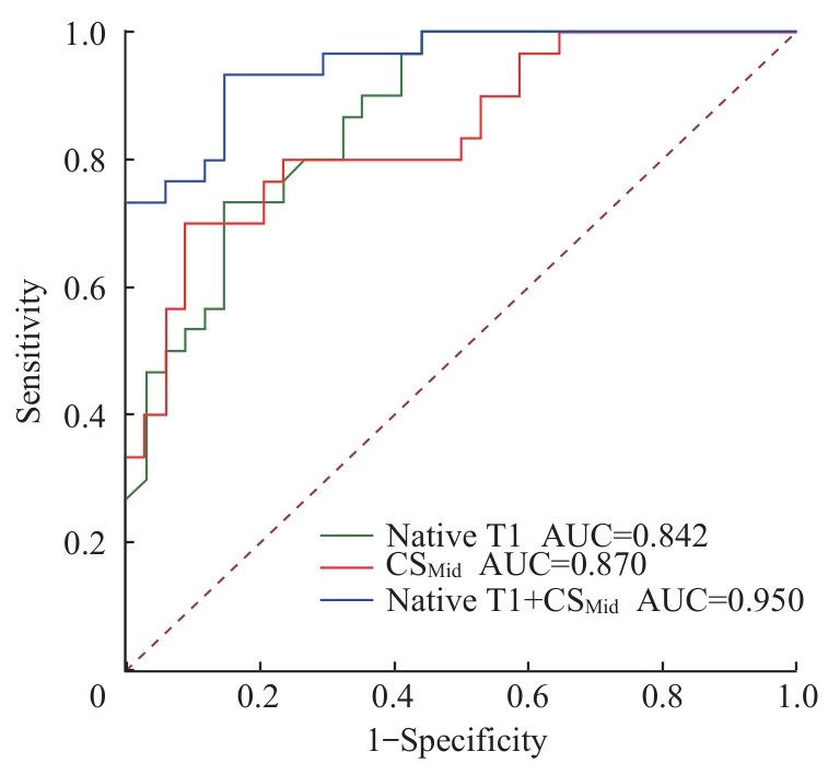
Fig 2 ROC curve analysis of the combined diagnostic model for COVID-19-associated myocardial injury
| [1] | Siddiqi R, Farhan S H, Shah S A, et al. National trends in heart failure and acute myocarditis-related death before and during the COVID-19 pandemic[J]. J Am Heart Assoc, 2025, 14(10): e038987. |
| [2] | Puntmann V O, Martin S, Shchendrygina A, et al. Long-term cardiac pathology in individuals with mild initial COVID-19 illness[J]. Nat Med, 2022, 28(10): 2117-2123. |
| [3] | Holby S N, Richardson T L Jr, Laws J L, et al. Multimodality cardiac imaging in COVID[J]. Circ Res, 2023, 132(10): 1387-1404. |
| [4] | Puntmann V O, Carerj M L, Wieters I, et al. Outcomes of cardiovascular magnetic resonance imaging in patients recently recovered from coronavirus disease 2019 (COVID-19)[J]. JAMA Cardiol, 2020, 5(11): 1265-1273. |
| [5] | Huang L X, Li X, Gu X Y, et al. Health outcomes in people 2 years after surviving hospitalisation with COVID-19: a longitudinal cohort study[J]. Lancet Respir Med, 2022, 10(9): 863-876. |
| [6] | Goerlich E, Chung T H, Hong G H, et al. Cardiovascular effects of the post-COVID-19 condition[J]. Nat Cardiovasc Res, 2024, 3(2): 118-129. |
| [7] | Davis H E, McCorkell L, Vogel J M, et al. Long COVID: major findings, mechanisms and recommendations[J]. Nat Rev Microbiol, 2023, 21(3): 133-146. |
| [8] | Steffen Johansson R, Loewenstein D, Lodin K, et al. Long-term coronary microvascular and cardiac dysfunction after severe COVID-19 hospitalization[J]. JAMA Netw Open, 2025, 8(6): e2514411. |
| [9] | Goerlich E, Minhas A S, Mukherjee M, et al. Multimodality imaging for cardiac evaluation in patients with COVID-19[J]. Curr Cardiol Rep, 2021, 23(5): 44. |
| [10] | Puntmann V O, Valbuena S, Hinojar R, et al. Society for Cardiovascular Magnetic Resonance (SCMR) expert consensus for CMR imaging endpoints in clinical research: part I - analytical validation and clinical qualification[J]. J Cardiovasc Magn Reson, 2018, 20(1): 67. |
| [11] | Carrick D, Haig C, Rauhalammi S, et al. Pathophysiology of LV remodeling in survivors of STEMI: inflammation, remote myocardium, and prognosis[J]. JACC Cardiovasc Imaging, 2015, 8(7): 779-789. |
| [12] | Nagel E, Kwong R Y, Chandrashekhar Y S. CMR in nonischemic myocardial inflammation: solving the problem of diagnosing myocarditis or still diagnostic ambiguity [J]. JACC Cardiovasc Imaging, 2020, 13(1 Pt 1): 163-166. |
| [13] | Shi S B, Qin M, Shen B, et al. Association of cardiac injury with mortality in hospitalized patients with COVID-19 in Wuhan, China[J]. JAMA Cardiol, 2020, 5(7): 802-810. |
| [14] | Kotecha T, Knight D S, Razvi Y, et al. Patterns of myocardial injury in recovered troponin-positive COVID-19 patients assessed by cardiovascular magnetic resonance[J]. Eur Heart J, 2021, 42(19): 1866-1878. |
| [15] | Ballering A V, van Zon S K R, Olde Hartman T C, et al. Persistence of somatic symptoms after COVID-19 in the Netherlands: an observational cohort study[J]. Lancet, 2022, 400(10350): 452-461. |
| [16] | Gluckman T J, Bhave N M, Allen L A, et al. 2022 ACC expert consensus decision pathway on cardiovascular sequelae of COVID-19 in adults: myocarditis and other myocardial involvement, post-acute sequelae of SARS-CoV-2 infection, and return to play a report of the American college of cardiology solution set oversight committee[J]. J Am Coll Cardiol, 2022, 79(17): 1717-1756. |
| [17] | Wan E Y F, Mathur S, Zhang R, et al. Association of COVID-19 with short- and long-term risk of cardiovascular disease and mortality: a prospective cohort in UK Biobank[J]. Cardiovasc Res, 2023, 119(8): 1718-1727. |
| [18] | Leitman M, Lysiansky M, Lysyansky P, et al. Circumferential and longitudinal strain in 3 myocardial layers in normal subjects and in patients with regional left ventricular dysfunction[J]. J Am Soc Echocardiogr, 2010, 23(1): 64-70. |
| [19] | Quinaglia T, Gongora C, Awadalla M, et al. Global circumferential and radial strain among patients with immune checkpoint inhibitor myocarditis[J]. JACC Cardiovasc Imaging, 2022, 15(11): 1883-1896. |
| [20] | Joy G, Artico J, Kurdi H, et al. Prospective case-control study of cardiovascular abnormalities 6 months following mild COVID-19 in healthcare workers[J]. JACC Cardiovasc Imaging, 2021, 14(11): 2155-2166. |
| [21] | Meindl C, Paulus M, Poschenrieder F, et al. Patients with acute myocarditis and preserved systolic left ventricular function: comparison of global and regional longitudinal strain imaging by echocardiography with quantification of late gadolinium enhancement by CMR[J]. Clin Res Cardiol, 2021, 110(11): 1792-1800. |
| [22] | Fu H, Zhang N, Zheng Y L, et al. Risk stratification of cardiac sequelae detected using cardiac magnetic resonance in late convalescence at the six-month follow-up of recovered COVID-19 patients[J]. J Infect, 2021, 83(1): 119-145. |
| [23] | Nensa F, Kloth J, Tezgah E, et al. Feasibility of FDG-PET in myocarditis: comparison to CMR using integrated PET/MRI[J]. J Nucl Cardiol, 2018, 25(3): 785-794. |
| [24] | Hanneman K, Houbois C, Kei T, et al. Multimodality cardiac imaging, cardiac symptoms, and clinical outcomes in patients who recovered from mild COVID-19[J]. Radiology, 2023, 308(1): e230767. |
| [25] | Kravchenko D, Isaak A, Zimmer S, et al. Cardiac MRI in patients with prolonged cardiorespiratory symptoms after mild to moderate COVID-19[J]. Radiology, 2021, 301(3): E419-E425. |
| [26] | Raman B, Cassar M P, Tunnicliffe E M, et al. Medium-term effects of SARS-CoV-2 infection on multiple vital organs, exercise capacity, cognition, quality of life and mental health, post-hospital discharge[J]. EClinicalMedicine, 2021, 31: 100683. |
| [27] | Sewanan LR, Di Tullio MR, Laine AF, et al. Absence of long-term structural and functional cardiac abnormalities on multimodality imaging in a multi-ethnic group of COVID-19 survivors from the early stage of the pandemic[J]. Eur Heart J Imaging Methods Pract, 2023, 1(2): qyad034. |
| [28] | Jerosch-Herold M, Rickers C, Petersen S E, et al. Myocardial tissue characterization in cardiac magnetic resonance studies of patients recovering from COVID-19: a meta-analysis[J]. J Am Heart Assoc, 2023, 12(6): e027801. |
| [29] | Siripanthong B, Nazarian S, Muser D, et al. Recognizing COVID-19-related myocarditis: the possible pathophysiology and proposed guideline for diagnosis and management[J]. Heart Rhythm, 2020, 17(9): 1463-1471. |
| [30] | Brandt Y, Lubrecht J M, Adriaans B P, et al. Quantification of left ventricular myocardial strain: comparison between MRI tagging, MRI feature tracking, and ultrasound speckle tracking[J]. NMR Biomed, 2024, 37(9): e5164. |
| [1] | Zhang Yeting, Zheng Yuxuan, Tao Pengjie, Zhou Wenqin, Lü Dongchao. Overview of new advances in the treatment of myocardial injury with circular RNA [J]. Journal of Shanghai Jiao Tong University (Medical Science), 2026, 46(3): 391-399. |
| [2] | LI Yaomin, XU Jianguo, YU Xia. Brugada phenocopy induced by heatstroke: a case report [J]. Journal of Shanghai Jiao Tong University (Medical Science), 2025, 45(4): 523-528. |
| [3] | LI Wenli, JIN Lixing, ZHAO Yichao, ZHONG Fangyuan, SHI Yao, LEI Jie, PU Jun, GE Heng. Impact of left ventricular myocardial strain injury on secondary tricuspid regurgitation in acute STEMI assessed by cardiac magnetic resonance [J]. Journal of Shanghai Jiao Tong University (Medical Science), 2025, 45(12): 1578-1588. |
| [4] | LI Xinxin, BIAN Yize, ZHAO Hang, JIANG Meng. Research progress in the artificial intelligence-assisted measurement of myocardial strain [J]. Journal of Shanghai Jiao Tong University (Medical Science), 2024, 44(6): 773-778. |
| [5] | LIU Qiming, LU Qifan, CHAI Yezi, JIANG Meng, PU Jun. Short-axis cine cardiac magnetic resonance images-derived radiomics for hypertrophic cardiomyopathy and healthy control classification [J]. Journal of Shanghai Jiao Tong University (Medical Science), 2024, 44(1): 79-86. |
| [6] | LIU Qiming, LU Qifan, CHAI Yezi, JIANG Meng, PU Jun. Radiomics-based left ventricular ejection fraction prediction: a feasibility study [J]. Journal of Shanghai Jiao Tong University (Medical Science), 2023, 43(9): 1162-1168. |
| [7] | ZHANG Yutang, JIN Yijie, ZHANG Fengchun, XU Yingchun. Exploration on rationalization of diagnosis and treatment of breast cancer patients combined with COVID-19 [J]. Journal of Shanghai Jiao Tong University (Medical Science), 2022, 42(12): 1745-1750. |
| [8] | Jiang YUE, Yong ZHOU, Hua XU, Wen LIU, Xiao-feng HAN, Qing MAO, Ji-dong ZHANG, Jing MA, Han-dong JIANG, Wei LIU. Characteristic analysis and comparison of glycolipid metabolism in patients with coronavirus disease 2019 in common condition and severe cases [J]. JOURNAL OF SHANGHAI JIAOTONG UNIVERSITY (MEDICAL SCIENCE), 2021, 41(3): 355-359. |
| [9] | Ze-hao FENG, Ye-zi CHAI, Xuan SU, Bao-hang-xing SUN, Qi-ming LIU, Meng JIANG, Jun PU. Association between body mass index and myocardial involvements in patients with systemic lupus erythematosus [J]. JOURNAL OF SHANGHAI JIAOTONG UNIVERSITY (MEDICAL SCIENCE), 2021, 41(2): 180-186. |
| [10] | Pei-kun HU, Jie HE, Lian-ming WU, Heng GE, Jian-rong XU, Jun PU. Effect of microvascular obstruction on left ventricle function and prognosis in patients with ST-segment elevation myocardial infarction [J]. JOURNAL OF SHANGHAI JIAOTONG UNIVERSITY (MEDICAL SCIENCE), 2021, 41(2): 173-179. |
| [11] | Huan LI, Pei-qiang YI, Jun SU, Pei-zhan CHEN, Cheng XU, Lu CAO, Jia-yi CHEN, Min LI. Protective effect of pituitary adenylate cyclase-activating polypeptide 38 on acute radiation-induced myocardial injury [J]. JOURNAL OF SHANGHAI JIAOTONG UNIVERSITY (MEDICAL SCIENCE), 2021, 41(2): 129-133. |
| [12] | Jian-xun DONG, Lai WEI, Jie HE, Ling-cong KONG, Heng GE, Jun PU. Progress of cardiac magnetic resonance in assessment of left ventricular mechanical dyssynchrony [J]. JOURNAL OF SHANGHAI JIAOTONG UNIVERSITY (MEDICAL SCIENCE), 2021, 41(12): 1698-1702. |
| [13] | Ya-jie GAO, Wen-kun MA, Cheng-jie GAO, Yi ZHOU, Jing-wei PAN. Exploration of the predictive value of myocardial strain on ventricular remodeling after acute ST-segment elevation myocardial infarction [J]. JOURNAL OF SHANGHAI JIAOTONG UNIVERSITY (MEDICAL SCIENCE), 2021, 41(11): 1478-1484. |
| [14] | SHI Da-ke, HU Wei-guo, YANG Zhi-tao, LIN Jing-sheng, WANG Xiao-ning, GUO Ying, QIAN Wen-jing, CAI Ming, XIANG Xiao-gang, LIANG Xiao-hong, ZHAI Rong-cheng, ZHANG Yi-bo, NI Yu-Xing. Experience of healthcare-acquired infection control against coronavirus disease 2019 by integrated medical team in Wuhan [J]. JOURNAL OF SHANGHAI JIAOTONG UNIVERSITY (MEDICAL SCIENCE), 2020, 40(8): 1009-1012. |
| [15] | ZHA Qiong-fang1, LI Hong-bo2, QIN Hui1. Research progress of interaction between coronavirus disease 2019 and cardiovascular system [J]. JOURNAL OF SHANGHAI JIAOTONG UNIVERSITY (MEDICAL SCIENCE), 2020, 40(7): 863-866. |
| Viewed | ||||||
|
Full text |
|
|||||
|
Abstract |
|
|||||