乳腺癌是女性发病率最高的恶性肿瘤,在女性高发的恶性肿瘤中其死亡率居第二位,严重威胁着女性的生命和健康[1]。研究[2]显示,乳腺癌的转移和复发是患者死亡的主要原因,其具体机制尚未被明确。本课题组的前期研究[3-5]发现,四次跨膜蛋白(tetraspanin,TSPAN)家族在乳腺癌的转移、耐药和干性维持中发挥了重要作用。TSPAN家族由33个成员组成(TSPAN1~33),其结构含有4个高度保守的疏水跨膜结构域。相关研究[6]显示,TSPAN家族蛋白可与整合素、主要组织相容性抗原、T细胞受体、生长因子受体等跨膜蛋白组成超模体结构TEM(tetraspanin-enriched microdomain),将胞外信号传导至胞内,以调节乳腺癌细胞的恶性表型;同时,该家族蛋白还可通过外泌体调节乳腺癌细胞与肿瘤微环境的相互作用,以促进乳腺癌的进展。
1 资料与方法
1.1 标本收集与临床资料获取
选择2020年1月—2021年12月的上海交通大学医学院附属第一人民医院的106例乳腺癌手术患者为研究对象,获取其癌组织及癌旁组织,并经石蜡包埋制成组织芯片。收集乳腺癌患者的临床特征信息,包括总TNM分期、M分期、N分期和病理学分级。
1.2 数据收集及分析
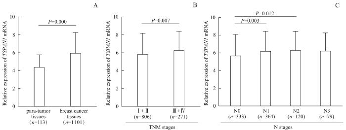
从癌症基因组图谱(The Cancer Genome Atlas,TCGA)数据库中下载乳腺癌组织和癌旁组织的转录组测序数据,采用R4.0.1软件对组织中TSPAN1 mRNA表达水平的差异进行分析;下载乳腺癌患者的临床特征信息(包括肿瘤总TNM分期和N分期),分析TSPAN1 mRNA的表达与该临床特征信息的相关性。
1.3 细胞、主要试剂及仪器
1.3.1 细胞
人Luminal A型乳腺癌细胞系MCF-7,人三阴性乳腺癌细胞系SUM149PT、SUM159PT、MDA-MB-468、MDA-MB-231,以及人表皮生长因子受体2过表达型乳腺癌细胞系SK-BR-3均购自中国科学院典型培养物保藏委员会细胞库。
1.3.2 主要试剂
免疫组化试剂盒(碧云天生物技术有限公司),DMEM培养基、青-链霉素混合双抗(Gibco,美国),转染试剂Lipofectamine 3000(Thermo Fisher Scientific,美国),TSPAN1 siRNA及阴性对照siRNA(广州瑞博生物公司),兔源TSPAN1抗体、鼠源β-actin抗体、兔源波形蛋白(vimentin)抗体、兔源E-钙黏蛋白(E-cadherin)抗体(Proteintech,美国),鼠源N-钙黏蛋白(N-cadherin)抗体(Cell Signaling Technology,美国),Transwell小室(Corning,美国)。
1.3.3 主要仪器
生物安全柜和细胞恒温培养箱(Thermo Fisher Scientific,美国),光学显微镜(Nikon,日本),水浴锅(拓赫机电有限公司),摇床振荡器(普瑞博仪器有限公司),离心机(Eppendorf,德国)。
1.4 实验方法
1.4.1 组织芯片中TSPAN1表达量检测及其与乳腺癌患者临床特征相关性分析
采用免疫组织化学染色(immunohistochemistry staining,IHC)检测组织芯片中TSPAN1的表达,即将石蜡包埋的组织经染色、脱水、封片后于光学显微镜下观察组织染色的情况。其中,一抗为TSPAN1抗体(工作浓度为1∶800),对应二抗的工作浓度为1∶1 000。
分别由2位实验人员采用双盲阅片的方式,对上述组织染色情况进行评分。于镜下随机选取每例组织的5个视野,按如下规则进行评分:① 阳性细胞占比:≤5%记为0分,6%~25%记为1分,26%~50%记为2分,51%~75%记为3分,>75%记为4分;该占比百分数仅取整数。② 染色强度:淡黄色记为1分,棕黄色记为2分,棕褐色记为3分。③ 将阳性细胞占比得分与染色强度得分相乘,记为免疫组化的总评分。
而后,对TSPAN1表达与乳腺癌患者的临床特征进行相关性分析。
1.4.2 细胞培养
于37 ℃、5%CO2恒温培养箱中,使用含10%胎牛血清和1%青-链霉素混合双抗的DMEM培养基分别培养MCF-7、SUM149PT、SUM159PT、MDA-MB-468、MDA-MB-231、SK-BR-3细胞,当其生长融合至70%~80%时使用胰酶消化并传代。
1.4.3 TSPAN1表达筛选
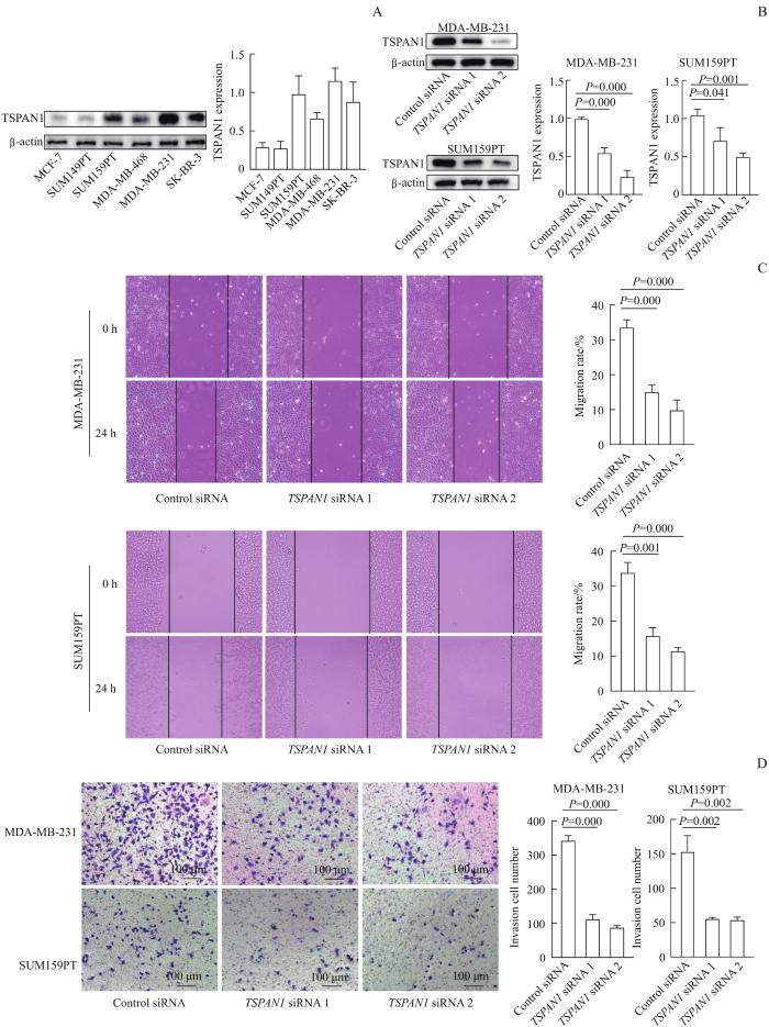
使用含1%蛋白酶抑制剂的细胞裂解液分别提取上述6种野生型细胞的总蛋白,采用蛋白质印迹法(Western blotting)检测TSPAN1的表达,并选取TSPAN1表达较高的2种细胞进行后续细胞实验。其中,使用的一抗为TSPAN1抗体,其工作浓度为1∶1 000,对应的荧光二抗的工作浓度为1∶5 000。
1.4.4 细胞转染
分别使用Control siRNA、TSPAN1 siRNA转染“1.4.3”部分获得的2种细胞。转染8 h后换液,继续培养48 h后提取上述4种细胞的总蛋白开展后续相关功能实验。转染实验的siRNA序列见表1。
表1 siRNA 序列
Tab 1
| siRNA | Forward (5′→3′) | |
|---|---|---|
| Control siRNA | UUCUCCGAACGUGUCACGUTT | ACGUGACACGUUCGGAGAATT |
| TSPAN1 siRNA 1 | CGUUUCGUCAAAGAGAAUATT | UAUUCUCUUUGACGAAACGTT |
| TSPAN1 siRNA 2 | GGCUCACGACCAAAAAGUATT | UACUUUUUGGUCGUGAGCCTT |
1.4.5 转染效率验证及EMT相关蛋白表达检测
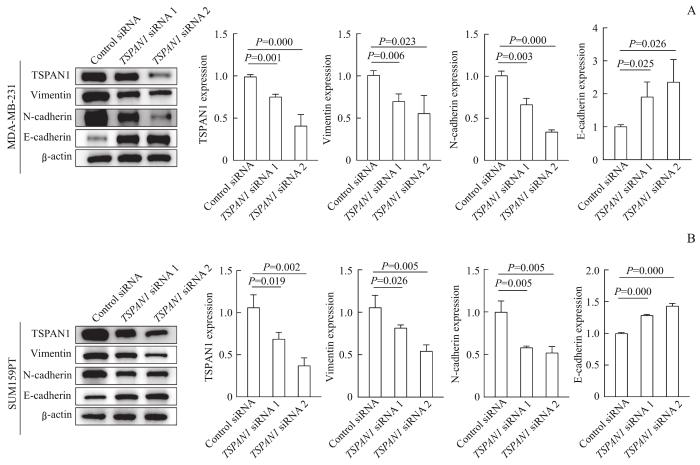
获取“1.4.4”部分中4种已转染细胞的总蛋白,采用Western blotting分别检测TSPAN1和EMT相关蛋白(E-钙黏蛋白、N-钙黏蛋白和波形蛋白)的表达,以验证转染效率以及下调TSPAN1对EMT相关蛋白表达的影响。其中,使用的一抗为TSPAN1抗体、E-钙黏蛋白抗体、N-钙黏蛋白抗体、波形蛋白抗体,其工作浓度分别为1∶1 000、1∶2 000、1∶800、1∶1 500,对应的荧光二抗的工作浓度均为1∶5 000。
1.4.6 细胞迁移能力检测
采用细胞划痕实验检测“1.4.4”部分获得的已转染细胞的迁移能力。待上述细胞生长融合至90%时,使用枪头刮划细胞产生划痕,更换无血清DMEM培养基后继续培养24 h,并于显微镜下拍照,观察细胞划痕的愈合率。
1.4.7 细胞侵袭能力检测
采用细胞侵袭实验检测“1.4.4”部分获得的已转染细胞的侵袭能力。在预先铺好基质胶的Transwell小室中分别加入2×10⁵个上述细胞,下室中加入500 μL 10%胎牛血清的DMEM培养基。24 h后取出小室,使用甲醇室温固定细胞15 min,结晶紫染色30 min,再用棉签擦去小室膜上侧的细胞并于显微镜下拍照,随机取3个视野进行细胞计数。
1.5 统计学方法
采用GraphPad Prism 8.0软件进行作图,并运用SPSS 26.0软件进行统计分析。符合正态分布的定量资料以x±s表示,使用t检验进行2组间比较,使用单因素方差分析(one-way ANOVA)进行多组间比较;不符合正态分布的定量资料以M(Q1,Q3)表示,使用Mann-Whitney U检验进行2组间比较。P<0.05表示差异具有统计学意义。
2 结果
2.1 TSPAN1在乳腺癌组织及癌旁组织的表达及其与患者临床特征的相关性
图1
图1
TSPAN1在乳腺癌组织及癌旁组织中的表达及其与患者临床特征的关系
Note: A. Detection of TSPAN1 expression in breast cancer tissues and para-tumor tissues by IHC. B. Statistical analysis of TSPAN1 expression in breast cancer tissues and para-tumor tissues. C. Statistical analysis of TSPAN1 expression in breast cancer tissues of TNM stages. D. Statistical analysis of TSPAN1 expression in breast cancer tissues of M stages. E. Statistical analysis of TSPAN1 expression in breast cancer tissues of N stages. F. Statistical analysis of TSPAN1 expression in breast cancer tissues of different pathological grades. Due to missing data of some cases, the total case number in fig C and F is less than 106 cases.
Fig 1
Expression of TSPAN1 in breast cancer tissues and para-tumor tissues and its relationship with clinical characteristics of patients
2.2 TCGA数据库中 TSPAN1 mRNA在乳腺癌组织及癌旁组织的表达及其与患者临床特征的相关性
图2
图2
TCGA数据库中 TSPAN1 mRNA在乳腺癌组织及癌旁组织中的表达及其与患者临床特征的相关性
Note: A. Relative expression of TSPAN1 mRNA in breast cancer tissues and para-tumor tissues in TCGA database. B. Relative expression of TSPAN1 mRNA in breast cancer tissues of TNM stages in TCGA database. C. Relative expression of TSPAN1 mRNA in breast cancer tissues of N0-N3 stages. In TCGA database, due to missing data of some cases, the total case number in fig B and C is less than 1 101 cases.
Fig 2
Expression of TSPAN1 mRNA in breast cancer tissues and para-tumor tissues and its correlation with patients' clinical characteristics in TCGA database
2.3 TSPAN1表达对乳腺癌细胞迁移和侵袭能力的影响
图3
图3
沉默 TSPAN1 对MDA-MB-231、SUM159PT细胞迁移、侵袭能力的影响
Note: A. Detection of the expression level of TSPAN1 in different breast cancer cells by Western blotting. B. Detection of the expression level of TSPAN1 in MDA-MB-231 and SUM159PT cells transfected with Control siRNA or TSPAN1 siRNA by Western blotting. C/D. Effects of TSPAN1 siRNA on the migration (C) and invasion (D) ability of MDA-MB-231 and SUM159PT cells.
Fig 3
Effects of silencing TSPAN1 on the migration and invasion ability of MDA-MB-231 and SUM159PT cells
分别采用细胞划痕实验、细胞侵袭实验检测上述已转染的MDA-MB-231和SUM159PT细胞的迁移和侵袭能力,结果(图3C、D)显示,转染TSPAN1 siRNA的乳腺癌细胞的迁移和侵袭能力均低于转染Control siRNA的乳腺癌细胞(均P<0.005)。
2.4 TSPAN1表达对乳腺癌细胞EMT相关蛋白的影响
使用Western blotting检测已转染的MDA-MB-231和SUM159PT细胞中EMT相关蛋白的表达,结果(图4)显示与转染Control siRNA相比,转染TSPAN1 siRNA的MDA-MB-231和SUM159PT细胞的上皮表型标志物E-钙黏蛋白的表达均较高,间质表型标志物N-钙黏蛋白、波形蛋白的表达均较低(均P<0.05)。
图4
图4
沉默 TSPAN1 后MDA-MB-231、SUM159PT细胞中EMT相关蛋白的表达
Note: A. MDA-MB-231 cells. B. SUM159PT cells.
Fig 4
Detection of EMT-related proteins expression in MDA-MB-231 and SUM159PT cells after silencing TSPAN1
3 讨论
本研究中,我们聚焦四次跨膜蛋白家族成员TSPAN1,研究其在乳腺癌中的作用。通过IHC对临床样本进行分析后发现,TSPAN1在乳腺癌组织中高表达,其表达水平与肿瘤的总TNM分期、M分期、N分期和病理学分级相关,继而提示TSPAN1可作为乳腺癌治疗的特异性靶点。随后,我们结合生物信息学对TSPAN1 mRNA在乳腺癌中的表达行进一步分析,结果显示TSPAN1 mRNA在乳腺癌组织中高表达,且其表达水平与肿瘤的总TNM分期、N分期相关。接着,我们使用TSPAN1 siRNA分别转染MDA-MB-231、SUM159PT细胞后发现,下调TSPAN1可抑制细胞的迁移和侵袭,同时上调E-钙黏蛋白的表达、下调N-钙黏蛋白和波形蛋白的表达,继而提示TSPAN1可促进乳腺癌细胞的迁移、侵袭及EMT,但其具体机制仍需进一步探究。
本研究尚存在一定的不足之处:① 选用细胞系种类不足,研究中未能涵盖乳腺癌全部病理类型。② 未开展有关TSPAN1促进乳腺癌细胞迁移、侵袭的作用机制研究。综上,本研究表明TSPAN1在乳腺癌组织中高表达,可促进乳腺癌细胞的迁移和侵袭,该结果或将为进一步探索乳腺癌的潜在治疗靶点提供参考。
作者贡献声明
李军建、朱灜、王红霞、曹源参与了实验设计,曹源开展了实验操作和生物信息学分析,李军建、曹源参与了论文的写作和修改。所有作者均阅读并同意了最终稿件的提交。
The study was designed by LI Junjian, ZHU Ying, WANG Hongxia and CAO Yuan. The experiments and bioinformatics analysis were carried out by CAO Yuan. The manuscript was drafted and revised by LI Junjian and CAO Yuan. All the authors have read the last version of paper and consented for submission.
利益冲突声明
所有作者声明不存在利益冲突。
All authors disclose no relevant conflict of interests.
参考文献


