目的·探究去唾液酸糖蛋白受体1(asialoglycoprotein receptor 1,ASGR1)在肝细胞癌(hepatocellular carcinoma,HCC)中的意义及潜在机制。方法·通过R语言分析癌症基因组图谱(The Cancer Genome Atlas,TCGA)数据库中ASGR1在肝癌患者中的表达情况并绘制相关生存曲线。利用人类蛋白质图谱(The Human Protein Atlas,HPA)数据库获得人体正常肝组织和肝癌组织的免疫组织化学(immunohistochemistry,IHC)数据来分析ASGR1的蛋白表达情况。利用流体动力学尾静脉注射(hydrodynamic tail vein injection,HTVI)递送方法,在免疫完全的小鼠肝脏中敲除Asgr1探究其在体内的致瘤功能,并通过蛋白免疫印迹法(Western blotting,WB)验证基因敲除效率。利用R语言进行京都基因与基因组百科全书(Kyoto Encyclopedia of Genes and Genomes,KEGG)通路富集分析及相关性分析,利用基因探针富集(Gene Set Enrichment Analysis,GSEA)软件进行GSEA hallmark相关通路分析,利用实时荧光定量PCR(quantitative real-time PCR,qPCR)在小鼠肝癌组织中验证糖酵解关键基因表达水平。结果·ASGR1在肝癌组织中显著低表达,在肝癌患者中ASGR1的低表达与患者较差的总体生存期(overall survival,OS)、无疾病间隔(disease free interval,DFI)、无进展间隔期(progression free interval,PFI)和疾病特异性生存期(disease specific survival,DSS)相关;肿瘤分级程度越高的肝癌患者ASGR1基因表达水平越低。人体正常肝组织ASGR1蛋白的表达显著高于肝癌组织。在免疫完全的肝细胞癌小鼠模型中,小鼠内源性Asgr1敲除可增加肝组织中肿瘤结节的大小和数量。TCGA数据库中ASGR1低表达组肝癌患者富集到多条癌症及代谢相关通路,ASGR1表达与部分糖酵解关键基因表达呈负相关,Asgr1敲除组的小鼠肝癌组织中糖酵解水平高于对照组,提示ASGR1低表达很可能促进肝癌的生长发展,加强代谢重编程促进肿瘤的合成代谢发展。结论·ASGR1在肝癌患者中表达显著降低,与患者的预后呈正相关;小鼠体内敲除Asgr1可促进肝细胞癌的发生发展;ASGR1可以作为肝癌预后不良的潜在生物标志物和潜在治疗新靶点。
关键词:去唾液酸糖蛋白受体1(ASGR1)
;
肝细胞癌
;
流体动力学尾静脉注射
;
治疗靶点
Abstract
Objective ·To explore the significance and mechanism of asialoglycoprotein receptor 1 (ASGR1) in hepatocellular carcinoma. Methods ·The expression of ASGR1 in patients with liver cancer in The Cancer Genome Atlas (TCGA) database was analyzed by R language and the related survival curves were drawn. The Human Protein Atlas (HPA) database was used to obtain the immunohistochemistry (IHC) data of normal human liver tissue and liver cancer tissue to analyze the protein expression of ASGR1. By using the hydrodynamic tail vein injection (HTVI) delivery method, Asgr1 was knocked out in the liver of fully immune mice to explore its tumorigenic function invivo. Gene knockout efficiency was verified by Western blotting (WB). The Kyoto Encyclopedia of Genes and Genomes (KEGG) pathway enrichment analysis and correlation analysis were performed by using R language. The GSEA hallmark correlation pathway analysis was performed by using Gene Set Enrichment Analysis (GSEA) software. The expression level of key genes of glycolysis in mouse liver cancer tissue was verified by quantitative real-time PCR (qPCR). Results ·ASGR1 was significantly low-expressed in liver cancer tissue, and the low expression of ASGR1 in liver cancer patients was associated with poorer overall survival (OS), disease-free interval (DFI), progression-free interval (PFI), and disease-specific survival (DSS). The higher the degree of tumor grade, the lower the expression level of ASGR1 in patients with liver cancer. Immunohistochemistry showed that the protein expression of ASGR1 in normal human liver tissue was significantly higher than that in liver cancer tissue. In an immunocompetent mouse model of hepatocellular carcinoma, knockout of endogenous Asgr1 in mice increased the size and number of tumor nodules in liver tissue. In the TCGA database, patients with liver cancer in the ASGR1 low-expression group were enriched in multiple cancer and metabolic pathways. The expression of ASGR1 was negatively correlated with some key genes of glycolysis. The level of glycolysis in liver cancer tissues of mice in the Asgr1 knockout group was higher than that in the control group. It was suggested that the low expression of ASGR1 be likely to promote the growth and development of liver cancer and strengthen metabolic reprogramming to promote the anabolic development of tumors. Conclusion ·The expression of ASGR1 is significantly reduced in patients with liver cancer, which is positively correlated with the prognosis of patients. Knocking out Asgr1 in mice can promote the occurrence and development of hepatocellular carcinoma. ASGR1 can be used as a potential biomarker for poor prognosis of liver cancer and a new target for potential treatment.
LI Qianyu, GUO Wenyun, QIAN Yifei, LI Songling, ZHU Zijun, LIU Yanfeng. Study on the significance and mechanism of ASGR1 in hepatocellular carcinoma. Journal of Shanghai Jiao Tong University (Medical Science)[J], 2023, 43(9): 1107-1114 doi:10.3969/j.issn.1674-8115.2023.09.005
通过UCSC Xena(xenabrowser.net)网站获取癌症基因组图谱(The Cancer Genome Atlas,TCGA)数据库的基因表达矩阵和患者临床数据。人体正常肝组织及肝癌组织相关的免疫组织化学(immunohistochemistry,IHC)数据来自人类蛋白质图谱(The Human Protein Atlas,HPA)数据库(https://www.proteinatlas.org/)。
1.2 生物信息学分析
使用R语言对基因表达矩阵进行癌和癌旁组织的表达量分析及肝癌患者不同基因表达之间的相关性分析。使用R语言survival包和survminer包进行生存分析。使用R语言limma包对基因表达矩阵按照ASGR1的中位值分为高低2组后进行差异基因分析,通过P值及错误发现率(false discovery rate,FDR)筛选差异基因,使用R语言ClusterProfiler包进行京都基因与基因组百科全书(Kyoto Encyclopedia of Genes and Genomes,KEGG)通路富集分析,使用基因探针富集(Gene Set Enrichment Analysis,GSEA)软件进行基因集富集分析。
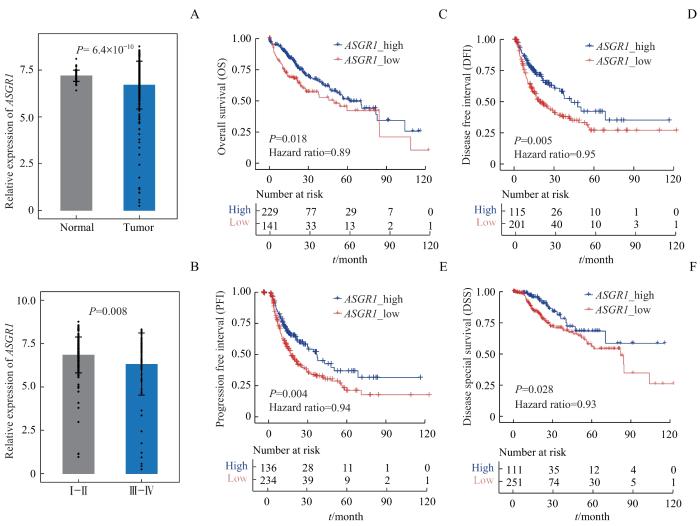
Note: A. Expression of ASGR1 in tumor and normal tissues of liver cancer patients from TCGA database. B. Expression of ASGR1 in different stages of liver cancer patients from TCGA database. C‒F. The overall survival (C), disease-free interval (D), progression-free interval (E) and disease-specific survival (F) curves of liver cancer patients with low or high ASGR1 expression from TCGA database.
Fig 1
Expression of ASGR1 in liver cancer samples and its correlation with prognosis
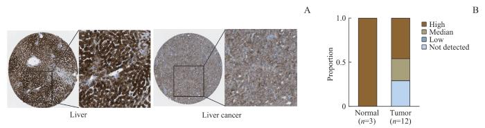
Note: A. Immunohistochemical pictures of ASGR1-related protein expression levels in normal human liver tissue and liver cancer tissue from the HPA database. B. The proportion of different degrees of ASGR1 staining in normal human liver tissue and liver cancer tissue.
Fig 2
Protein expression level of ASGR1 in normal human liver tissue and liver cancer tissue
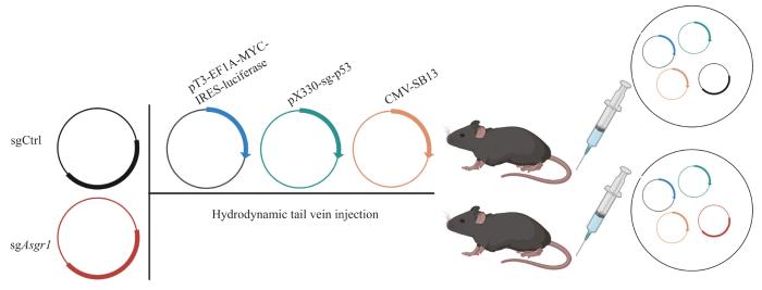
Note: Delivery via HTVI in fully immune mice by injection of pT3-EF1A-MYC-IRES-luciferase, pX330-sg-p53, CMV-SB13, lenti-CRISPR sgAsgr1 (sgAsgr1 group) or lenti-CRISPR v2 (sgCtrl group).
Fig 3
Schematic diagram of mouse hydrodynamic tail vein injection model
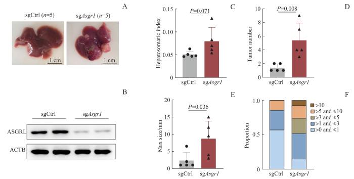
Note: A. Representative pictures of hepatocellular carcinoma in sgCtrl and sgAsgr1 group. B. Protein expression of ASGR1 and ACTB in liver tumor tissues of mice in sgCtrl and sgAsgr1 group. C‒F. Liver weight ratio (C), tumor number (D), max size (E) and proportion of tumor numbers (F) with different sizes in sgCtrl and sgAsgr1 group.
Fig 4
Validation of Asgr1 knockout and formation of hepatocellular carcinoma in mice
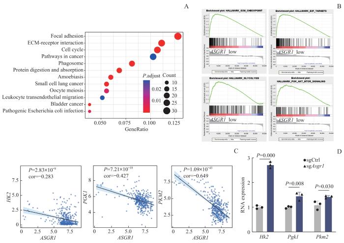
Note: A. KEGG pathway enrichment analysis of genes negatively correlated with ASGR1. B. GSEA analysis of hallmark pathway enrichment between ASGR1 low and high group. C. Correlation analysis of expression of ASGR1 and glycolysis-related genes. D. The mRNA expression levels of glycolysis-related genes in mouse liver cancer tissue.
Fig 5
Bioinformatics analysis of ASGR1 expression in TCGA liver cancer database and verification of mRNA expression levels in mouse liver cancer tissue
The study was designed by LI Qianyu and GUO Wenyun. The manuscript was drafted and revised by LI Qianyu and QIAN Yifei. The document was searched by LI Songling and ZHU Zijun. The research was guided by LIU Yanfeng. All the authors have read the last version of paper and consented for submission.
利益冲突声明
所有作者声明不存在利益冲突。
COMPETING INTERESTS
All authors disclose no relevant conflict of interests.
BRAY F, FERLAY J, SOERJOMATARAM I, et al. Global cancer statistics 2018: globocan estimates of incidence and mortality worldwide for 36 cancers in 185 countries[J]. CA Cancer J Clin, 2018, 68(6): 394-424.
SUNG H, FERLAY J, SIEGEL R L, et al. Global cancer statistics 2020: globocan estimates of incidence and mortality worldwide for 36 cancers in 185 countries[J]. CA Cancer J Clin, 2021, 71(3): 209-249.
SINGH M K, DAS B K, CHOUDHARY S, et al. Diabetes and hepatocellular carcinoma: a pathophysiological link and pharmacological management[J]. Biomed Pharmacother, 2018, 106: 991-1002.
TANOWITZ M, HETTRICK L, REVENKO A, et al. Asialoglycoprotein receptor 1 mediates productive uptake of N-acetylgalactosamine-conjugated and unconjugated phosphorothioate antisense oligonucleotides into liver hepatocytes[J]. Nucleic Acids Res, 2017, 45(21): 12388-12400.
XIA M D, LIAO W, XIANG Q, et al. Research progress of asialoglycoprotein receptor 1 in atherosclerosis[J]. Chinese Journal of Arteriosclerosis, 2022, 30(6): 541-545.
DE GALARRETA M R, BRESNAHAN E, MOLINA-SÁNCHEZ P, et al. β-catenin activation promotes immune escape and resistance to anti-PD-1 therapy in hepatocellular carcinoma[J]. Cancer Discov, 2019, 9(8): 1124-1141.
RAHIB L, SMITH B D, AIZENBERG R, et al. Projecting cancer incidence and deaths to 2030: the unexpected burden of thyroid, liver, and pancreas cancers in the United States[J]. Cancer Res, 2014, 74(11): 2913-2921.
ZHOU J, SUN H C, WANG Z, et al. Guidelines for the diagnosis and treatment of hepatocellular carcinoma (2019 edition)[J]. Liver Cancer, 2020, 9(6): 682-720.
SUGAHARA K, TOGASHI H, TAKAHASHI K, et al. Separate analysis of asialoglycoprotein receptors in the right and left hepatic lobes using Tc-GSA SPECT[J]. Hepatology, 2003, 38(6): 1401-1409.
RIGOPOULOU E I, ROGGENBUCK D, SMYK D S, et al. Asialoglycoprotein receptor (ASGPR) as target autoantigen in liver autoimmunity: lost and found[J]. Autoimmun Rev, 2012, 12(2): 260-269.
GU D, JIN H, JIN G, et al. The asialoglycoprotein receptor suppresses the metastasis of hepatocellular carcinoma via LASS2-mediated inhibition of V-ATPase activity[J]. Cancer Lett, 2016, 379(1): 107-116.
ZHU X X, SONG G Y, ZHANG S Y, et al. Asialoglycoprotein receptor 1 functions as a tumor suppressor in liver cancer via inhibition of STAT3[J]. Cancer Res, 2022, 82(21): 3987-4000.
TONG M, WONG T L, ZHAO H, et al. Loss of tyrosine catabolic enzyme HPD promotes glutamine anaplerosis through mTOR signaling in liver cancer[J]. Cell Rep, 2021, 36(8): 109617.
YEH Y C, HO H L, WU Y C, et al. AKT1 internal tandem duplications and point mutations are the genetic hallmarks of sclerosing pneumocytoma[J]. Mod Pathol, 2020, 33(3): 391-403.
CAI J, SUN X H, GUO H, et al. Non-metabolic role of UCK2 links EGFR-AKT pathway activation to metastasis enhancement in hepatocellular carcinoma[J]. Oncogenesis, 2020, 9(12): 103.
ICARD P, SIMULA L, WU Z R, et al. Why may citrate sodium significantly increase the effectiveness of transarterial chemoembolization in hepatocellular carcinoma?[J]. Drug Resist Updat, 2021, 59: 100790.
BUONTEMPO F, ERSAHIN T, MISSIROLI S, et al. Inhibition of Akt signaling in hepatoma cells induces apoptotic cell death independent of Akt activation status[J]. Invest New Drugs, 2011, 29(6): 1303-1313.


