上海交通大学学报(医学版), 2024, 44(2): 161-168 doi: 10.3969/j.issn.1674-8115.2024.02.002

论著 · 基础研究

牙龈素提取物对口腔鳞癌细胞HN6生物学特性的影响

李虎虓,1, 李笑甜1, 赵旭日2, 张桓瑜1, 周薇2, 宋忠臣,1

1.上海交通大学医学院附属第九人民医院牙周病科,上海交通大学口腔医学院,国家口腔医学中心,国家口腔疾病临床医学研究中心,上海市口腔医学重点实验室,上海市口腔医学研究所,上海 200011

2.上海交通大学医学院附属第九人民医院口腔微生态与系统性疾病实验室,上海交通大学口腔医学院,国家口腔医学中心,国家口腔疾病临床医学研究中心,上海市口腔医学重点实验室,上海 200125

Effects of gingipain extract on the biological characteristics of oral squamous cell carcinoma cell HN6

LI Huxiao,1, LI Xiaotian1, ZHAO Xuri2, ZHANG Huanyu1, ZHOU Wei2, SONG Zhongchen,1

1.Department of Periodontology, Shanghai Ninth People's Hospital, Shanghai Jiao Tong University School of Medicine; College of Stomatology, Shanghai Jiao Tong University; National Center for Stomatology; National Clinical Research Center for Oral Diseases; Shanghai Key Laboratory of Stomatology, Shanghai Research Institute of Stomatology, Shanghai 200011, China

2.Laboratory of Oral Microbiota and Systemic Disease, Shanghai Ninth People's Hospital, Shanghai Jiao Tong University School of Medicine; College of Stomatology, Shanghai Jiao Tong University; National Center for Stomatology; National Clinical Research Center for Oral Diseases; Shanghai Key Laboratory of Stomatology, Shanghai 200125, China

通讯作者: 宋忠臣,电子信箱:szhongchen@sina.com

编委: 张慧俊

收稿日期: 2023-09-20   接受日期: 2023-12-26  

基金资助: 国家自然科学基金.  82071112
上海交通大学医学院附属第九人民医院“交叉基金”.  JYJC202005

Corresponding authors: SONG Zhongchen, E-mail:szhongchen@sina.com.

Received: 2023-09-20   Accepted: 2023-12-26  

作者简介 About authors

李虎虓(1994—),男,硕士,住院医师;电子信箱:tentigerli@163.com。 E-mail:tentigerli@163.com

摘要

目的·观察牙龈素提取物对口腔鳞状细胞癌(oral squamous cell carcinoma,OSCC)细胞HN6生物学特性的影响。方法·选取人OSCC细胞系HN6,加入牙龈卟啉单胞菌(Porphyromonas gingivalisP. gingivalis)牙龈素提取物进行培养。根据牙龈素提取物的蛋白浓度不同分为对照组(control组),及牙龈素提取物蛋白浓度3.125 μg/mL组、6.25 μg/mL组、12.5 μg/mL组、25 μg/mL组、50 μg/mL组和100 μg/mL组,培养24、48 h后,采用细胞计数试剂盒8(cell counting kit-8,CCK-8)检测牙龈素提取物对HN6细胞增殖活性的影响。后续其他实验,分为control组、25 μg/mL组和50 μg/mL组进行。通过流式细胞术检测牙龈素提取物对细胞周期的影响,划痕实验和Transwell实验检测细胞迁移和侵袭能力,实时荧光定量PCR(real-time PCR,RT-PCR)和Western blotting检测细胞E-cadherin和N-cadherin蛋白和基因的表达。结果·在牙龈素提取物刺激HN6细胞24 h时,相比较control组,牙龈素提取物蛋白浓度25 μg/mL组(P=0.025),50 μg/mL组(P=0.000)和100 μg/mL组(P=0.049)的HN6细胞增殖活性显著增加;当刺激48 h时,6.25 μg/mL组(P=0.024)、12.5 μg/mL组(P=0.006)、25 μg/mL组(P=0.000)、50 μg/mL组(P=0.000)和100 μg/mL组(P=0.000)均较control组HN6细胞增殖活性有显著增加。细胞周期检测结果显示,与control组相比,经牙龈素提取物刺激24 h,HN6细胞G1期比例下降,S+G2期比例显著性上升(25 μg/mL组:P=0.024;50 μg/mL组:P=0.001)。细胞迁移实验结果显示,与control组相比,随着牙龈素提取物浓度升高,划痕愈合的百分比显著增加(P=0.001)。细胞Transwell侵袭实验结果显示,与control组相比,随着牙龈素提取物浓度升高,细胞穿过小室底部的数量显著性增加。RT-PCR和Western blotting实验结果显示,与control组相比,随着牙龈素提取物浓度增加,HN6细胞中N-cadherin mRNA和蛋白表达量显著性增加,E-cadherin mRNA和蛋白表达量显著性减少。结论·牙龈素提取物对OSCC细胞HN6的增殖、迁移和侵袭有促进作用。

关键词: 牙龈素 ; 牙周炎 ; 口腔鳞状细胞癌 ; 细胞增殖 ; 细胞迁移 ; 细胞侵袭

Abstract

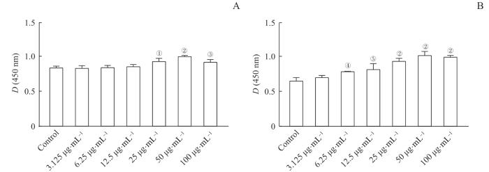
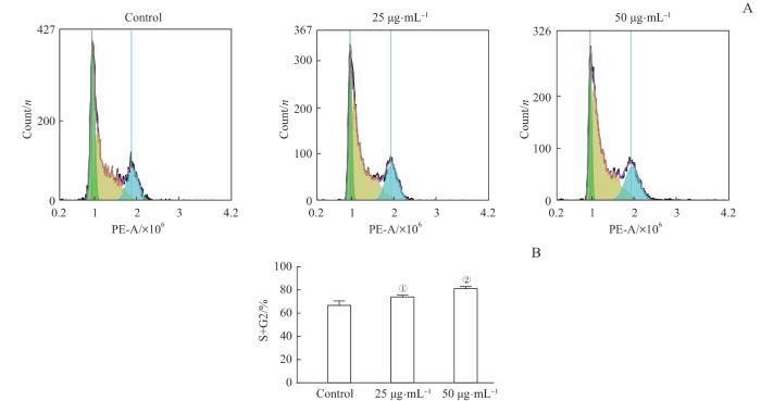
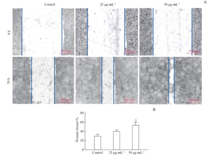
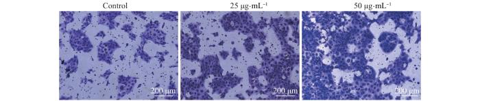
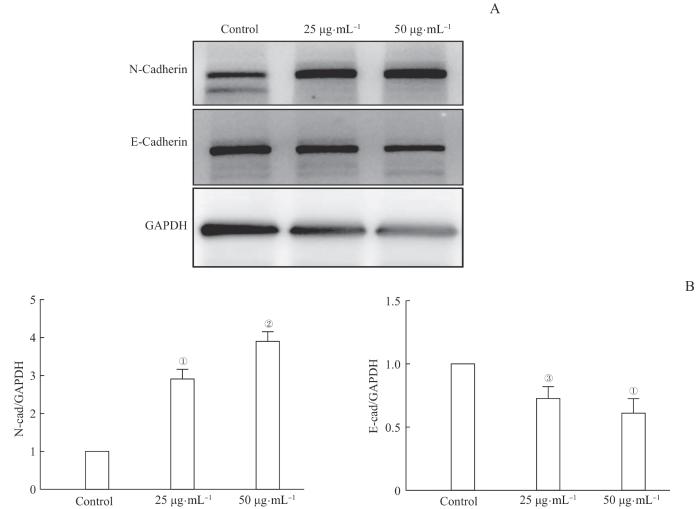
Objective ·To observe the effects of gingipain extract on the biological characteristics of oral squamous cell carcinoma cell HN6. Methods ·The HN6 cell line was selected, cultivated, and divided into different groups based on the protein concentration of gingipain extract from Porphyromonas gingivalis: control group, 3.125 μg/mL group, 6.25 μg/mL group, 12.5 μg/mL group, 25 μg/mL group, 50 μg/mL group, and 100 μg/mL group. After 24 and 48 h of cultivation, CCK-8 assay was used to detect the effects of gingipain extract on HN6 cell proliferation activity. Subsequent experiments were divided into control group, 25 μg/mL group and 50 μg/mL group. Flow cytometry was used to examine the effects of gingipain extract on cell cycle. Scratch assay and Transwell assay were performed to evaluate cell migration and invasion ability. Real-time PCR (RT-PCR) and Western blotting were used to measure the expression of E-cadherin and N-cadherin proteins and genes in cells. Results ·Stimulated with gingipain extract for 24 h, the HN6 cells showed significantly increased proliferation activity in the 25 μg/mL (P=0.025), 50 μg/mL (P=0.000), and 100 μg/mL (P=0.049) groups compared to the control group. After 48 h, proliferation activity was significantly higher in the 6.25 μg/mL(P=0.024), 12.5 μg/mL (P=0.006), 25 μg/mL (P=0.000), 50 μg/mL (P=0.000), and 100 μg/mL (P=0.000) groups compared to the control group. Cell cycle analysis revealed that, after 24 h of gingipain stimulation, the proportion of HN6 cells in the G1 phase decreased, while the proportion in the S+G2 phase significantly increased compared to the control group (25 μg/mL group: P=0.024; 50 μg/mL group: P=0.001). Compared to the control group, the scratch assay demonstrated a significant increase in the percentage of scratch closure as the concentration of gingipain extract increased (P=0.001). Compared to the control group, the Transwell invasion assay showed a significant increase in the number of cells passing through the bottom of the chamber as the concentration of gingipain extract increased. RT-PCR and Western blotting results indicated that as the concentration of gingipain extract increased, the expression levels of N-cadherin mRNA and protein in HN6 cells significantly increased, while the expression levels of E-cadherin mRNA and protein significantly decreased compared to the control group. Conclusion ·Gingipain extract could promote proliferation, migration, and invasion of oral squamous cell carcinoma HN6 cells.

Keywords: gingipain ; periodontitis ; oral squamous cell carcinoma (OSCC) ; cell proliferation ; cell migration ; cell invasion

PDF (3960KB) 元数据 多维度评价 相关文章 导出 EndNote| Ris| Bibtex  收藏本文

本文引用格式

李虎虓, 李笑甜, 赵旭日, 张桓瑜, 周薇, 宋忠臣. 牙龈素提取物对口腔鳞癌细胞HN6生物学特性的影响. 上海交通大学学报(医学版)[J], 2024, 44(2): 161-168 doi:10.3969/j.issn.1674-8115.2024.02.002

LI Huxiao, LI Xiaotian, ZHAO Xuri, ZHANG Huanyu, ZHOU Wei, SONG Zhongchen. Effects of gingipain extract on the biological characteristics of oral squamous cell carcinoma cell HN6. Journal of Shanghai Jiao Tong University (Medical Science)[J], 2024, 44(2): 161-168 doi:10.3969/j.issn.1674-8115.2024.02.002

牙周炎是一种慢性感染性疾病,临床表现为牙龈红肿出血、牙周袋形成、牙槽骨吸收、牙齿松动1。第四次全国口腔流行病学调查2显示,在35~64岁居民中,存在牙周疾病的人群高达93%,牙周健康率仅为7%。研究3-4发现,牙周炎与口腔鳞状细胞癌(oral squamous cell carcinoma,OSCC)有着密切的联系。鳞状细胞癌在我国口腔颌面部的恶性肿瘤中是最常见的,发病率可达到口腔颌面部恶性肿瘤的80%,且其发病率还在不断攀升,而OSCC的发病年龄多为40~60岁成人,这就意味着大部分的OSCC患者,往往同时患有牙周炎5

牙龈卟啉单胞菌(Porphyromonas gingivalisP. gingivalis)是牙周炎的主要致病菌之一,越来越多的研究6-7表明,P. gingivalis与OSCC存在着相关性。有研究8发现P. gingivalis对人类永生化口腔上皮细胞有促增殖和侵袭的作用。而最近的一项临床研究9显示,P. gingivalis中的牙龈素(gingipain)与OSCC的预后相关。牙龈素是P. gingivalis的主要毒力因子之一,是由精氨酸和赖氨酸特异性半胱氨酸蛋白酶组成的10。目前关于牙龈素与OSCC细胞的直接相互作用还未有报道,本研究旨在探究从P. gingivalis中提取的牙龈素对OSCC细胞HN6生物学特性的影响。

1 材料和方法

1.1 细胞和试剂

人OSCC细胞系HN6由上海交通大学医学院附属第九人民医院口腔颌面-头颈肿瘤科提供,用含10%胎牛血清的DMEM培养基培养于37 ℃恒温培养箱中。牙龈素的提取采用课题组前期研究使用的方法,提取后进行鉴定并检测蛋白浓度11

细胞计数试剂盒8(cell counting kit-8,CCK-8)、细胞周期检测试剂盒购自上海碧云天生物技术股份有限公司,Transwell小室购自美国Falcon公司,E-cadherin和N-cadherin抗体购自美国Abbkine公司。

1.2 方法

1.2.1 细胞增殖实验

将HN6细胞按3×10³个/孔种于96孔板中,分为对照组(control组)和牙龈素提取物蛋白浓度3.125 μg/mL组、6.25 μg/mL组、12.5 μg/mL组、25 μg/mL组、50 μg/mL组、100 μg/mL组。培养24和48 h后换液,根据CCK-8试剂盒说明书,以培养基10%比例加入CCK-8试剂,于酶标仪(Bio-Tek,美国)检测在450 nm波长处的吸光度即D(450 nm)。

1.2.2 细胞周期实验

取对数生长期的细胞,以3×10⁵个/孔接种于6孔板内,分为对照组(control组)和牙龈素提取物蛋白浓度25 μg/mL组、50 μg/mL组。培养24 h后用胰酶消化,201×g离心5 min,收集细胞沉淀,弃上清,用预冷PBS洗涤2次,加入预冷75%乙醇,于4 ℃固定4 h以上。固定后,201×g离心5 min,弃上清,根据细胞周期检测试剂盒说明书加入检测试剂,37 ℃避光孵育300 min,流式细胞仪(NovoCyte,美国安捷伦)上机检测。

1.2.3 细胞迁移实验

用马克笔在24孔板底部画横线标记,将细胞按6×10⁴个/孔接种,分为对照组(control组)和牙龈素提取物蛋白浓度25 μg/mL组、50 μg/mL组,培养24 h待细胞铺满板底。用1 mL枪头垂直于孔板划痕,尽量保证各个划痕宽度一致,划痕后吸去细胞培养液,用PBS冲洗孔板3次,分别在0和24 h拍照记录。用0 h的划痕面积减去24 h剩余的划痕面积,可得出各组的划痕愈合面积进行比较。

1.2.4 Transwell细胞侵袭实验

将细胞按照5×10³个/孔,每孔200 µL无血清培养基加入细胞小室,并分为对照组(control组)和牙龈素提取物蛋白浓度25 μg/mL组、50 μg/mL组,同时在下室加入含10%胎牛血清的DMEM培养基500 µL,放入37 ℃培养箱培养24 h。取出后,吸去上室多余液体,用PBS清洗2次,而后用棉棒在上室中轻轻转动,吸干水分并擦去膜内侧的细胞。再在上室中加入结晶紫染液,染色5 min,回收染液,用流水缓缓冲去染液。然后再次用棉棒在上室中轻轻转动,吸干水分,于显微镜下观察拍照。

1.2.5 实时荧光定量PCR检测mRNA表达

在6孔板内将细胞按3×10⁵个/孔种入,分为对照组(control组)和牙龈素提取物蛋白浓度25、50 μg/mL组。培养24 h后,弃上清,Trizol裂解法提取RNA,测定所得RNA浓度,根据20 μL反应体系反转录RNA,再通过PCR仪(LightCycler 480,瑞士罗氏)进行实时荧光定量PCR(real-time PCR,RT-PCR)反应,所得结果根据相对定量2-ΔΔCT法计算目的基因的相对表达量。PCR扩增引物序列见表1

表1   RT-PCR引物序列

Tab 1  Primer sequences for real-time PCR

GeneForward primer (5´→3´)Reverse primer (5´→3´)
GAPDHGAAGGTGAAGGTCGGTCGAAGATGGTGATGGGATTTC
N-cadherinCAGGGACCAGTTGAAGCACTTGCCGTGGCCTTAAAGTTAT
E-cadherinCGAAGATGTAAACGAAGCCGCCATTTCCAGTGACAATC

Note:GAPDH—glyceraldehyde 3-phosphate dehydrogenase.

新窗口打开| 下载CSV


1.2.6 Western blotting检测蛋白表达

将细胞按3×10⁵个/孔在6孔板内种入,分为对照组(control组)和牙龈素提取物蛋白浓度25 μg/mL组、50 μg/mL组。培养48 h后,弃上清,用Ripa裂解液提取蛋白,二辛可宁酸法(bicinchoninic acid,BCA)检测蛋白浓度。制备分离胶和浓缩胶,电泳分离蛋白,并转至聚偏二氟乙烯(polyvinylidene fluoride,PVDF)膜,用5%脱脂奶粉封闭1 h。于4 ℃ E-cadherin(φ=1∶1 000)、N-cadherin(φ=1∶1 000)、GAPDH(φ=1∶1 000)一抗孵育过夜,洗膜后,再在室温下用山羊抗兔二抗(1∶2 000)中孵育1 h,加显影液显影,曝光拍照。

1.3 统计学分析

采用 Graphpad Prism 7.0 软件进行统计分析。符合正态分布的定量数据以x±s表示,采用ANOVA单因素方差分析进行组间比较,使用Tukey's多重t检验进行两两比较。P<0.05表示差异具有统计学意义。

2 结果

2.1 牙龈素提取物对HN6细胞增殖的影响

经牙龈素提取物刺激24 h时,HN6细胞增殖活性随牙龈素提取物蛋白浓度增加有显著上升趋势,25 μg/mL组、50 μg/mL组、100 μg/mL组与对照组相比差异均具有统计学意义(均P<0.05)。当刺激48 h时,与对照组相比,HN6细胞增殖活性在蛋白浓度6.25 μg/mL组、12.5 μg/mL组、25 μg/mL组、50 μg/mL组、100 μg/mL组有显著上升趋势(均P<0.05),且细胞增殖活性在蛋白浓度为50 μg/mL时达到最高。结果说明,牙龈素提取物对OSCC细胞HN6有促进增殖作用,且其促增殖作用具有浓度依赖性(图1)。

图1

图1   牙龈素提取物对HN6细胞增殖的影响

Note: A/B. The effects of gingipain extract on HN6 cell proliferation after 24 h (A) and 48 h (B). P=0.025, P=0.000, P=0.049, P=0.024, P=0.006, compared with the control group.

Fig 1   Effects of gingipain extract on HN6 cell proliferation


2.2 牙龈素提取物对HN6细胞周期的影响

细胞周期检测结果显示如图2所示。与对照组相比,HN6细胞经牙龈素提取物刺激24 h,牙龈素提取物蛋白浓度25 μg/mL组和50 μg/mL组的G1期比例下降;S+G2期比例上升,与对照组相比,差异有统计学意义(25 μg/mL组:P=0.024;50 μg/mL组:P=0.001)。这表明经牙龈素提取物刺激后,HN6细胞增殖活性增强。

图2

图2   牙龈素提取物对HN6细胞周期的影响

Note: A. The effects of gingipain extract on HN6 cell cycle detected by flow cytometry. B. The effects of gingipain extract on S+G2 of HN6 cells.P=0.024, P=0.001, compared with the control group.

Fig 2   Effects of gingipain extract on HN6 cell cycle


2.3 牙龈素提取物对HN6细胞迁移作用的影响

细胞迁移实验结果显示如图3所示。与对照组相比,随着牙龈素提取物蛋白浓度升高,划痕愈合的百分比增加,差异有统计学意义(P=0.001)。表明牙龈素提取物对HN6细胞迁移有促进作用。

图3

图3   牙龈素提取物对HN6细胞迁移作用的影响

Note: A. Effects of gingipain extract on HN6 cell migration. B. The percentage of scratch closure. P=0.001, compared with the control group.

Fig 3   Effects of gingipain extract on HN6 cell migration


2.4 牙龈素提取物对HN6细胞侵袭的影响

细胞Transwell侵袭实验结果如图4所示。结果表明,与对照组相比,随着牙龈素提取物浓度升高,细胞穿过小室底部的数量增加,提示牙龈素提取物对HN6细胞侵袭有促进作用。

图4

图4   牙龈素对HN6细胞侵袭的影响(×100

Fig 4   Effects of gingipain extract on HN6 cell invasion (×100)


2.5 牙龈素提取物对HN6细胞侵袭相关蛋白基因表达的影响

RT-PCR实验结果如图5所示。结果显示,在牙龈素提取物刺激HN6细胞24 h后,与对照组相比,25 μg/mL组和50 μg/mL组N-cadherin mRNA相对表达量增加,E-cadherin mRNA相对表达量减少,差异有统计学意义(均P<0.05)。表明牙龈素提取物增强了HN6的侵袭和转移能力。

图5

图5   牙龈素提取物对HN6细胞侵袭相关基因表达的影响

NoteP=0.000, P=0.005, P=0.001, compared with the control group.

Fig 5   Effects of gingipain extract on invasion-associated genes of HN6 cells


2.6 牙龈素提取物对HN6细胞侵袭相关蛋白表达的影响

Western blotting结果显示,在牙龈素提取物刺激HN6细胞48 h,与对照组相比,25 μg/mL组和50 μg/mL组的N-cadherin蛋白表达量增加,而E-cadherin蛋白表达量减少,差异有统计学意义(均P<0.05)。表明牙龈素提取物对HN6的侵袭和转移有促进作用(图6)。

图6

图6   牙龈素对HN6细胞侵袭相关蛋白表达的影响

Note: A/B. Western blotting (A) and grayscale analysis (B) of invasion-related protein expression in HN6 cells

Fig 6   Effects of gingipain extract on invasion-associated proteins of HN6 cells


3 讨论

牙周炎作为一种慢性感染性疾病,其致病菌对全身的影响一直受到广泛关注。其主要致病菌之一的P. gingivalis与包括结直肠癌、食管癌、胰腺癌等在内的多种恶性肿瘤存在关联12-16。而OSCC作为发生在头颈部的恶性肿瘤,可直接影响患者的发音和进食,严重降低患者的生活质量,也给患者的生命造成巨大威胁。近年来,P. gingivalis与OSCC的相关作用机制也开始受到关注。P. gingivalis可通过微RNA(microRNA,miRNA)21/程序性细胞死亡因子4(programmed cell death factor 4,PDCD4)/激活蛋白-1(activator protein-1,AP-1)负反馈信号通路调节细胞周期蛋白D1(cyclin D1,CCND1)的表达,从而增加OSCC细胞的增殖17。部分化学治疗(化疗)药物通过诱导肿瘤细胞的凋亡发生作用,而在给小鼠移植OSCC细胞形成的肿瘤中,感染了P. gingivalis的肿瘤通过激活Notch1信号转导显示出对化疗药物紫杉醇的耐药性18。此外,P. gingivalis还可调节巨噬细胞的免疫活性,帮助OSCC细胞逃避免疫反应以存活19P. gingivalis对OSCC可能产生作用的毒力因子有菌毛蛋白(fimbriae,FimA)、脂多糖(lipopolysaccharide,LPS)和牙龈素等7,其中牙龈素在肿瘤的发生发展中发挥了重要作用。早在2014年,就有学者20发现牙龈素刺激蛋白酶活化受体2(protease-activated receptor 2,PAR2)和PAR4,通过细胞外信号调节蛋白激酶(extracellular signal-related kinases,ERK)1/2-E26转录因子1(E26 transformation specific-1,Ets1)、p38/热休克蛋白27(heat shock protein 27,HSP27)和核因子κB(nuclear factor κB,NF-κB)等通路增加信号转导,诱导基质金属蛋白酶酶原促进OSCC细胞的侵袭。OSCC的转移作用与上皮-间充质转化(epithelial-mesenchymal transition,EMT)密切相关。在一项研究中,有学者发现P. gingivalis可以上调一种控制EMT和炎症反应的转录因子ZEB2,而敲除牙龈素的P. gingivalis菌株没有表现出上调作用21。最近还有研究发现牙龈素可以通过对树突细胞的调控加重口腔癌22。这些都提示牙龈素可能在OSCC的发生发展中扮演着重要角色。而目前尚未见牙龈素直接作用于OSCC细胞的相关报道。

OSCC细胞增殖越快,侵袭能力越强,其恶性程度越高,患者的预后越差。因此,研究OSCC细胞的增殖、侵袭能力有着重要意义。本研究在课题组前期基础上,从P. gingivalis中提取牙龈素,观察牙龈素提取物对OSCC细胞HN6增殖、侵袭等相关生物学特性的直接影响。先通过CCK-8实验检测发现牙龈素提取物对HN6细胞的增殖活性有促进作用,且在一定浓度范围内,其促增殖作用有浓度依赖性。流式细胞术细胞周期结果检测同样发现,经牙龈素提取物刺激24 h后,G2+S期的细胞比例增多,说明HN6细胞有更多的细胞处于增殖状态。

在肿瘤的发展过程中,肿瘤细胞进入循环系统的能力与其恶性程度高度相关,可通过Transwell实验和划痕实验观察肿瘤细胞的侵袭和迁移能力。在本实验中,经过牙龈素提取物的刺激,HN6细胞划痕的愈合面积明显升高,从小室侵袭至另一侧的细胞也明显增多,在细胞行为学上说明了牙龈素对OSCC细胞HN6侵袭和迁移能力的促进作用。

为了更进一步观察牙龈素提取物对肿瘤侵袭作用的影响机制,我们初步研究了其对EMT的作用。EMT是胚胎正常发育的重要过程之一,指细胞从上皮细胞失去极性后向间质细胞转化23-25。当肿瘤细胞进行EMT过程时,能使细胞间的黏附性降低,N-cadherin蛋白取代原有的上皮细胞黏附蛋白,使得细胞更加灵活,运动性增强,最终导致肿瘤细胞具有较高的迁移与侵袭性26-27。因此,在细胞发生EMT过程中,E-cadherin作为上皮标志物表达减少,而N-cadherin作为间质标志物表达增加28。已有研究29-30发现,牙龈素可能对E-cadherin产生裂解作用,从而导致E-cadherin的减少。在本次研究中,牙龈素提取物刺激后,E-cadherin的基因表达和蛋白表达量都减少,而N-cadherin基因和蛋白表达增加,表明牙龈素增强了HN6细胞的迁移和侵袭性。

综上所述,牙龈素提取物对OSCC细胞HN6的增殖、迁移和侵袭能力有着促进作用,其侵袭能力的增强可能与牙龈素促进EMT有关。牙龈素作为P. gingivalis的主要毒力因子之一,可能对OSCC的发生发展产生重要作用。提示在OSCC患者中,应控制牙周炎,防止P. gingivalis对OSCC的产生不利影响。在后续的研究中,考虑用牙龈素刺激不同的OSCC细胞,发现牙龈素对不同OSCC细胞作用的共同点,以发现牙龈素对OSCC的作用机制。本研究仅在体外细胞层面观察了牙龈素提取物的影响,后续计划进行动物实验。如在小鼠口内成瘤模型、裸鼠成瘤模型中,加入牙龈素的刺激,验证牙龈素在体内对肿瘤的促进作用,进一步探究牙龈素在体内对OSCC的作用机制。

作者贡献声明

宋忠臣、李虎虓、周薇参与实验设计;李虎虓、李笑甜、赵旭日、张桓瑜、周薇、宋忠臣参与论文的写作和修改。所有作者均阅读并同意了最终稿件的提交。

AUTHOR's CONTRIBUTIONS

The study was designed by SONG Zhongchen, LI Huxiao and ZHOU Wei. The manuscript was drafted and revised by LI Huxiao, LI Xiaotian, ZHAO Xuri, ZHANG Huanyu, ZHOU Wei and SONG Zhongchen. All the authors have read the last version of paper and consented for submission.

利益冲突声明

所有作者声明不存在利益冲突。

COMPETING INTERESTS

All authors disclose no relevant conflict of interests.

参考文献

KOMLÓS G, CSURGAY K, HORVÁTH F, et al. Periodontitis as a risk for oral cancer: a case-control study[J]. BMC Oral Health, 2021, 21(1): 640.

[本文引用: 1]

SANZ M, MARCO DEL CASTILLO A, JEPSEN S, et al. Periodontitis and cardiovascular diseases: consensus report[J]. J Clin Periodontol, 2020, 47(3): 268-288.

[本文引用: 1]

王兴. 第四次全国口腔健康流行病学调查报告[M]. 北京: 人民卫生出版社, 2018.

[本文引用: 1]

WANG X. Report on the Fourth National Epidemiological Survey of Oral Health[M]. Beijing: People's Health Publishing House, 2018.

[本文引用: 1]

KAVARTHAPU A, GURUMOORTHY K. Linking chronic periodontitis and oral cancer: a review[J]. Oral Oncol, 2021, 121: 105375.

[本文引用: 1]

ALLON I, ABBA M, KAPLAN I, et al. Oral variant of acantholytic squamous cell carcinoma: histochemical and immunohistochemical features[J]. Acta Histochem, 2019, 121(8): 151443.

[本文引用: 1]

MORRISON A G, SARKAR S, UMAR S, et al. The contribution of the human oral microbiome to oral disease: a review[J]. Microorganisms, 2023, 11(2): 318.

[本文引用: 1]

LAMONT R J, FITZSIMONDS Z R, WANG H Z, et al. Role of Porphyromonas gingivalis in oral and orodigestive squamous cell carcinoma[J]. Periodontol 2000, 2022, 89(1): 154-165.

[本文引用: 2]

GENG F X, LIU J C, GUO Y, et al. Persistent exposure to Porphyromonas gingivalis promotes proliferative and invasion capabilities, and tumorigenic properties of human immortalized oral epithelial cells[J]. Front Cell Infect Microbiol, 2017, 7: 57.

[本文引用: 1]

KYLMÄ A K, SORSA T, JOUHI L, et al. Prognostic role of Porphyromonas gingivalis gingipain rgp and matrix metalloproteinase 9 in oropharyngeal squamous cell carcinoma[J]. Anticancer Res, 2022, 42(11): 5415-5430.

[本文引用: 1]

GUO K, LIU Y K, ZHOU H J, et al. Involvement of protein kinase C β-extracellular signal-regulating kinase 1/2/p38 mitogen-activated protein kinase-heat shock protein 27 activation in hepatocellular carcinoma cell motility and invasion[J]. Cancer Sci, 2008, 99(3): 486-496.

[本文引用: 1]

张桓瑜, 江旖婷, 朱晓晨, 等. 牙龈素提取物对小鼠脑神经炎症的影响[J]. 上海交通大学学报(医学版), 2022, 42(5): 570-577.

[本文引用: 1]

ZHANG H Y, JIANG Y T, ZHU X C, et al. Effects of gingipain extracts on brain neuroinflammation in mice[J]. Journal of Shanghai Jiao Tong University (Medical Science), 2022, 42(5): 570-577.

[本文引用: 1]

KOLIARAKIS I, MESSARITAKIS I, NIKOLOUZAKIS T K, et al. Oral bacteria and intestinal dysbiosis in colorectal cancer[J]. Int J Mol Sci, 2019, 20(17): 4146.

[本文引用: 1]

MALINOWSKI B, WĘSIERSKA A, ZALEWSKA K, et al. The role of Tannerella forsythia and Porphyromonas gingivalis in pathogenesis of esophageal cancer[J]. Infect Agent Cancer, 2019, 14: 3.

TAN Q, MA X, YANG B, et al. Periodontitis pathogen Porphyromonas gingivalis promotes pancreatic tumorigenesis via neutrophil elastase from tumor-associated neutrophils[J]. Gut Microbes, 2022, 14(1): 2073785.

GAO S G, LI S G, MA Z K, et al. Presence of Porphyromonas gingivalis in esophagus and its association with the clinicopathological characteristics and survival in patients with esophageal cancer[J]. Infect Agent Cancer, 2016, 11: 3.

LIU Y W, YUAN X, CHEN K S, et al. Clinical significance and prognostic value of Porphyromonas gingivalis infection in lung cancer[J]. Transl Oncol, 2021, 14(1): 100972.

[本文引用: 1]

CHANG C R, WANG H Y, LIU J C, et al. Porphyromonas gingivalis infection promoted the proliferation of oral squamous cell carcinoma cells through the miR-21/PDCD4/AP-1 negative signaling pathway[J]. ACS Infect Dis, 2019, 5(8): 1336-1347.

[本文引用: 1]

WOO B H, KIM D J, CHOI J I, et al. Oral cancer cells sustainedly infected with Porphyromonas gingivalis exhibit resistance to Taxol and have higher metastatic potential[J]. Oncotarget, 2017, 8(29): 46981-46992.

[本文引用: 1]

LIU S Y, ZHOU X D, PENG X, et al. Porphyromonas gingivalis promotes immunoevasion of oral cancer by protecting cancer from macrophage attack[J]. J Immunol, 2020, 205(1): 282-289.

[本文引用: 1]

INABA H, SUGITA H, KUBONIWA M, et al. Porphyromonas gingivalis promotes invasion of oral squamous cell carcinoma through induction of proMMP9 and its activation[J]. Cell Microbiol, 2014, 16(1): 131-145.

[本文引用: 1]

OHSHIMA J, WANG Q, FITZSIMONDS Z R, et al. Streptococcus gordonii programs epithelial cells to resist ZEB2 induction by Porphyromonas gingivalis[J]. Proc Natl Acad Sci USA, 2019, 116(17): 8544-8553.

[本文引用: 1]

REN J L, HAN X, LOHNER H, et al. P. gingivalis infection upregulates PD-L1 expression on dendritic cells, suppresses CD8+ T-cell responses, and aggravates oral cancer[J]. Cancer Immunol Res, 2023, 11(3): 290-305.

[本文引用: 1]

WHEELOCK M J, JOHNSON K R. Cadherins as modulators of cellular phenotype[J]. Annu Rev Cell Dev Biol, 2003, 19: 207-235.

[本文引用: 1]

CHRISTOFORI G. Changing neighbours, changing behaviour: cell adhesion molecule-mediated signalling during tumour progression[J]. EMBO J, 2003, 22(10): 2318-2323.

LAMOUILLE S, XU J, DERYNCK R. Molecular mechanisms of epithelial-mesenchymal transition[J]. Nat Rev Mol Cell Biol, 2014, 15(3): 178-196.

[本文引用: 1]

XU Z L, CHEN Z M, PENG M S, et al. MicroRNA miR-490-5p suppresses pancreatic cancer through regulating epithelial-mesenchymal transition via targeting MAGI2 antisense RNA 3[J]. Bioengineered, 2022, 13(2): 2673-2685.

[本文引用: 1]

PIERCEALL W E, WOODARD A S, MORROW J S, et al. Frequent alterations in E-cadherin and α- and β-catenin expression in human breast cancer cell lines[J]. Oncogene, 1995, 11(7): 1319-1326.

[本文引用: 1]

DERYCKE L D M, BRACKE M E. N-cadherin in the spotlight of cell-cell adhesion, differentiation, embryogenesis, invasion and signalling[J]. Int J Dev Biol, 2004, 48(5/6): 463-476.

[本文引用: 1]

GRABOWSKA M M, DAY M L. Soluble E-cadherin: more than a symptom of disease[J]. Front Biosci (Landmark Ed), 2012, 17(5): 1948-1964.

[本文引用: 1]

KATZ J, YANG Q B, ZHANG P, et al. Hydrolysis of epithelial junctional proteins by Porphyromonas gingivalis gingipains[J]. Infect Immun, 2002, 70(5): 2512-2518.

[本文引用: 1]

/