Promotion of Nd:YAP laser biostimulation on the proliferation and osteogenic differentiation of human periodontal ligament cells through WNT/β-catenin signaling pathway
XU Muxin, LIU Xian, JIANG Lishan, SUN Qing,
Department of Periodontics, Zhonglou Branch of Changzhou Traditional Chinese Medicine Hospital/Changzhou Stomatological Hospital, Jiangsu Province, Changzhou 213003, China
Objective ·To study the effect of neodymium-doped yttrium aluminum perovskite (Nd:YAP) laser biostimulation on the proliferation and osteogenic differentiation of human periodontal ligament cells (hPDLCs) and its possible mechanism. Methods ·Five premolars removed for orthodontic reasons were collected from Changzhou Stomatological Hospital, and the periodontal ligament tissues from the middle 1/3 of the roots were taken to culture hPDLCs in vitro. The cells were irradiated with the biostimulation function [G (-) mode] of the Nd:YAP laser. According to the irradiation time, the cells were divided into a control group (without laser irradiation), and groups irradiated for 5 s, 10 s, 15 s, 20 s and 30 s. The cell counting kit-8 (CCK-8) method was used to detect the proliferation of hPDLCs in each group. After osteogenic differentiation was induced, the alkaline phosphatase (ALP) content and activity level of the cells were detected using an ALP staining kit and an ALP activity detection kit. The calcium salt level of the cells was evaluated by alizarin red S staining and calcium quantitative analysis. The expression of genes and proteins related to the WNT/β-catenin signaling pathway, including dickkopf-related protein 1 (DKK-1), β-catenin, and runt-related transcription factor 2 (RUNX2), was analyzed by quantitative real-time PCR (qPCR) and Western blotting. Results ·The results of CCK-8 showed that the proliferation level of cells in the 10 s, 15 s, 20 s, and 30 s groups was enhanced from 3 d after irradiation (all P<0.05). After induction of osteogenic differentiation, ALP content, activity, and calcium salt level in the laser irradiation groups increased with the extension of irradiation time (all P<0.05). The results of qPCR and Western blotting analysis showed that the expression levels of the DKK-1 gene and protein in the laser irradiation groups decreased with the extension of irradiation time. However, the expression levels of β-catenin and RUNX2 genes and proteins increased significantly with the extension of irradiation time; there were statistically significant differences between the 15 s, 20 s, and 30 s groups and the control group (all P<0.05). Conclusion ·Nd:YAP laser biostimulation may promote proliferation and osteogenic differentiation of hPDLCs through WNT/β-catenin signaling pathway.
XU Muxin, LIU Xian, JIANG Lishan, SUN Qing. Promotion of Nd:YAP laser biostimulation on the proliferation and osteogenic differentiation of human periodontal ligament cells through WNT/β-catenin signaling pathway. Journal of Shanghai Jiao Tong University (Medical Science)[J], 2025, 45(5): 562-569 doi:10.3969/j.issn.1674-8115.2025.05.004
DMEM培养基(江苏凯基生物技术股份有限公司),胎牛血清(南京生航生物技术有限公司),细胞计数试剂盒-8(cell counting kit-8,CCK-8)、细胞RNA快速提取试剂盒、反转录试剂盒、2×SYBR Green qPCR混合物、RIPA裂解液、极超敏化学发光试剂盒(山东思科捷生物技术有限公司),地塞米松、抗坏血酸(上海源叶生物科技有限公司),β-甘油磷酸钠(Sigma,美国),碱性磷酸酶(alkaline phosphatase,ALP)活性检测试剂盒、ALP染色试剂盒(上海碧云天生物技术有限公司),茜素红S(alizarin red S,ARS)染色液(Cyagen,美国),氯化十六烷基吡啶(北京索莱宝科技有限公司),PVDF膜(Bio-Rad,美国),dickkopf相关蛋白1(dickkopf-related protein 1,DKK-1)抗体(ab109416)、β-catenin抗体(ab32572)、runt相关转录因子2(runt-related transcription factor 2,RUNX2)抗体(ab236639)、β-肌动蛋白(β-actin)抗体(ab8227;Abcam,美国),羊抗兔二抗(武汉三鹰生物技术有限公司)。
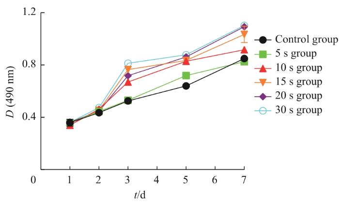
Note: Compared with the control group, the proliferation levels of the 10 s group (P<0.001), 15 s group (P<0.001), 20 s group (P<0.001), and 30 s group (P<0.001) were significantly different (n=3).
Fig 1
Effect of Nd:YAP laser biostimulation on proliferation of hPDLCs
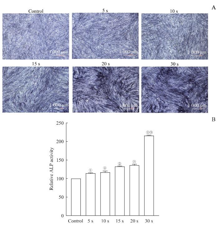
Note: A. ALP staining after osteogenic differentiation for 7 d (×40). B. ALP activity detection after osteogenic differentiation for 7 d (n=3). ①P=0.002, ②P<0.001, compared with the control group; ③P<0.001, compared with the 20 s group.
Fig 2
Effect of Nd:YAP laser biostimulation on ALP content and activity of hPDLCs during early osteogenic differentiation
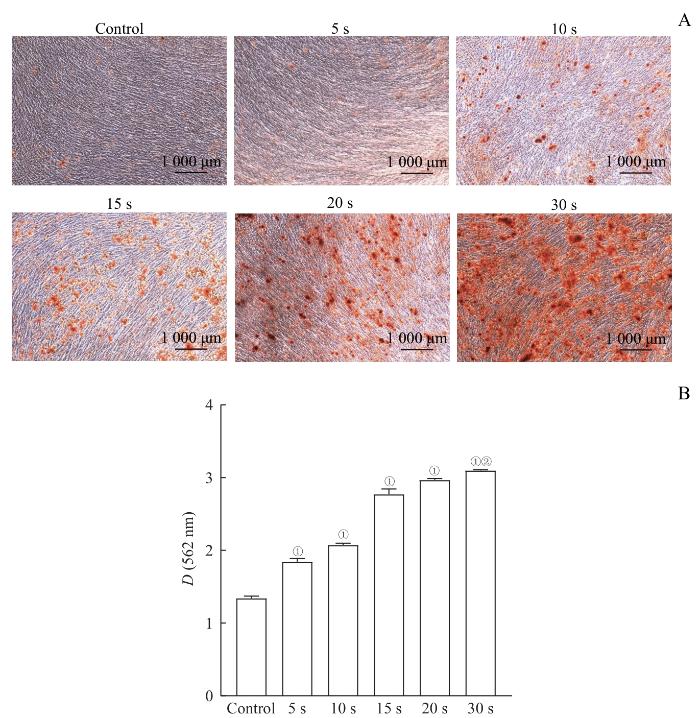
Note: A. ARS staining after osteogenic differentiation for 21 d (×40). B. Quantitative determination of calcium levels after osteogenic differentiation for 21 d (n=3). ①P<0.001, compared with the control group; ②P=0.028, compared with the 20 s group.
Fig 3
Effect of Nd:YAP laser biostimulation on calcium level of hPDLCs during osteogenic differentiation
Note: A. Protein bands in Western blotting. B. Quantitative analysis. ①P<0.001, compared with the control group; ②P<0.001, compared with the 15 s group; ③P<0.001, compared with the 10 s group; ④P<0.001, compared with the 20 s group. (n=3)
Fig 5
Effect of Nd:YAP laser biostimulation on expression of DKK-1, β-catenin and RUNX2 proteins in hPDLCs
XU Muxin participated in the design of the study, and the drafting and revision of the paper. SUN Qing was responsible for the overall research design and implementation, data analysis, and the drafting and revision of the paper. LIU Xian participated in the design of the study. JIANG Lishan participated in the implementation of the experiment and the drafting of the paper. All authors have read the final version of paper and consented to submission.
利益冲突声明
所有作者声明不存在利益冲突。
COMPETING INTERESTS
All authors declare no relevant conflict of interests.
SOLAKOGLU Ö, HEYDECKE G, AMIRI N, et al. The use of plasma rich in growth factors (PRGF) in guided tissue regeneration and guided bone regeneration. A review of histological, immunohistochemical, histomorphometrical, radiological and clinical results in humans[J]. Ann Anat, 2020, 231: 151528.
GE L H, SUN M J, SHU R. Study on the effect of Nd:YAG water-cooled laser on the surface of titanium implant[J]. Journal of Shanghai Jiao Tong University (Medical Science), 2020, 40(10): 1371-1375.
SHEN Y, ZHANG Y, CHEN N J, et al. Clinical and microbiological evaluation of Nd:YAP laser for adjuvant treatment of periodontitis[J]. Stomatology, 2022, 42(5): 446-450, 461.
WANG T, ZHU Y Q. Research status of application of Nd:YAP laser and Er:YAG laser in oral medicine[J]. Chinese Journal of Dental Materials and Devices, 2018, 27(4): 225-228.
LIN T C, YU C C, LIU C M, et al. Er:YAG laser promotes proliferation and wound healing capacity of human periodontal ligament fibroblasts through galectin-7 induction[J]. J Formos Med Assoc, 2021, 120(1 Pt 2): 388-394.
CHOI E J, YIM J Y, KOO K T, et al. Biological effects of a semiconductor diode laser on human periodontal ligament fibroblasts[J]. J Periodontal Implant Sci, 2010, 40(3): 105-110.
FU Q Y, LAN X M, XU R W, et al. Effects of different signaling pathways on osteogenic differentiation of periodontal ligament stem cells[J]. Chinese Journal of Tissue Engineering Research, 2023, 27(24): 3910-3919.
JIANG J Q, WANG Z C, LI C H, et al. Primary culture of human periodontal ligament cells using three methods[J]. Journal of Clinical Rehabilitative Tissue Engineering Research, 2010, 14(23): 4290-4294.
WANG Y P, LIN Z K, SHU R. Short-term clinical efficacy observation of adjunctive Er,Cr:YSGG laser application following subgingival scaling in patients with severe periodontitis[J]. Journal of Shanghai Jiao Tong University (Medical Science), 2019, 39(4): 378-382.
PAVONE C, PERUSSI L R, DE OLIVEIRA G J P L, et al. Effect of Er,Cr:YSGG laser application in the treatment of experimental periodontitis[J]. Lasers Med Sci, 2015, 30(3): 993-999.
AMBROSINI P, MILLER N, BRIANÇON S, et al. Clinical and microbiological evaluation of the effectiveness of the Nd:Yap laser for the initial treatment of adult periodontitis. A randomized controlled study[J]. J Clin Periodontol, 2005, 32(6): 670-676.
LIU T, HUANG Z Q, JU Y Y, et al. Bactericidal efficacy of three parameters of Nd:YAP laser irradiation against Enterococcus faecalis compared with NaOCl irrigation[J]. Lasers Med Sci, 2019, 34(2): 359-366.
ZHANG L, WANG X Y. Efficiency and temperature rise of file ablation by neodymium: yttrium-aluminum-perovskite laser in vitro[J]. J Endod, 2021, 47(6): 982-988.
FORNAINI C, BRULAT-BOUCHARD N, MEDIONI E, et al. Nd:Yap laser in the treatment of dentinal hypersensitivity: an ex vivo study[J]. J Photochem Photobiol B, 2020, 203: 111740.
OHSUGI Y, NIIMI H, SHIMOHIRA T, et al. In vitro cytological responses against laser photobiomodulation for periodontal regeneration[J]. Int J Mol Sci, 2020, 21(23): 9002.
PONNAIYAN D, RUGHWANI R R, SHETTY G, et al. The effect of adjunctive LASER application on periodontal ligament stem cells[J]. Front Cell Dev Biol, 2023, 11: 1341628.
WU Y, ZHU T T, YANG Y Y, et al. Irradiation with red light-emitting diode enhances proliferation and osteogenic differentiation of periodontal ligament stem cells[J]. Lasers Med Sci, 2021, 36(7): 1535-1543.
WANG L Y, LIU C, WU F. Low-level laser irradiation enhances the proliferation and osteogenic differentiation of PDLSCs via BMP signaling[J]. Lasers Med Sci, 2022, 37(2): 941-948.
RUAN Y R, KATO H, TAGUCHI Y, et al. Irradiation by high-intensity red light-emitting diode enhances human bone marrow mesenchymal stem cells osteogenic differentiation and mineralization through Wnt/β-catenin signaling pathway[J]. Lasers Med Sci, 2021, 36(1): 55-65.
SAMIEI M, JANJIĆ K, CVIKL B, et al. The role of sclerostin and dickkopf-1 in oral tissues: a review from the perspective of the dental disciplines[J]. F1000Res, 2019, 8: 128.
HIE M, IITSUKA N, OTSUKA T, et al. Insulin-dependent diabetes mellitus decreases osteoblastogenesis associated with the inhibition of Wnt signaling through increased expression of Sost and Dkk1 and inhibition of Akt activation[J]. Int J Mol Med, 2011, 28(3): 455-462.
LIAN J B, JAVED A, ZAIDI S K, et al. Regulatory controls for osteoblast growth and differentiation: role of Runx/Cbfa/AML factors[J]. Crit Rev Eukaryot Gene Expr, 2004, 14(1/2): 1-41.
BIAN Y, XIANG J. Salvianolic acid B promotes the osteogenic differentiation of human periodontal ligament cells through Wnt/β-catenin signaling pathway[J]. Arch Oral Biol, 2020, 113: 104693.
QUAN H, DAI X P, LIU M Y, et al. Luteolin supports osteogenic differentiation of human periodontal ligament cells[J]. BMC Oral Health, 2019, 19(1): 229.
SANZ M, HERRERA D, KEBSCHULL M, et al. Treatment of stage Ⅰ‒Ⅲ periodontitis: the EFP S3 level clinical practice guideline[J]. J Clin Periodontol, 2020, 47(Suppl 22): 4-60.


