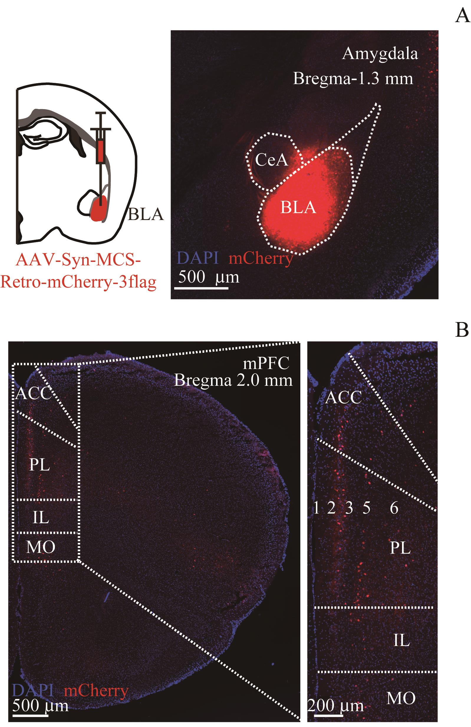
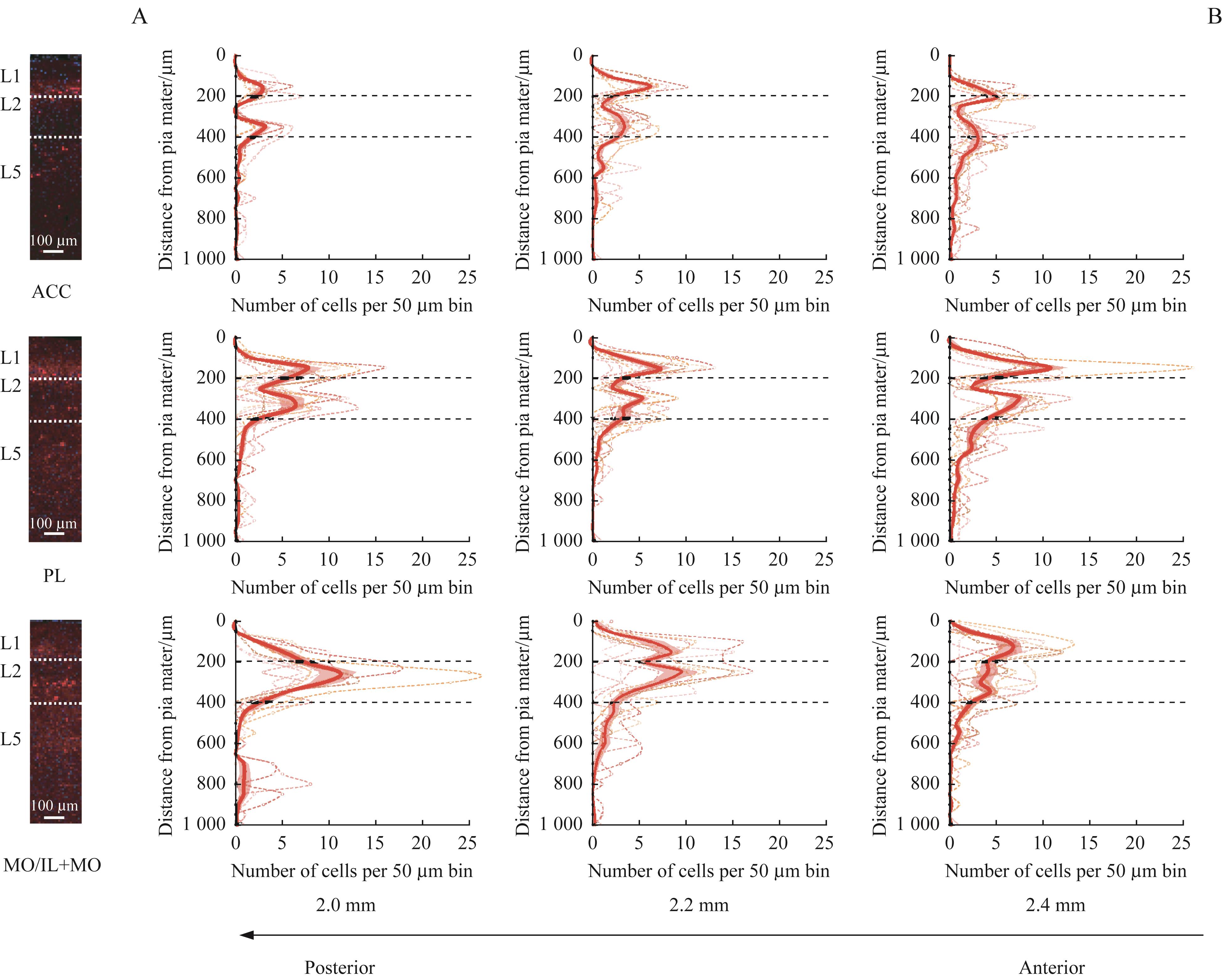
| 1 |
MILLER E K, COHEN J D. An integrative theory of prefrontal cortex function[J]. Annu Rev Neurosci, 2001, 24: 167-202.
|
| 2 |
ROBBINS T W, ARNSTEN A F T. The neuropsychopharmacology of fronto-executive function: monoaminergic modulation[J]. Annu Rev Neurosci, 2009, 32: 267-287.
|
| 3 |
LAUBACH M, AMARANTE L M, SWANSON K, et al. What, if anything, is rodent prefrontal cortex? [J]. eNeuro, 2018, 5(5): ENEURO.0315-18.2018.
|
| 4 |
ANASTASIADES P G, CARTER A G. Circuit organization of the rodent medial prefrontal cortex[J]. Trends Neurosci, 2021, 44(7): 550-563.
|
| 5 |
SCHUMAN B, MACHOLD R P, HASHIKAWA Y, et al. Four unique interneuron populations reside in neocortical layer 1[J]. J Neurosci, 2019, 39(1): 125-139.
|
| 6 |
ABS E, POORTHUIS R B, APELBLAT D, et al. Learning-related plasticity in dendrite-targeting layer 1 interneurons[J]. Neuron, 2018, 100(3): 684-699.e6.
|
| 7 |
ANASTASIADES P G, COLLINS D P, CARTER A G. Mediodorsal and ventromedial thalamus engage distinct L1 circuits in the prefrontal cortex[J]. Neuron, 2021, 109(2): 314-330.e4.
|
| 8 |
JANAK P H, TYE K M. From circuits to behaviour in the amygdala[J]. Nature, 2015, 517(7534): 284-292.
|
| 9 |
BLAIR H T, HUYNH V K, VAZ V T, et al. Unilateral storage of fear memories by the amygdala[J]. J Neurosci, 2005, 25(16): 4198-4205.
|
| 10 |
SAH P, FABER E S L, LOPEZ DE ARMENTIA M, et al. The amygdaloid complex: anatomy and physiology[J]. Physiol Rev, 2003, 83(3): 803-834.
|
| 11 |
FERRARA N C, TRASK S, ROSENKRANZ J A. Maturation of amygdala inputs regulate shifts in social and fear behaviors: a substrate for developmental effects of stress[J]. Neurosci Biobehav Rev, 2021, 125: 11-25.
|
| 12 |
GANGOPADHYAY P, CHAWLA M, DAL MONTE O, et al. Prefrontal-amygdala circuits in social decision-making[J]. Nat Neurosci, 2021, 24(1): 5-18.
|
| 13 |
ARRUDA-CARVALHO M, CLEM R L. Pathway-selective adjustment of prefrontal-amygdala transmission during fear encoding[J]. J Neurosci, 2014, 34(47): 15601-15609.
|
| 14 |
BUKALO O, PINARD C R, SILVERSTEIN S, et al. Prefrontal inputs to the amygdala instruct fear extinction memory formation[J]. Sci Adv, 2015, 1(6): e1500251.
|
| 15 |
BLOODGOOD D W, SUGAM J A, HOLMES A, et al. Fear extinction requires infralimbic cortex projections to the basolateral amygdala[J]. Transl Psychiatry, 2018, 8(1): 60.
|
| 16 |
HUANG W C, ZUCCA A, LEVY J, et al. Social behavior is modulated by valence-encoding mPFC-amygdala sub-circuitry[J]. Cell Rep, 2020, 32(2): 107899.
|
| 17 |
FRANKLIN K B J, PAXINOS G. The mouse brain in stereotaxic coordinates [M]. 3rd ed. San Diego: Academic Press, 2008.
|
| 18 |
GERFEN C R, PALETZKI R, HEINTZ N. GENSAT BAC Cre-recombinase driver lines to study the functional organization of cerebral cortical and basal ganglia circuits[J]. Neuron, 2013, 80(6): 1368-1383.
|
| 19 |
MATHO K S, HUILGOL D, GALBAVY W, et al. Genetic dissection of the glutamatergic neuron system in cerebral cortex[J]. Nature, 2021, 598(7879): 182-187.
|
| 20 |
HE M, TUCCIARONE J, LEE S, et al. Strategies and tools for combinatorial targeting of GABAergic neurons in mouse cerebral cortex[J]. Neuron, 2016, 91(6): 1228-1243.
|
| 21 |
BOUWMEESTER H, SMITS K, VAN REE J M. Neonatal development of projections to the basolateral amygdala from prefrontal and thalamic structures in rat[J]. J Comp Neurol, 2002, 450(3): 241-255.
|
| 22 |
MCDONALD A J, MASCAGNI F, GUO L. Projections of the medial and lateral prefrontal cortices to the amygdala: a Phaseolus vulgaris leucoagglutinin study in the rat[J]. Neuroscience, 1996, 71(1): 55-75.
|
| 23 |
GABBOTT P L A, WARNER T A, JAYS P R L, et al. Prefrontal cortex in the rat: projections to subcortical autonomic, motor, and limbic centers[J]. J Comp Neurol, 2005, 492(2): 145-177.
|
| 24 |
HARRIS K D, SHEPHERD G M G. The neocortical circuit: themes and variations[J]. Nat Neurosci, 2015, 18(2): 170-181.
|
 ), 黄东萍1,2(