上海交通大学学报(医学版) ›› 2025, Vol. 45 ›› Issue (11): 1527-1535.doi: 10.3969/j.issn.1674-8115.2025.11.012
侯森林1,2, 邓翔天1,2, 郑庭佳2, 韩奕菲2, 刘珅1,2( )
)
收稿日期:2025-02-24
接受日期:2025-07-06
出版日期:2025-11-28
发布日期:2025-12-03
通讯作者:
刘 珅,研究员,博士;电子信箱:liushensjtu@126.com。作者简介:第一联系人:为共同第一作者 (Co-first authors)。
基金资助:
HOU Senlin1,2, DENG Xiangtian1,2, ZHENG Tingjia2, HAN Yifei2, LIU Shen1,2()
Received:2025-02-24
Accepted:2025-07-06
Online:2025-11-28
Published:2025-12-03
Contact:
LIU Shen, E-mail: liushensjtu@126.com.Supported by:摘要:
组织修复是机体组织损伤后的重要生理过程,常受组织纤维化、氧化应激等问题干扰,并伴随细胞衰老现象。细胞衰老指细胞在不利刺激下出现的增殖停滞、功能衰退现象,在组织修复中具有双向调节作用。在伤口愈合早期,细胞衰老能限制纤维化、诱导细胞可塑性,从而促进修复;但衰老细胞的长期积累会干扰细胞正常增殖分化,阻碍修复进程。随着组织学和细胞生物学的不断发展,组织修复和细胞衰老机制研究不断深入,为抗衰老材料在组织修复中的应用提供了理论依据。抗衰老材料负载抗衰药物、能量补充剂或抗氧化剂等成分,后者通过诱导细胞凋亡、激活自噬、逆转衰老进程等多种途径发挥积极作用。这些抗衰材料的应用为解决慢性创面的组织纤维化等临床难题提供了新思路,有望成为干预组织损伤修复的有效手段。近年来,抗衰老材料广泛应用于组织再生与修复,对此展开综述能够促进更多基础研究的转化应用,为攻克临床上组织修复难题提供参考。
中图分类号:
侯森林, 邓翔天, 郑庭佳, 韩奕菲, 刘珅. 抗衰老材料在组织修复中应用的研究进展[J]. 上海交通大学学报(医学版), 2025, 45(11): 1527-1535.
HOU Senlin, DENG Xiangtian, ZHENG Tingjia, HAN Yifei, LIU Shen. Progress in applications of anti-senescence materials for tissue repair[J]. Journal of Shanghai Jiao Tong University (Medical Science), 2025, 45(11): 1527-1535.
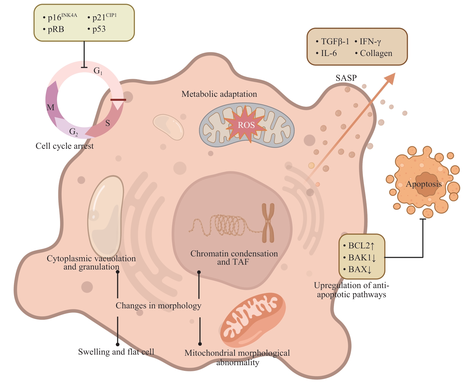
图1 衰老细胞的表型
Fig 1 Phenotypes of senescent cells
| Feature | Acute wound | Chronic wound |
|---|---|---|
| SASP secretory pattern | Short-term, mainly promoting repair | Long-term, mainly promoting inflammation |
| SASP secretory substances | Growth factors such as TGF-β, ECM remodeling enzymes such as MMPs, anti-inflammatory cytokines such as IL-10, etc | Proinflammatory factors such as IL-1β and TNF-α, and growth inhibitors such as sFRP |
| Oxidative stress level | Controllable, with a transient rise in ROS | Persistent high-level, causing accumulation of DNA damage |
| Microenvironment remediation ability | Dynamic balance (orderly ECM remodeling) | Imbalance (excessive ECM degradation or fibrosis) |
表1 细胞衰老在急性创面和慢性创面中的作用比较
Tab 1 Comparison of the roles of cellular senescence is acute and chronic wounds
| Feature | Acute wound | Chronic wound |
|---|---|---|
| SASP secretory pattern | Short-term, mainly promoting repair | Long-term, mainly promoting inflammation |
| SASP secretory substances | Growth factors such as TGF-β, ECM remodeling enzymes such as MMPs, anti-inflammatory cytokines such as IL-10, etc | Proinflammatory factors such as IL-1β and TNF-α, and growth inhibitors such as sFRP |
| Oxidative stress level | Controllable, with a transient rise in ROS | Persistent high-level, causing accumulation of DNA damage |
| Microenvironment remediation ability | Dynamic balance (orderly ECM remodeling) | Imbalance (excessive ECM degradation or fibrosis) |
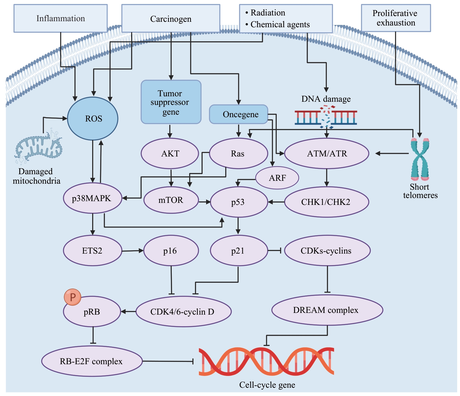
图2 细胞衰老原因及信号通路示意图
Fig 2 Schematic diagram of the causes and signaling pathways of cellular senescence
| Anti-senescence material | Injury type | Mechanism of action | Biocompatibility | Experimental stage and evaluation | Reference |
|---|---|---|---|---|---|
| Dasatinib + quercetin environment-responsive hydrogel | Myocardial injury | Senolytic | High biocompatibility, low toxicity, and no obvious side effects | Clinical randomized controlled trial | [ |
| Metformin + zinc ion hydrogel | Traumatic skin defects, burns | Activating autophagy | High biocompatibility, low toxicity, and easy metabolism | Mouse experiment | [ |
| Resveratrol + angiopoietin 2 hydrogel | Bone tissue injury | Activating autophagy | High biocompatibility, low toxicity, and no obvious side effects | Rat experiment | [ |
| Yap1 protein exosome hydrogel | Tendon injury | Inhibiting SASP | High biocompatibility, low toxicity, and no obvious side effects | Rat experiment | [ |
| Microneedle patches loaded with taurine | Soft tissue injury | Inhibiting SASP | High biocompatibility, low toxicity, and no obvious side effects | Rat experiment | [ |
| Dynamic self-healing hydrogel loaded with melatonin | Intervertebral disc annulus fibrosus injury | Enhancing energy supply | High biocompatibility, low toxicity, and no obvious side effects | Rat experiment | [ |
| Hydrogel loaded with myelinated mesenchymal stem cells | Bone tissue injury | Enhancing energy supply | High biocompatibility, low toxicity, and no obvious side effects | Mouse experiment | [ |
| Nano-thylakoid membrane system | Cartilage injury | Enhancing energy supply | High biocompatibility, low toxicity, and no obvious side effects | Animal experiment (without specifying species) | [ |
| PLGA-PEG nanodelivery system loaded with ginsentriol | Osteoarthritis | Reversing senescent cells | Lower side effects than systemic administration, reduced risk of infection from intra-articular injection | Mouse and human tissue in vitro experiment | [ |
| Pha-encapsulated CaSi₂ nanoparticles releasing H₂ | Bone defect | Inhibiting oxygenated stress | High biocompatibility, low toxicity, and easy metabolism | Mouse experiment | [ |
| Hydrogel loaded with synovial mesenchymal stem cells | Cartilage injury | Reversing senescent cells | High biocompatibility, low toxicity, and easy metabolism | Rat experiment | [ |
| Biological heterojunctions releasing gaseous H₂Se | Stalled healing of infectious wounds | Inhibiting oxygenated stress | High biocompatibility, low toxicity, and easy metabolism | Rat experiment | [ |
| PEG liposomes targeting CD9 | Skin injury | Reversing senescent cells | High targeting, efficient drug utilization, and low toxicity | Cell in vitro experiment | [ |
| Honokiol | Silicosis | Inhibiting oxygenated stress | High biocompatibility, low toxicity, and easy metabolism | Mouse experiment | [ |
表2 各种抗衰老材料信息汇总
Tab 2 Information summary of various anti-senescence materials
| Anti-senescence material | Injury type | Mechanism of action | Biocompatibility | Experimental stage and evaluation | Reference |
|---|---|---|---|---|---|
| Dasatinib + quercetin environment-responsive hydrogel | Myocardial injury | Senolytic | High biocompatibility, low toxicity, and no obvious side effects | Clinical randomized controlled trial | [ |
| Metformin + zinc ion hydrogel | Traumatic skin defects, burns | Activating autophagy | High biocompatibility, low toxicity, and easy metabolism | Mouse experiment | [ |
| Resveratrol + angiopoietin 2 hydrogel | Bone tissue injury | Activating autophagy | High biocompatibility, low toxicity, and no obvious side effects | Rat experiment | [ |
| Yap1 protein exosome hydrogel | Tendon injury | Inhibiting SASP | High biocompatibility, low toxicity, and no obvious side effects | Rat experiment | [ |
| Microneedle patches loaded with taurine | Soft tissue injury | Inhibiting SASP | High biocompatibility, low toxicity, and no obvious side effects | Rat experiment | [ |
| Dynamic self-healing hydrogel loaded with melatonin | Intervertebral disc annulus fibrosus injury | Enhancing energy supply | High biocompatibility, low toxicity, and no obvious side effects | Rat experiment | [ |
| Hydrogel loaded with myelinated mesenchymal stem cells | Bone tissue injury | Enhancing energy supply | High biocompatibility, low toxicity, and no obvious side effects | Mouse experiment | [ |
| Nano-thylakoid membrane system | Cartilage injury | Enhancing energy supply | High biocompatibility, low toxicity, and no obvious side effects | Animal experiment (without specifying species) | [ |
| PLGA-PEG nanodelivery system loaded with ginsentriol | Osteoarthritis | Reversing senescent cells | Lower side effects than systemic administration, reduced risk of infection from intra-articular injection | Mouse and human tissue in vitro experiment | [ |
| Pha-encapsulated CaSi₂ nanoparticles releasing H₂ | Bone defect | Inhibiting oxygenated stress | High biocompatibility, low toxicity, and easy metabolism | Mouse experiment | [ |
| Hydrogel loaded with synovial mesenchymal stem cells | Cartilage injury | Reversing senescent cells | High biocompatibility, low toxicity, and easy metabolism | Rat experiment | [ |
| Biological heterojunctions releasing gaseous H₂Se | Stalled healing of infectious wounds | Inhibiting oxygenated stress | High biocompatibility, low toxicity, and easy metabolism | Rat experiment | [ |
| PEG liposomes targeting CD9 | Skin injury | Reversing senescent cells | High targeting, efficient drug utilization, and low toxicity | Cell in vitro experiment | [ |
| Honokiol | Silicosis | Inhibiting oxygenated stress | High biocompatibility, low toxicity, and easy metabolism | Mouse experiment | [ |
| [1] | PEÑA O A, MARTIN P. Cellular and molecular mechanisms of skin wound healing[J]. Nat Rev Mol Cell Biol, 2024, 25(8): 599-616. |
| [2] | XIONG Y, MI B B, LIN Z, et al. The role of the immune microenvironment in bone, cartilage, and soft tissue regeneration: from mechanism to therapeutic opportunity[J]. Mil Med Res, 2022, 9(1): 65. |
| [3] | 方邵一涵,刘德伍. 细胞衰老在慢性创面愈合中的作用研究进展[J]. 中华烧伤与创面修复杂志, 2023, 39(8): 795-800. |
| FANG S Y H, LIU D W. Research advances on the role of cell senescence in chronic wound healing[J]. Chinese Journal of Burns and Wounds, 2023, 39(8): 795-800. | |
| [4] | HUANG W, HICKSON L J, EIRIN A, et al. Cellular senescence: the good, the bad and the unknown[J]. Nat Rev Nephrol, 2022, 18(10): 611-627. |
| [5] | 刘晓南,张培林,刘勇敢. 细胞衰老相关分子机制及鉴定的研究进展[J]. 中华实验外科杂志, 2024, 41(2): 420-424. |
| LIU X N, ZHANG P L, LIU Y G. Research progress in the molecular mechanisms and identification of cellular senescence[J]. Chinese Journal of Experimental Surgery, 2024, 41(2): 420-424. | |
| [6] | LIU X N, GU Y R, KUMAR S, et al. Oxylipin-PPARγ-initiated adipocyte senescence propagates secondary senescence in the bone marrow[J]. Cell Metab, 2023, 35(4): 667-684.e6. |
| [7] | ZHU J, WU C Y, YANG L D. Cellular senescence in Alzheimer′s disease: from physiology to pathology[J]. Transl Neurodegener, 2024, 13(1): 55. |
| [8] | SURYADEVARA V, HUDGINS A D, RAJESH A, et al. SenNet recommendations for detecting senescent cells in different tissues[J]. Nat Rev Mol Cell Biol, 2024, 25(12): 1001-1023. |
| [9] | FRANCO A C, AVELEIRA C, CAVADAS C. Skin senescence: mechanisms and impact on whole-body aging[J]. Trends Mol Med, 2022, 28(2): 97-109. |
| [10] | LI X, LUO X, HE Y, et al. Micronano titanium accelerates mesenchymal stem cells aging through the activation of senescence-associated secretory phenotype[J]. ACS Nano, 2023, 17(22): 22885-22900. |
| [11] | WANG C, TANIZAWA H, HILL C, et al. METTL3-mediated chromatin contacts promote stress granule phase separation through metabolic reprogramming during senescence[J]. Nat Commun, 2024, 15(1): 5410. |
| [12] | CORADDUZZA D, CONGIARGIU A, CHEN Z, et al. Ferroptosis and senescence: a systematic review[J]. Int J Mol Sci, 2023, 24(4): 3658. |
| [13] | MAUS M, LÓPEZ-POLO V, MATEO L, et al. Iron accumulation drives fibrosis, senescence and the senescence-associated secretory phenotype[J]. Nat Metab, 2023, 5(12): 2111-2130. |
| [14] | YU B, MA J, LI J, et al. Mitochondrial phosphatase PGAM5 modulates cellular senescence by regulating mitochondrial dynamics[J]. Nat Commun, 2020, 11(1): 2549. |
| [15] | CARUSILLO A, MUSSOLINO C. DNA damage: from threat to treatment[J]. Cells, 2020, 9(7): E1665. |
| [16] | WANG Y, ZHANG Z, MI X, et al. Elevation of effective p53 expression sensitizes wild-type p53 breast cancer cells to CDK7 inhibitor THZ1[J]. Cell Commun Signal, 2022, 20(1): 96. |
| [17] | ZHU Y K, LIU X W, DING X L, et al. Telomere and its role in the aging pathways: telomere shortening, cell senescence and mitochondria dysfunction[J]. Biogerontology, 2019, 20(1): 1-16. |
| [18] | MUÑOZ-ESPÍN D, SERRANO M. Cellular senescence: from physiology to pathology[J]. Nat Rev Mol Cell Biol, 2014, 15(7): 482-496. |
| [19] | ZHU H, BLAKE S, KUSUMA F K, et al. Oncogene-induced senescence: from biology to therapy[J]. Mech Ageing Dev, 2020, 187: 111229. |
| [20] | CHAPPLE I L C, HIRSCHFELD J, KANTARCI A, et al. The role of the host: neutrophil biology[J]. Periodontol 2000, 2023: prd.12490. |
| [21] | MATIAS I, DINIZ L P, DAMICO I V, et al. Loss of lamin-B1 and defective nuclear morphology are hallmarks of astrocyte senescence in vitro and in the aging human hippocampus[J]. Aging Cell, 2022, 21(1): e13521. |
| [22] | DE MAGALHÃES J P. Cellular senescence in normal physiology[J]. Science, 2024, 384(6702): 1300-1301. |
| [23] | YUN M H. Cellular senescence in tissue repair: every cloud has a silver lining[J]. Int J Dev Biol, 2018, 62(6/7/8): 591-604. |
| [24] | LIU X, CHAI Y, LIU G, et al. Osteoclasts protect bone blood vessels against senescence through the angiogenin/plexin-B2 axis[J]. Nat Commun, 2021, 12(1): 1832. |
| [25] | LI X S, CHEN M H, CHEN X, et al. TRAP1 drives smooth muscle cell senescence and promotes atherosclerosis via HDAC3-primed histone H4 lysine 12 lactylationFree[J]. Eur Heart J, 2024, 45(39): 4219-4235. |
| [26] | RITSCHKA B, KNAUER-MEYER T, GONÇALVES D S, et al. The senotherapeutic drug ABT-737 disrupts aberrant p21 expression to restore liver regeneration in adult mice[J]. Genes Dev, 2020, 34(7/8): 489-494. |
| [27] | WILKINSON H N, HARDMAN M J. Cellular senescence in acute and chronic wound repair[J]. Cold Spring Harb Perspect Biol, 2022, 14(11): a041221. |
| [28] | LEWIS-MCDOUGALL F C, RUCHAYA P J, DOMENJO-VILA E, et al. Aged-senescent cells contribute to impaired heart regeneration[J]. Aging Cell, 2019, 18(3): e12931. |
| [29] | GATHER L, NATH N, FALCKENHAYN C, et al. Macrophages are polarized toward an inflammatory phenotype by their aged microenvironment in the human skin[J]. J Invest Dermatol, 2022, 142(12): 3136-3145.e11. |
| [30] | LI C, SHEN Y, HUANG L, et al. Senolytic therapy ameliorates renal fibrosis postacute kidney injury by alleviating renal senescence[J]. FASEB J, 2021, 35(1): e21229. |
| [31] | KISSELEVA T, BRENNER D. Molecular and cellular mechanisms of liver fibrosis and its regression[J]. Nat Rev Gastroenterol Hepatol, 2021, 18(3): 151-166. |
| [32] | PARAMOS-DE-CARVALHO D, MARTINS I, CRISTÓVÃO A M, et al. Targeting senescent cells improves functional recovery after spinal cord injury[J]. Cell Rep, 2021, 36(1): 109334. |
| [33] | WILKINSON H N, HARDMAN M J. Senescence in wound repair: emerging strategies to target chronic healing wounds[J]. Front Cell Dev Biol, 2020, 8: 773. |
| [34] | HEJAZIAN S M, HEJAZIAN S S, MOSTAFAVI S M, et al. Targeting cellular senescence in kidney diseases and aging: a focus on mesenchymal stem cells and their paracrine factors[J]. Cell Commun Signal, 2024, 22(1): 609. |
| [35] | MARINO F, SCALISE M, SALERNO N, et al. Diabetes-induced cellular senescence and senescence-associated secretory phenotype impair cardiac regeneration and function independently of age[J]. Diabetes, 2022, 71(5): 1081-1098. |
| [36] | LIU Z W, TANG W Z, LIU J Y, et al. A novel sprayable thermosensitive hydrogel coupled with zinc modified metformin promotes the healing of skin wound[J]. Bioact Mater, 2023, 20: 610-626. |
| [37] | FAN D, LIU H, ZHANG Z, et al. Resveratrol and angiogenin-2 combined with PEGDA/TCS hydrogel for the targeted therapy of hypoxic bone defects via activation of the autophagy pathway[J]. Front Pharmacol, 2021, 12: 618724. |
| [38] | LU J W, YANG X H, HE C F, et al. Rejuvenation of tendon stem/progenitor cells for functional tendon regeneration through platelet-derived exosomes loaded with recombinant Yap1[J]. Acta Biomater, 2023, 161: 80-99. |
| [39] | ZHU W B, LIU Q, ZHANG Z H, et al. Photothermal microneedle hydrogel patch for refractory soft tissue injuries through thermosensitized anti-inflammaging modulation[J]. Small Struct, 2024, 5(5): 2470023. |
| [40] | HU X Y, TIAN X, YANG C J, et al. Melatonin-loaded self-healing hydrogel targets mitochondrial energy metabolism and promotes annulus fibrosus regeneration[J]. Mater Today Bio, 2023, 23: 100811. |
| [41] | QU X, XIE Z, ZHANG J, et al. Regulating mitochondrial aging via targeting the gut-bone axis in BMSCs with oral hydrogel microspheres to inhibit bone loss[J]. Small, 2025, 21(4): e2409936. |
| [42] | CHEN P, LIU X, GU C, et al. A plant-derived natural photosynthetic system for improving cell anabolism[J]. Nature, 2022, 612(7940): 546-554. |
| [43] | KUANG B, GENG N N, YI M, et al. Panaxatriol exerts anti-senescence effects and alleviates osteoarthritis and cartilage repair fibrosis by targeting UFL1[J]. J Adv Res, 2025, 74: 493-511. |
| [44] | CHEN S, YU Y, XIE S, et al. Local H2 release remodels senescence microenvironment for improved repair of injured bone[J]. Nat Commun, 2023, 14(1): 7783. |
| [45] | SUN Y, YOU Y Q, WU Q, et al. Senescence-targeted microRNA/Organoid composite hydrogel repair cartilage defect and prevention joint degeneration via improved chondrocyte homeostasis[J]. Bioact Mater, 2024, 39: 427-442. |
| [46] | YANG F, SHU R, DAI W Y, et al. H2Se-evolving bio-heterojunctions promote cutaneous regeneration in infected wounds by inhibiting excessive cellular senescence[J]. Biomaterials, 2024, 311: 122659. |
| [47] | NGUYEN H T, THAPA R K, SHIN B S, et al. CD9 monoclonal antibody-conjugated PEGylated liposomes for targeted delivery of rapamycin in the treatment of cellular senescence[J]. Nanotechnology, 2017, 28(9): 095101. |
| [48] | ZHOU Q, YI G, CHANG M Y, et al. Activation of Sirtuin3 by honokiol ameliorates alveolar epithelial cell senescence in experimental silicosis via the cGAS-STING pathway[J]. Redox Biol, 2024, 74: 103224. |
| [49] | WEI X, LI M, ZHENG Z, et al. Senescence in chronic wounds and potential targeted therapies[J]. Burns Trauma, 2022, 10: tkab045. |
| [50] | XING X, HUANG H, GAO X, et al. Local elimination of senescent cells promotes bone defect repair during aging[J]. ACS Appl Mater Interfaces, 2022, 14(3): 3885-3899. |
| [51] | TOMBULTURK F K, SOYDAS T, KANIGUR-SULTUYBEK G. Metformin as a modulator of autophagy and hypoxia responses in the enhancement of wound healing in diabetic rats[J]. Inflammation, 2025, 48(3): 1391-1402. |
| [52] | PARSAMANESH N, ASGHARI A, SARDARI S, et al. Resveratrol and endothelial function: a literature review[J]. Pharmacol Res, 2021, 170: 105725. |
| [53] | LAGOUMTZI S M, CHONDROGIANNI N. Senolytics and senomorphics: natural and synthetic therapeutics in the treatment of aging and chronic diseases[J]. Free Radic Biol Med, 2021, 171: 169-190. |
| [54] | COVARRUBIAS A J, PERRONE R, GROZIO A, et al. NAD+ metabolism and its roles in cellular processes during ageing[J]. Nat Rev Mol Cell Biol, 2021, 22(2): 119-141. |
| [55] | LEI J X, WANG L, YANG C, et al. Dasatinib and erianin co-loaded ion-responsive in situ hydrogel for effective treatment of corneal neovascularization[J]. J Control Release, 2024, 376: 94-107. |
| [56] | DENG J Y, COHEN D J, SABALEWSKI E L, et al. Semaphorin 3A delivered by a rapidly polymerizing click hydrogel overcomes impaired implant osseointegration in a rat type 2 diabetes model[J]. Acta Biomater, 2023, 157: 236-251. |
| [57] | FREEDMAN B R, HWANG C, TALBOT S, et al. Breakthrough treatments for accelerated wound healing[J]. Sci Adv, 2023, 9(20): eade7007. |
| [58] | WANG X F, LU W Z, XIA X Y, et al. Selenomethionine mitigate PM2.5-induced cellular senescence in the lung via attenuating inflammatory response mediated by cGAS/STING/NF-κB pathway[J]. Ecotoxicol Environ Saf, 2022, 247: 114266. |
| [1] | 张星语, 李若谷. 主动脉瘤单细胞转录组的系统性分析与探索[J]. 上海交通大学学报(医学版), 2025, 45(6): 735-744. |
| [2] | 杨淑, 崔文国, 魏杰, 蔡正伟. 自愈合可注射性透明质酸水凝胶的构建及促进血管生成的研究[J]. 上海交通大学学报(医学版), 2023, 43(12): 1480-1492. |
| [3] | 夏一如,谢玉峰,束蓉. 人牙龈成纤维细胞复制性衰老模型的构建[J]. 上海交通大学学报(医学版), 2017, 37(5): 578-. |
| [4] | 姚 敏, 方 勇, 王 莹. 激光医学与组织修复[J]. , 2010, 30(12): 1451-. |
| 阅读次数 | ||||||
|
全文 |
|
|||||
|
摘要 |
|
|||||