JOURNAL OF SHANGHAI JIAOTONG UNIVERSITY (MEDICAL SCIENCE) ›› 2022, Vol. 42 ›› Issue (1): 70-76.doi: 10.3969/j.issn.1674-8115.2022.01.010
• Clinical research • Previous Articles Next Articles
Cui CHEN( ), Ye JIN, Lin WANG, Hongli LI, Caifeng WAN(), Lixin JIANG()
Received:2021-07-23
Online:2022-01-28
Published:2022-02-18
Contact:
Caifeng WAN,Lixin JIANG
E-mail:sjtuchencui1989@163.com;wancaifengky@sina.com;jinger_28@sina.com
Supported by:CLC Number:
Cui CHEN, Ye JIN, Lin WANG, Hongli LI, Caifeng WAN, Lixin JIANG. Comparative analysis of 30 cases of metaplastic carcinoma of the breast[J]. JOURNAL OF SHANGHAI JIAOTONG UNIVERSITY (MEDICAL SCIENCE), 2022, 42(1): 70-76.
Add to citation manager EndNote|Ris|BibTeX
URL: https://xuebao.shsmu.edu.cn/EN/10.3969/j.issn.1674-8115.2022.01.010
| Item | Squamous cell carcinoma (n=18) | Carcinoma with mesenchymal differentiation (n=6) | Spindle cell carcinoma (n=2) | Myoepithelial carcinoma (n=1) | Mixed metaplastic carcinoma (n=3) | Total/n(%) |
|---|---|---|---|---|---|---|
| Shape | ||||||
| Regular | 2 | 3 | 0 | 1 | 1 | 7 (23.3) |
| Irregular | 16 | 3 | 2 | 0 | 2 | 23 (76.7) |
| Margin | ||||||
| Clear | 7 | 5 | 1 | 1 | 2 | 16 (53.3) |
| Not clear | 11 | 1 | 1 | 0 | 1 | 14 (46.7) |
| Echo | ||||||
| Hypoechoic | 14 | 3 | 1 | 0 | 1 | 19 (63.3) |
| Hybrid echo | 4 | 3 | 1 | 1 | 2 | 11 (36.7) |
| Calcification | ||||||
| Tiny | 5 | 2 | 0 | 0 | 0 | 7 (23.3) |
| Coarse | 0 | 0 | 0 | 0 | 0 | 0 (0) |
| None | 13 | 4 | 2 | 1 | 3 | 23 (76.7) |
| Axillary lymph node | ||||||
| Swollen | 5 | 0 | 0 | 0 | 0 | 5 (16.7) |
| Normal | 13 | 6 | 2 | 1 | 3 | 25 (83.3) |
Tab 1 Ultrasound features of MBC subtypes
| Item | Squamous cell carcinoma (n=18) | Carcinoma with mesenchymal differentiation (n=6) | Spindle cell carcinoma (n=2) | Myoepithelial carcinoma (n=1) | Mixed metaplastic carcinoma (n=3) | Total/n(%) |
|---|---|---|---|---|---|---|
| Shape | ||||||
| Regular | 2 | 3 | 0 | 1 | 1 | 7 (23.3) |
| Irregular | 16 | 3 | 2 | 0 | 2 | 23 (76.7) |
| Margin | ||||||
| Clear | 7 | 5 | 1 | 1 | 2 | 16 (53.3) |
| Not clear | 11 | 1 | 1 | 0 | 1 | 14 (46.7) |
| Echo | ||||||
| Hypoechoic | 14 | 3 | 1 | 0 | 1 | 19 (63.3) |
| Hybrid echo | 4 | 3 | 1 | 1 | 2 | 11 (36.7) |
| Calcification | ||||||
| Tiny | 5 | 2 | 0 | 0 | 0 | 7 (23.3) |
| Coarse | 0 | 0 | 0 | 0 | 0 | 0 (0) |
| None | 13 | 4 | 2 | 1 | 3 | 23 (76.7) |
| Axillary lymph node | ||||||
| Swollen | 5 | 0 | 0 | 0 | 0 | 5 (16.7) |
| Normal | 13 | 6 | 2 | 1 | 3 | 25 (83.3) |
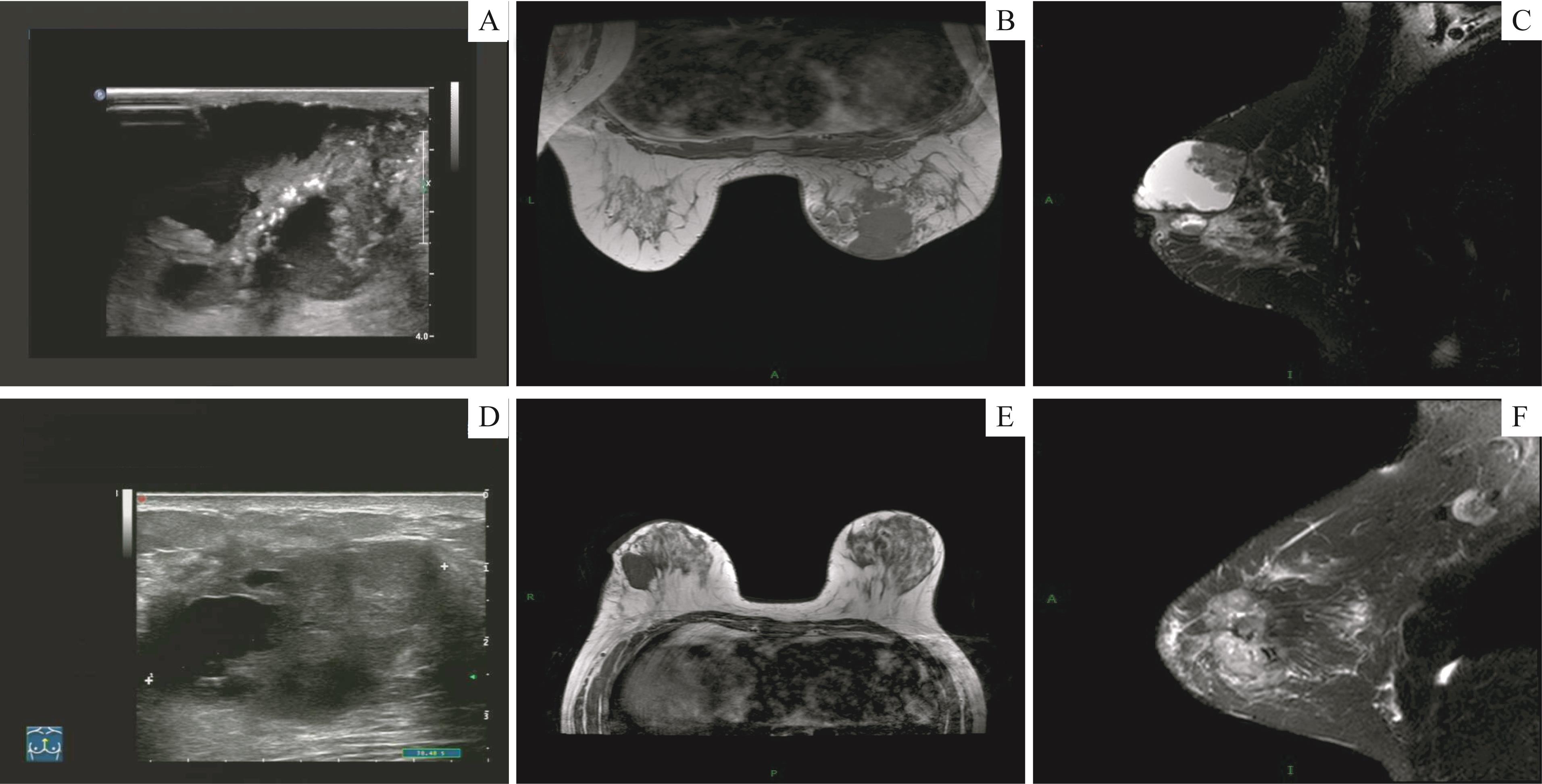
Fig 1 Ultrasound and MRI imaging features about two cases of squamous cell carcinoma
| Subtype | Shape, signal and echo | Margin | Calcification | BI-RADS category | Swollen axillary lymph node | ||||||||||
|---|---|---|---|---|---|---|---|---|---|---|---|---|---|---|---|
| CR | MRI | Ultrasound | CR | MRI | Ultrasound | CR | MRI | Ultrasound | CR | MRI | Ultrasound | CR | MRI | Ultrasound | |
| Squamous cell carcinoma | Irregularly high density | Cyst-like mass with thick wall, low T1WI and T2WI signals; multiregional non-mass-like enhancement | Irregular shape, hybrid echoic | Not clear | Not clear | Not clear | Multiple and tiny | None | Multiple and tiny | 5 | 5 | 5 | Both sides | Both sides | Right side |
| Squamous cell carcinoma | Irregularly high density | Module-like, low T1WI and T2WI signals; unevenly enhancement | Irregular shape, hypoechoic | Not clear | Not clear | Not clear | None | None | None | 4c | 5 | 5 | None | None | None |
| Squamous cell carcinoma | Irregularly high density | Module-like, low T1WI and T2WI signals; early enhancement | Irregular shape, hypoechoic | Not clear | Not clear | Not clear | None | None | None | 0 | 5 | 5 | None | None | None |
| Spindle cell carcinoma | Irregularly high density | Module-like, low T1WI and T2WI signals; unevenly enhancement | Irregular shape, hypoechoic | Not clear | Not clear | Not clear | Tiny | None | Multiple and tiny | 4c | 5 | 4a | None | None | None |
Tab 2 CR, MRI and ultrasound features of four MBC cases
| Subtype | Shape, signal and echo | Margin | Calcification | BI-RADS category | Swollen axillary lymph node | ||||||||||
|---|---|---|---|---|---|---|---|---|---|---|---|---|---|---|---|
| CR | MRI | Ultrasound | CR | MRI | Ultrasound | CR | MRI | Ultrasound | CR | MRI | Ultrasound | CR | MRI | Ultrasound | |
| Squamous cell carcinoma | Irregularly high density | Cyst-like mass with thick wall, low T1WI and T2WI signals; multiregional non-mass-like enhancement | Irregular shape, hybrid echoic | Not clear | Not clear | Not clear | Multiple and tiny | None | Multiple and tiny | 5 | 5 | 5 | Both sides | Both sides | Right side |
| Squamous cell carcinoma | Irregularly high density | Module-like, low T1WI and T2WI signals; unevenly enhancement | Irregular shape, hypoechoic | Not clear | Not clear | Not clear | None | None | None | 4c | 5 | 5 | None | None | None |
| Squamous cell carcinoma | Irregularly high density | Module-like, low T1WI and T2WI signals; early enhancement | Irregular shape, hypoechoic | Not clear | Not clear | Not clear | None | None | None | 0 | 5 | 5 | None | None | None |
| Spindle cell carcinoma | Irregularly high density | Module-like, low T1WI and T2WI signals; unevenly enhancement | Irregular shape, hypoechoic | Not clear | Not clear | Not clear | Tiny | None | Multiple and tiny | 4c | 5 | 4a | None | None | None |
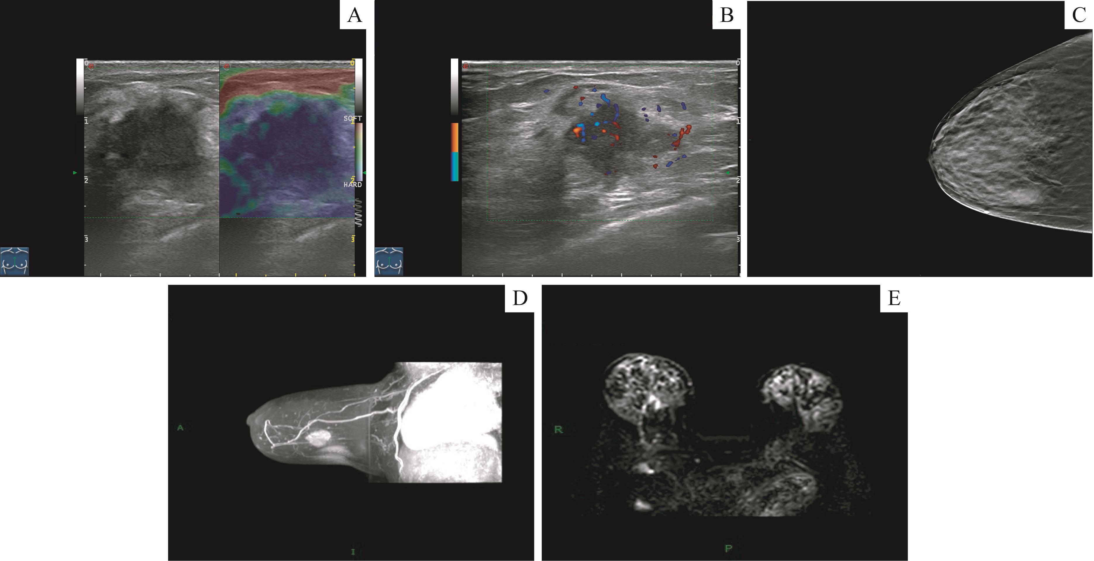
Fig 2 Ultrasound, CR and MRI imaging features of one squamous cell carcinoma case
| 1 | ZEIN DEI, HUGHES M, KUMAR S, et al. Metaplastic carcinoma of the breast is more aggressive than triple-negative breast cancer: a study from a single institution and review of literature[J]. Clin Breast Cancer, 2017, 17(5): 382-391. |
| 2 | LAKHANI S R. WHO classification of tumours of the breast[M]. 4th. Lyon: International Agency for Research on Cancer, 2012. |
| 3 | ONG C T, CAMPBELL B M, THOMAS S M, et al. Metaplastic breast cancer treatment and outcomes in 2500 patients: a retrospective analysis of a national oncology database[J]. Ann Surg Oncol, 2018, 25(8): 2249-2260. |
| 4 | 颜红菊, 许晓静, 谭艳娟, 等. 化生性乳腺癌的多模态超声特征[J]. 医学影像学杂志, 2018, 28(6): 945-949. |
| 5 | ADLER D D, CARSON P L, RUBIN J M, et al. Doppler ultrasound color flow imaging in the study of breast cancer: preliminary findings[J]. Ultrasound Med Biol, 1990, 16(6): 553-559. |
| 6 | 中国抗癌协会乳腺癌专业委员会. 中国抗癌协会乳腺癌诊治指南与规范(2019年版)[J]. 中国癌症杂志, 2019, 29(8): 609-680. |
| 7 | HUVOS A G, LUCAS J C JR, FOOTE F W JR. Metaplastic breast carcinoma. Rare form of mammary cancer[J]. N Y State J Med, 1973, 73(9): 1078-1082. |
| 8 | HARDY B M, CORTINA C S, JAVIDIPARSIJANI S, et al. Hypercalcemia in metaplastic squamous cell carcinoma of the breast[J]. Am J Case Rep, 2019, 20: 366-369. |
| 9 | GREENBERG D, MCINTYRE H, BIERRE T. Metaplastic breast cancer[J]. Australas Radiol, 2004, 48(2): 243-247. |
| 10 | LANGLANDS F, CORNFORD E, RAKHA E, et al. Imaging overview of metaplastic carcinomas of the breast: a large study of 71 cases[J]. Br J Radiol, 2016, 89(1064): 20140644. |
| 11 | CHOI B B, SHU K S. Metaplastic carcinoma of the breast: multimodality imaging and histopathologic assessment[J]. Acta Radiol Stock Swed, 2012, 53(1): 5-11. |
| 12 | 姜珊珊, 张乃千, 佟凌霞. 乳腺化生性癌的超声表现与临床病理特点[J]. 中国实验诊断学, 2020, 24(10): 1637-1639. |
| 13 | BAE S Y, LEE S K, KOO M Y, et al. The prognoses of metaplastic breast cancer patients compared to those of triple-negative breast cancer patients[J]. Breast Cancer Res Treat, 2011, 126(2): 471-478. |
| 14 | 贾懿, 詹维伟, 朱樱. 化生性乳腺癌的影像学诊断研究[J]. 医学影像学杂志, 2019, 29(5): 779-782. |
| 15 | RAKHA E A, TAN P H, VARGA Z, et al. Prognostic factors in metaplastic carcinoma of the breast: a multi-institutional study[J]. Br J Cancer, 2015, 112(2): 283-289. |
| 16 | LUINI A, AGUILAR M, GATTI G, et al. Metaplastic carcinoma of the breast, an unusual disease with worse prognosis: the experience of the European Institute of Oncology and review of the literature[J]. Breast Cancer Res Treat, 2007, 101(3): 349-353. |
| 17 | ZHANG Y Q, LV F, YANG Y L, et al. Clinicopathological features and prognosis of metaplastic breast carcinoma: experience of a major Chinese Cancer Center[J]. PLoS One, 2015, 10(6): e0131409. |
| 18 | TZANNINIS I G, KOTTEAS E A, NTANASIS-STATHOPOULOS I, et al. Management and outcomes in metaplastic breast cancer[J]. Clin Breast Cancer, 2016, 16(6): 437-443. |
| 19 | SALEMIS NS. Metaplastic carcinoma of the breast with mesenchymal differentiation (carcinosarcoma). A unique presentation of an aggressive malignancy and literature review[J]. Breast Dis, 2018, 37(3): 169-175. |
| 20 | HU Q, CHEN W X, ZHONG S L, et al. Current progress in the treatment of metaplastic breast carcinoma[J]. Asian Pac J Cancer Prev, 2013, 14(11): 6221-6225. |
| 21 | KRINGS G, CHEN Y Y. Genomic profiling of metaplastic breast carcinomas reveals genetic heterogeneity and relationship to ductal carcinoma[J]. Mod Pathol, 2018, 31(11): 1661-1674. |
| [1] | Yihuan WANG, Ruokun LI, Huanhuan CHONG, Fuhua YAN. Research progress of Gd-EOB-DTPA-enhanced magnetic resonance imaging in the evaluation of biological behavior of hepatocellular carcinoma [J]. JOURNAL OF SHANGHAI JIAOTONG UNIVERSITY (MEDICAL SCIENCE), 2022, 42(1): 130-134. |
| [2] | Yan-jie JI, Hao LUO, Hai-yan CAI, Xin-yu LIU, Shi-jia JIN, Shen-yue SU, Han-zhang XU, Hu LEI, Ying-li WU. Inhibition of CDDO-ME on ubiquitin-specific protease 2a activity and cell proliferation in triple negative breast cancer cells [J]. JOURNAL OF SHANGHAI JIAOTONG UNIVERSITY (MEDICAL SCIENCE), 2021, 41(8): 1025-1032. |
| [3] | YANG Tao, CHEN Jun, FANG Yi-ru. Advances in magnetic resonance imaging study of bipolar Ⅰdisorder [J]. JOURNAL OF SHANGHAI JIAOTONG UNIVERSITY (MEDICAL SCIENCE), 2020, 40(12): 1660-1664. |
| [4] | YUE Xiu-hui, KONG Wei-dan, REN Ji-liang, YUAN Ying#, TAO Xiao-feng#. Value of 3.0-T MR diffusion-weighted imaging combined with dynamic contrast-enhanced imaging in differentiating benign and malignant thyroid nodules [J]. JOURNAL OF SHANGHAI JIAOTONG UNIVERSITY (MEDICAL SCIENCE), 2020, 40(10): 1393-1397. |
| [5] | LI Xiao-min1, QU Yang1, WU Wen2, ZHAO Liang3, ZHANG Shao-ting3, HAO Yong-qiang2, DAI Ke-rong2, AI Song-tao1. Preliminary application of MR imaging-pathology co-localization by 3D printing box in pelvic tumor assessment [J]. JOURNAL OF SHANGHAI JIAOTONG UNIVERSITY (MEDICAL SCIENCE), 2020, 40(10): 1408-1413. |
| [6] | WANG Tao, ZHANG Chen-cheng, LI Dian-you, SUN Bo-min, FU Meng. Imaging law of postoperative electrode locations in deep brain stimulation for Parkinsons disease [J]. , 2020, 40(1): 64-. |
| [7] | JI Ying-ying, XUE Bin, HUANG Yue, ZHANG Jian-wei. Efficacy and safety of oral midazolam in combination with intranasal dexmedetomidine for paediatric magnetic resonance imaging sedation [J]. JOURNAL OF SHANGHAI JIAOTONG UNIVERSITY (MEDICAL SCIENCE), 2020, 40(08): 1098-1102. |
| [8] | RUAN Jing-jing, LU Qing, TANG Hui, ZHU Zhen-ya, FAN Yu, ZHAO Xin-xin, NIU Xiao-yin. Value of multi-parameter magnetic resonance imaging of cartilage in evaluating efficacy of adipose-derived mesenchymal progenitor cells on knee osteoarthritis [J]. , 2019, 39(12): 1409-. |
| [9] | LI Ruo-kun, QIANG Hu-ming, YAN Fu-hua, REN Xin-ping, WANG Tao, CHEN Wei-bo. T1ρ magnetic resonance imaging for liver fibrosis detection and staging [J]. , 2017, 37(11): 1470-. |
| [10] | XU Lian, SUN Xiao-guang, LIU Jian-jun, ZHOU Ming-ge, JIANG Zhou . Comparison of 99mTc-MIBI SPECT/CT and ultrasonography in diagnosis of hyperparathyroidism [J]. , 2017, 37(04): 496-. |
| [11] | WANG Cheng, ZHUANG Zhi-gang, SHAN Ming, XU Ming . Mechanisms of inhibiting the proliferation of triple negative breast cancer cell lines by silencing the CD147 gene [J]. , 2017, 37(03): 305-. |
| [12] | LU Jun, ZHANG Ji-chen, YOU Wen, YU Xue-mei, GU Ming-jun. Construction of predictive models for lower limb artery stenosis in patients with type 2 diabetes [J]. , 2016, 36(07): 1018-. |
| [13] | GU Wei, ZHOU Lei-ping, LIN Jing, et al. Value of indexes of uterine artery blood flow for prediction of preeclampsia [J]. , 2015, 35(2): 223-. |
| [14] | LIU Yi, XIA Jian-guo, LI Feng-hua, et al. Analysis of risk factors of bleeding complication of cytological biopsy of thyroid nodules under guidance of ultrasonography [J]. , 2015, 35(12): 1842-. |
| [15] | HU Feng, SHAO Chong-fei, HUANG Wei-qiang. Diagnostic value of ultrasonography and modified Alvarado score for elderly patients with acute appendicitis [J]. , 2014, 34(6): 939-. |
| Viewed | ||||||
|
Full text |
|
|||||
|
Abstract |
|
|||||