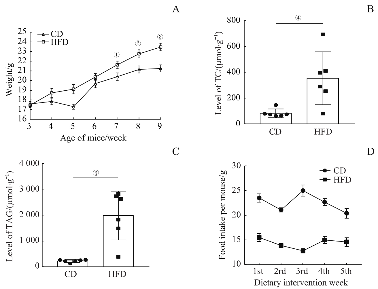
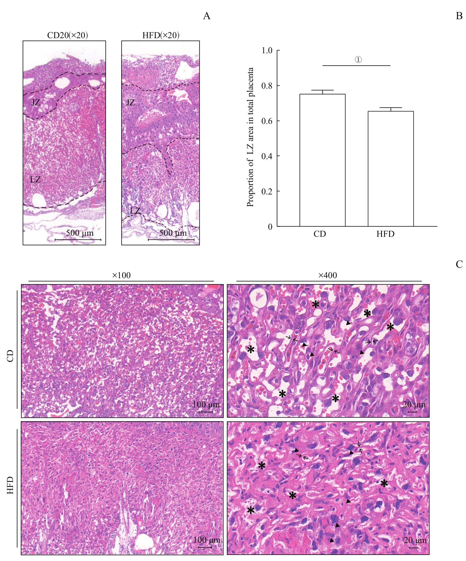
| 1 |
HOFFMAN D J, REYNOLDS R M, HARDY D B. Developmental origins of health and disease: current knowledge and potential mechanisms[J]. Nutr Rev, 2017, 75(12): 951-970.
|
| 2 |
ALFARADHI M Z, OZANNE S E. Developmental programming in response to maternal overnutrition[J]. Front Genet, 2011, 2: 27.
|
| 3 |
PANERA N, MANDATO C, CRUDELE A, et al. Genetics, epigenetics and transgenerational transmission of obesity in children[J]. Front Endocrinol (Lausanne), 2022, 13: 1006008.
|
| 4 |
CAO B G, LIU C X, ZHANG Q R, et al. Maternal high-fat diet leads to non-alcoholic fatty liver disease through upregulating hepatic SCD1 expression in neonate rats[J]. Front Nutr, 2020, 7: 581723.
|
| 5 |
GUDE N M, ROBERTS C T, KALIONIS B, et al. Growth and function of the normal human placenta[J]. Thromb Res, 2004, 114(5/6): 397-407.
|
| 6 |
MESTAN K, YU Y X, MATOBA N, et al. Placental inflammatory response is associated with poor neonatal growth: preterm birth cohort study[J]. Pediatrics, 2010, 125(4): e891-e898.
|
| 7 |
BURTON G J, FOWDEN A L, THORNBURG K L. Placental origins of chronic disease[J]. Physiol Rev, 2016, 96(4): 1509-1565.
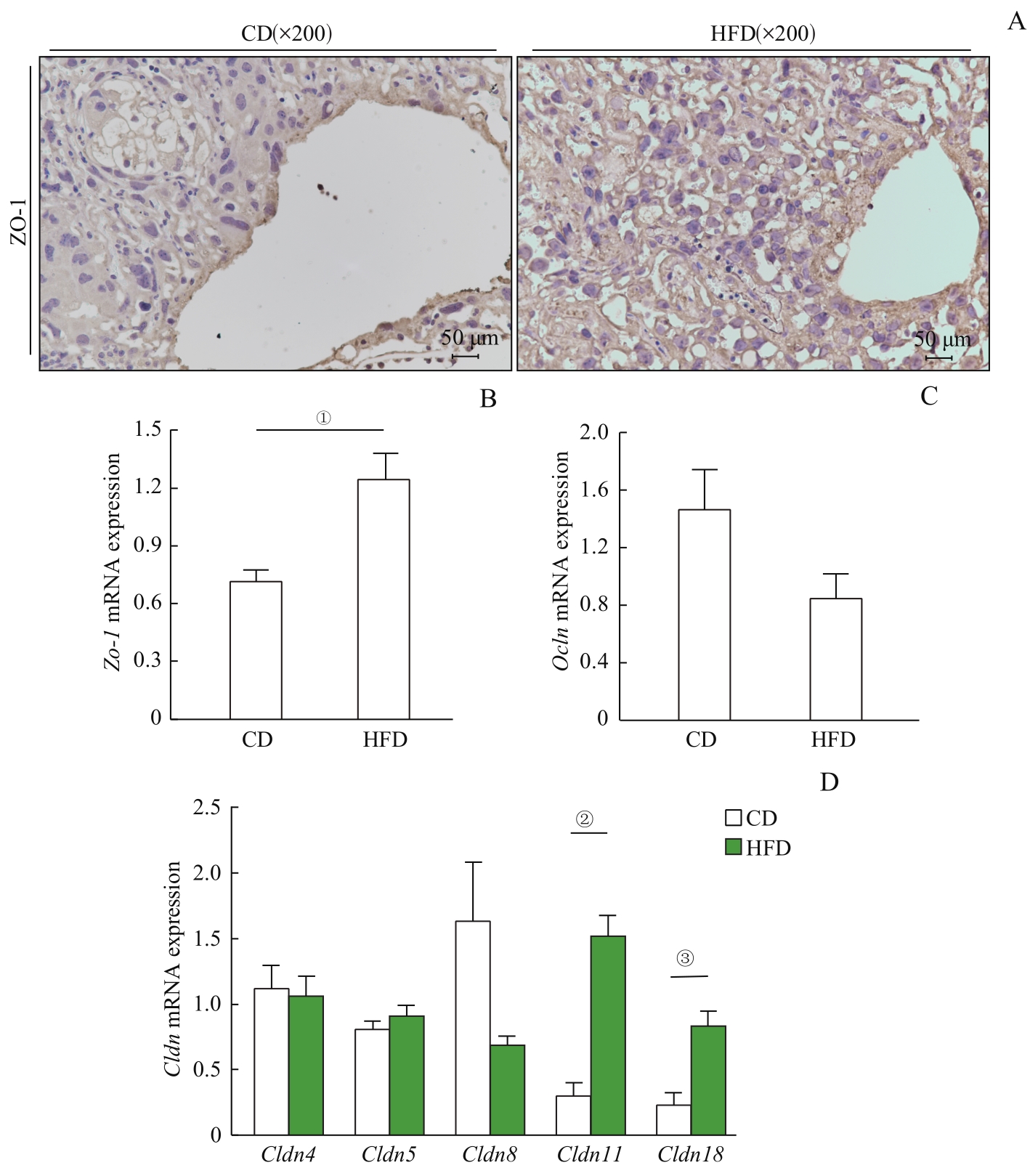
|
| 8 |
BARKER D J P. The fetal and infant origins of disease[J]. Eur J Clin Investig, 1995, 25(7): 457-463.
|
| 9 |
HUANG Y H, YE T T, LIU C X, et al. Maternal high-fat diet during pregnancy and lactation affects hepatic lipid metabolism in early life of offspring rat[J]. J Biosci, 2017, 42(2): 311-319.
|
| 10 |
ZHANG Q, YE L, XIN F, et al. Milk fat globule membrane supplementation during suckling ameliorates maternal high fat diet-induced hepatic steatosis in adult male offspring of mice[J]. J Nutr, 2021, 151(6): 1487-1496.
|
| 11 |
GODFREY K M, REYNOLDS R M, PRESCOTT S L, et al. Influence of maternal obesity on the long-term health of offspring[J]. Lancet Diabetes Endocrinol, 2017, 5(1): 53-64.
|
| 12 |
MALTEPE E, FISHER S J. Placenta: the forgotten organ[J]. Annu Rev Cell Dev Biol, 2015, 31: 523-552.
|
| 13 |
ROCHA V Z, LIBBY P. Obesity, inflammation, and atherosclerosis[J]. Nat Rev Cardiol, 2009, 6(6): 399-409.
|
| 14 |
CHALLIER J C, BASU S, BINTEIN T, et al. Obesity in pregnancy stimulates macrophage accumulation and inflammation in the placenta[J]. Placenta, 2008, 29(3): 274-281.
|
| 15 |
陈镇燕, 王琪, 黄光英. 鼠类胎盘结构、血液循环及其来源[J]. 解剖学杂志, 2010, 33(2): 256-259.
|
|
CHEN Z Y, WANG Q, HUANG G Y. Placenta structure, blood circulation and its source in rodents[J]. Chinese Journal of Anatomy, 2010, 33(2): 256-259.
|
| 16 |
郑婉珊, 胡晓倩, 王雁玲, 等. 胎盘屏障建立与维持的机制[J]. 生理学报, 2020, 72(1): 115-124.
|
|
ZHENG W S, HU X Q, WANG Y L, et al. Mechanism for establishment of the placental defensive barrier[J]. Acta Physiologica Sinica, 2020, 72(1): 115-124.
|
| 17 |
FURUKAWA S, TSUJI N, SUGIYAMA A. Morphology and physiology of rat placenta for toxicological evaluation[J]. J Toxicol Pathol, 2019, 32(1): 1-17.
|
| 18 |
WANG Y W, YU H R, TIAO M M, et al. Maternal obesity related to high fat diet induces placenta remodeling and gut microbiome shaping that are responsible for fetal liver lipid dysmetabolism[J]. Front Nutr, 2021, 8: 736944.
|
| 19 |
KRETSCHMER T, TURNWALD E M, JANOSCHEK R, et al. Maternal high fat diet-induced obesity affects trophoblast differentiation and placental function in mice[J]. Biol Reprod, 2020, 103(6): 1260-1274.
|
| 20 |
MARZIONI D, BANITA M, FELICI A, et al. Expression of ZO-1 and occludin in normal human placenta and in hydatidiform moles[J]. Mol Hum Reprod, 2001, 7(3): 279-285.
|
| 21 |
OTANI T, FURUSE M. Tight junction structure and function revisited[J]. Trends Cell Biol, 2020, 30(10): 805-817.
|
 ), ZHANG Qianren1, LU Xingyu1, DONG Yan1,2(